Diagnostic Imaging · Year 4 · Medical University of Sofia

01

Atom theory. Radiation

Free notes for topic 01 of the Diagnostic Imaging syllabus, open without an account. Written by a senior student against the syllabus question and checked line by line by a second student before publishing. How content is made

Updated

Introduction to atomic structure and radiation

note

This topic covers the structure of the atom, what radiation is and how it is divided into ionising and non-ionising forms, radioactivity and the particles it emits, the units used to measure radiation dose, the natural and human-made sources of exposure, and the biological effects that radiation can produce. Every later topic on X-ray imaging depends on these ideas, so they are set out first in full.

1. Structure of the atom

note

Matter is made of atoms. The structure of the atom underlies most of the techniques used in radiology, because the way radiation interacts with matter depends on the atoms it meets.

The nucleus

Nearly all the mass of an atom is concentrated in the central nucleus. Most of the volume of an atom is empty space. The nucleus is made of nucleons, which are protons and neutrons. The total number of nucleons is the mass number, symbol A. The number of protons is the atomic number, symbol Z. Protons carry a positive charge and neutrons carry no charge. Each proton and neutron has a relative mass of one.

Each proton is made of two up quarks and one down quark, and each neutron of two down quarks and one up quark, together with gluons that bind them.

The atomic number defines the element. An atom with six protons is carbon, whatever the number of neutrons it has. Atoms of the same element with different numbers of neutrons are called isotopes. Carbon, for example, may have six or eight neutrons and is still carbon, with six protons in each case.

Electron shells

Electrons orbit the nucleus in shells named K, L, M, N and so on from the centre outwards. They are much lighter than nucleons and carry a negative charge. In a neutral atom the number of electrons equals the number of protons.

The maximum number of electrons in a shell is 2n², where n is the number of the shell. The table gives the first three shells.

ShellNumber (n)Maximum electrons
K12
L28
M318

The innermost shell is filled first because it has the lowest energy, and it must be full before the next shell begins to fill. The outermost shell is the valence shell, and it may be only partly full. Electrons in the inner shells are bound most strongly, and this binding energy matters for X-ray production (topic DI.02).

2. Radiation and ionisation

note

What radiation is

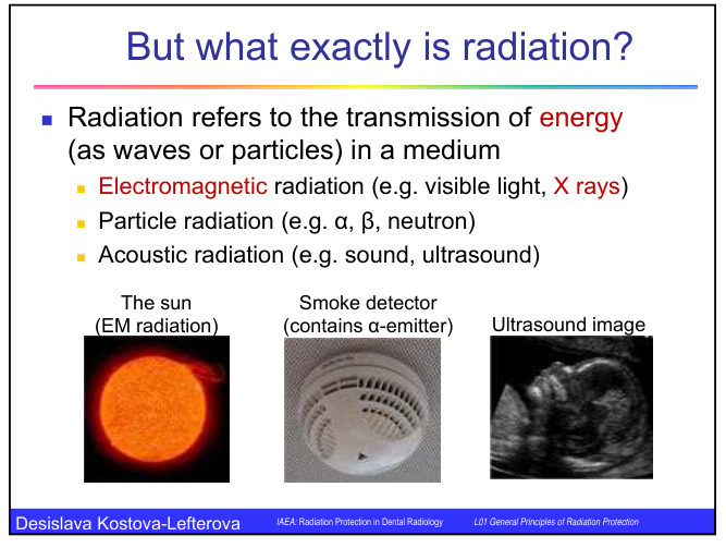
Radiation is the transmission of energy through space or a medium as waves or particles. It is grouped in three ways:

  • Electromagnetic radiation, for example visible light and X-rays.
  • Particle radiation, for example alpha particles, beta particles and neutrons.
  • Acoustic radiation, for example sound and ultrasound.

Electromagnetic radiation is shown with examples in the figure below: the sun emits it, a smoke detector contains an alpha emitter, and an ultrasound image is formed with acoustic radiation.

The sun as an electromagnetic radiation source, a smoke detector containing an alpha emitter, and an ultrasound image, shown as examples of radiation in the three groups

Ionising and non-ionising radiation

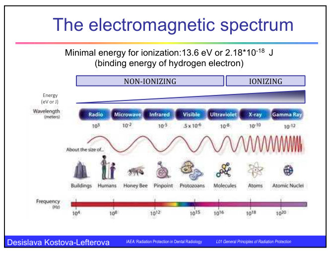
Radiation is called ionising when it carries enough energy to remove electrons from atoms in the material it passes through, forming positive ions. The energy needed to ionise an atom is measured in electronvolts. The minimum energy for ionisation is 13.6 eV, which is the binding energy of the electron in a hydrogen atom.

Most of the electromagnetic spectrum is non-ionising. This includes most ultraviolet wavelengths, visible light, infrared, microwaves and radio waves. X-rays and gamma rays are ionising.

FeatureIonising radiationNon-ionising radiation
EnergyEnough to remove electrons from atomsNot enough to ionise atoms
ExamplesX-rays, gamma rays, alpha and beta particlesVisible light, infrared, microwaves, radio waves, most ultraviolet
Medical useRadiography, computed tomography, nuclear medicine, radiotherapyUltrasonography (sound), magnetic resonance (radio waves and magnetic field)

Ultrasonography and magnetic resonance should be preferred wherever they answer the clinical question, because they involve no ionising radiation.

Direct and indirect ionising radiation

Ionising radiation is divided into two groups by the way it causes ionisation.

  • Directly ionising radiation consists of electrically charged particles, such as electrons, positrons, alpha particles, protons and heavy nuclei. These particles ionise and excite atoms directly, through collisions with them. Their ability to ionise depends on their mass, charge and energy.
  • Indirectly ionising radiation consists of electrically neutral particles or photons, such as neutrons and electromagnetic radiation (X-rays and gamma rays). Their energy is first transferred to charged particles, which then ionise the atoms.

X-rays and gamma rays are the most common forms of indirect ionising radiation.

Ionisation and excitation

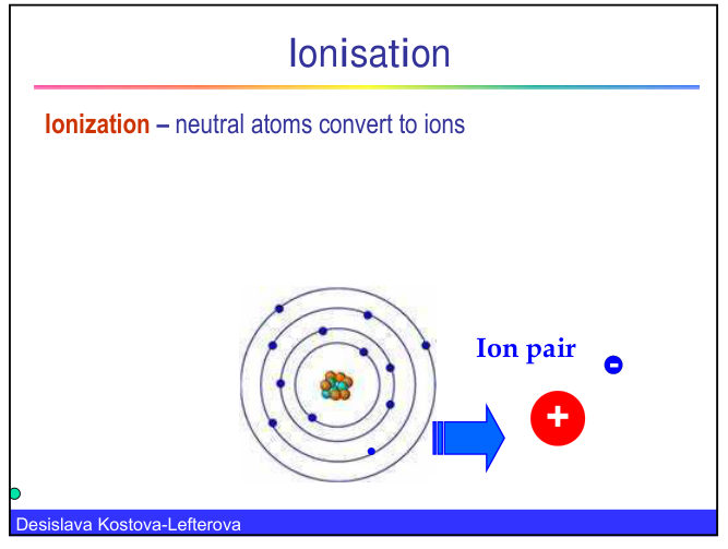
An atom that loses an electron becomes an ion. A neutral atom converted to an ion gives an ion pair: the negative electron and the positive atom that remains.

Ionisation, showing a neutral atom with an electron removed to form an ion pair consisting of a positive ion and a free electron

Ionising radiation dissipates its energy in tissue in three ways:

  1. Ionisation, the removal of electrons from atoms.
  2. Excitation, where electrons are raised to a higher energy level without leaving the atom.
  3. Thermal heating.

The energy is deposited randomly and very quickly, in less than 10⁻¹⁰ seconds, at the atomic and molecular level. Only a small fraction of the deposited energy causes chemical change. Most of it becomes heat, which has little biological significance. The energy that does cause chemical change can damage important molecules such as DNA. The resulting biological effects may appear only after a latent period, which can range from minutes to weeks, years or a lifetime.

Radiation in medicine

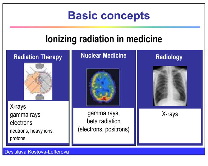
Ionising radiation is used in several branches of medicine.

  • Radiology uses X-rays.
  • Nuclear medicine uses gamma rays and beta radiation (electrons and positrons).
  • Radiation therapy uses X-rays, gamma rays and electrons, and also neutrons, heavy ions and protons.
Ionising radiation in medicine, with three panels: radiation therapy using X-rays, gamma rays, electrons, neutrons, heavy ions and protons; nuclear medicine using gamma rays and beta radiation; and radiology using X-rays

3. Electromagnetic spectrum and X-ray energy

note

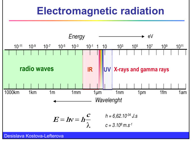
All electromagnetic radiation travels at the speed of light, c, which is about 3 × 10⁸ metres per second. Its energy is proportional to its frequency and inversely proportional to its wavelength. The relationship is E = hν = hc/λ, where h is Planck's constant, 6.62 × 10⁻³⁴ joule seconds.

The spectrum runs from radio waves, with wavelengths of kilometres, through microwaves, infrared, visible light and ultraviolet, to X-rays and gamma rays, with wavelengths below a nanometre. Energy rises as wavelength falls.

The spectrum is divided into non-ionising and ionising parts, with the boundary at the energy needed to remove an electron from hydrogen.

Electromagnetic spectrum diagram with a bar marking the non-ionising region from radio waves to ultraviolet and the ionising region of X-rays and gamma rays, with frequency and wavelength scales and the image of an electron energy of 13.6 eV for ionisation
Electromagnetic radiation chart showing energy in electronvolts, wavelength from 1000 km to 1 am, and the regions of radio waves, infrared, ultraviolet and X-rays and gamma rays

X-rays cover these ranges:

QuantityFull rangeDiagnostic radiology
Wavelength10 nm to 3 pm124 pm to 6 pm
Energy124 eV to 420 keV10 keV to 150 keV

Energy is measured in electronvolts (eV), where one electronvolt is the kinetic energy an electron gains when it moves through a potential difference of one volt, equal to 1.6 × 10⁻¹⁹ joule. One kiloelectronvolt (keV) is 1000 eV and one megaelectronvolt (MeV) is 1 000 000 eV. The SI unit of energy is the joule.

4. Radioactivity and particles

note

Discovery

Henri Becquerel discovered radioactivity in 1896. Radioactivity is the spontaneous change in the mass, electrical charge or energy of atomic nuclei, accompanied by the emission of alpha, beta or gamma rays. It is also called radioactive decay. Marie and Pierre Curie shared the Nobel Prize in Physics in 1903 for their work on radioactivity.

Types of emission

  • Alpha particles are nuclei of helium-4, which are heavy particles. Alpha emission occurs mainly from heavy elements such as polonium, radium, radon, thorium, uranium and plutonium, but also from some lighter natural emitters such as samarium-147 and gadolinium-152.
  • Beta particles are electrons and positrons. They are about 8000 times lighter than alpha particles. Beta emission occurs from light and heavy nuclides, for example tritium (3H), sodium-22 (22Na), potassium-40 (40K), iodine-131 (131I), iridium-192 (192Ir) and uranium-237 (237U).
  • Gamma rays are electromagnetic radiation with no mass or charge.

Natural and artificial radionuclides

About 60 natural radionuclides are known, mostly isotopes of heavy elements after bismuth-209. Light natural radionuclides include potassium-40, rubidium-87, samarium-147 and gadolinium-152. Artificial radionuclides are made by nuclear reactions. Irène and Frédéric Joliot-Curie produced the first artificial radionuclides in 1934, and they shared the Nobel Prize in Chemistry in 1935.

Behaviour of particles in matter

Light charged particles, such as beta particles, produce far more ion pairs per centimetre than heavy charged particles, such as alpha particles, and they travel much further. Alpha particles produce dense ionisation along a very short track.

ParticleIon pairs per cm of airRange in airRange in muscle
Alpha particle100 to 4001 to 10 cm50 to 130 micrometres
Beta particle, 200 keV20 000 to 60 00031 cm1 mm
Beta particle, 1 MeV20 000 to 60 0003 m5 mm

External and internal exposure

  • External exposure to alpha particles is not dangerous, because they cannot penetrate the outer dead layer of the skin. Beta particles are stopped by a plastic screen of 1 to 2 cm.
  • Internal exposure to alpha particles, for example after they are inhaled or swallowed, causes much greater injury than the same exposure to beta particles, because all their energy is deposited in the surrounding tissue.

5. Units of radiation dose

note

Absorbed dose

The absorbed dose, D, is the energy absorbed per unit mass at a given point. Its unit is the gray, Gy, which equals one joule per kilogram. It is named after Harold Gray (1905 to 1965). The older unit is the rad, where 1 rad equals 0.01 Gy and 1 Gy equals 100 rad.

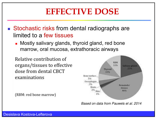
Absorbed dose is usually considered for an organ or tissue. For most medical exposures, which are not whole-body exposures, the dose to an organ depends on the fraction of that organ that is exposed. In dental radiography the highest absorbed doses to whole organs or tissues are found in the thyroid gland, the salivary glands, the oral mucosa and the extrathoracic airways.

Some typical absorbed doses:

  • one radiotherapy treatment, 1.5 to 8 Gy;
  • entrance skin dose for a chest X-ray, less than 1 mGy;
  • a lethal acute whole-body dose is about 5 Gy.

Dose rate

The dose rate is the dose absorbed per unit time, in Gy/s, Gy/h or mGy/h. The biological effect depends on the absorbed dose and on the time over which it is absorbed. Examples:

  • natural background in Sofia, about 0.1 microgray per hour;
  • at 1 m from the patient during fluoroscopy, about 1 mGy/h;
  • on the patient's skin during fluoroscopy, about 1 Gy/h.

Radiation weighting factor

The biological effect also depends on the type and energy of the radiation. The radiation weighting factor, wR, is dimensionless and allows for this.

RadiationwR
X-rays, gamma rays, beta particles, accelerated electrons1
Protons2
Neutronsa function of energy, between 2 and 20
Alpha particles20

Equivalent dose

The equivalent dose, HT, is the absorbed dose multiplied by the radiation weighting factor: HT = wR × DT. Its unit is the sievert, Sv. It is used for tissues and organs. A higher wR means a more damaging radiation.

Two examples show why. Gamma radiation with an absorbed dose of 1 Gy and electrons with an absorbed dose of 1 Gy have the same biological effect, so both give 1 Sv. Alpha particles with an absorbed dose of 1 Gy have a biological effect 20 times higher, so they give 20 Sv.

Effective dose

The effective dose, E, is the sum of the equivalent doses to all tissues, each multiplied by its tissue weighting factor, wT: E = Σ wT × HT. Its unit is also the sievert. It allows the stochastic risk from low doses, and from different types of exposure, to be compared, whether the exposure is internal or external and whether it is uniform or uneven.

TissuewT
Red bone marrow, colon, lung, stomach, breast, remainder tissues (together)0.12 each
Gonads0.08
Bladder, oesophagus, liver, thyroid0.04 each
Bone surface, brain, salivary glands, skin0.01 each

The sum of all tissue weighting factors is 1.

Effective dose slide showing the formula E = w1 H1 + w2 H2 + ..., the tissue weighting factors for each organ, and the statement that the sum of the weighting factors is 1

Some examples of effective dose:

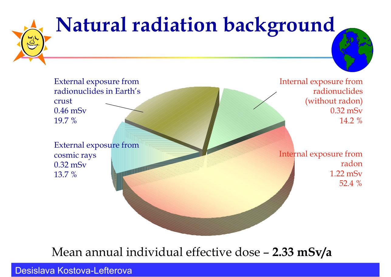
  • the mean natural background dose is 2.33 mSv per year (see section 6);
  • the mean effective dose of medical staff is about 1.2 mSv per year, well below the occupational dose limit of 20 mSv per year;
  • an effective dose of about 7 mSv is received from a barium enema.

The energy involved in a lethal acute dose is small. A dose of 5 Gy is 5 J/kg, so a 70 kg adult receives about 350 J, which is roughly the heat from drinking 4 ml of hot coffee.

6. Natural radiation background

note

People are exposed to radiation every day from natural sources. These fall into four groups: cosmic rays, natural radionuclides in the Earth's crust, radon, and natural radionuclides in food, water and the body.

Pie chart of the mean annual individual effective dose from natural background, 2.33 mSv per year: internal exposure from radon 1.22 mSv (52.4 per cent), external exposure from radionuclides in the Earth's crust 0.46 mSv (19.7 per cent), external exposure from cosmic rays 0.32 mSv (13.7 per cent), internal exposure from radionuclides other than radon 0.32 mSv (14.2 per cent)
SourceMean annual effective doseShare of total
Radon, internal (inhaled)1.22 mSv52.4 per cent
Radionuclides in the Earth's crust, external0.46 mSv19.7 per cent
Cosmic rays, external0.32 mSv13.7 per cent
Radionuclides other than radon, internal0.32 mSv14.2 per cent
Total2.33 mSv100 per cent

Cosmic rays

Cosmic rays come from the galaxy and the sun as primary radiation: protons of different energies, alpha particles and others. When they reach the atmosphere they produce secondary radiation, including neutrons, protons and electrons, and cosmogenic radionuclides such as tritium, beryllium-7, carbon-14 and sodium-22. The mean external dose from cosmic rays is 0.32 mSv per year.

The dose depends on latitude, being higher near the North and South Poles and lower at the equator. It also depends on altitude, rising at higher altitudes. Sofia lies at about 560 m, and the lecture contrasts this with sea level and with much higher altitudes such as 12 000 m and the summit of Everest at 8848 m. The lecture also notes that a flight from Sofia to Brussels gives about 10 µSv from cosmic rays.

Radionuclides in the Earth's crust

Natural radionuclides in the Earth's crust, such as potassium-40, rubidium-87 and the decay chains of uranium-238 and thorium-232, give both external and internal exposure.

  • External exposure from gamma radiation is about 0.01 to 0.2 µSv/h outdoors in most of the world. It is much higher in some places, up to 90 µSv/h in parts of Kerala (India) and Guarapari (Brazil). In Bulgaria the outdoor dose rate is about 0.06 to 0.13 µSv/h in normal areas, rising to 0.25 to 0.30 µSv/h over magmatic rocks. Indoors the values are 0.057 to 0.093 µSv/h. The mean external dose from the crust is 0.46 mSv per year.
  • Internal exposure is by ingestion and inhalation. Potassium-40 gives 0.17 mSv per year. Uranium and thorium decay products, such as radium-226, radium-228, lead-210 and polonium-210, give 0.15 mSv per year. Together the internal non-radon exposure averages 0.32 mSv per year.

Radon

Radon is a gas produced by the decay chains of uranium and thorium, which are present in soil, building materials and water. Radon-222 comes from the uranium-238 series, through radium-226. Radon-220 comes from the thorium-232 series. Radon and its decay products are inhaled and are the largest single part of natural background dose, averaging 1.22 mSv per year. Radon also increases the risk of lung cancer, and that risk is higher in smokers than in non-smokers.

Scale of common doses

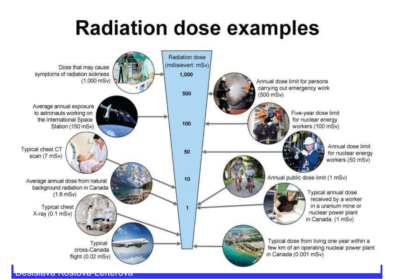
The lecture presents the following scale of doses. The values are in millisieverts.

Dose (mSv)Example
1000Dose that may cause symptoms of radiation sickness
500Annual dose limit for persons carrying out emergency work
150Average annual exposure of astronauts working on the International Space Station
100Five-year dose limit for nuclear energy workers
50Annual dose limit for nuclear energy workers
7Typical chest CT scan
1.8Average annual dose from natural background in Canada
1Annual public dose limit
1Typical annual dose for a worker in a uranium mine or nuclear power plant in Canada
0.1Typical chest X-ray
0.02Typical cross-Canada flight
0.001Typical dose from living one year within a few kilometres of an operating nuclear power plant in Canada
Radiation dose examples chart in millisieverts, with photographs of a medical CT scan, astronauts, nuclear workers, a chest X-ray and an aeroplane beside the matching dose levels

A popular way of illustrating radiation dose is the banana equivalent dose. Eating one banana gives a dose of about 1 µSv, from its naturally occurring potassium-40. Acute death from radiation would need a dose of about 10 Sv, which would mean eating about ten million bananas at once. The lecture adds that the dose makes the poison: a substance is harmful only at a dose high enough to cause harm, which is the principle that Paracelsus expressed in the sixteenth century.

7. Human-made sources of exposure

note

Nuclear weapons tests

Nuclear weapons tests were made in the atmosphere and underground from 1945 to 1998, with over 2000 tests in total. The Partial Test Ban Treaty limited signatories to underground testing, but atmospheric tests continued in some countries after that.

  • Local fallout falls within about 100 km of the explosion.
  • Global fallout spreads through the stratosphere, at 10 to 50 km altitude, and the troposphere, at 0 to 17 km.
  • Fallout radionuclides include strontium-90 and caesium-137, with half-lives of 29 and 30 years, and carbon-14, with a half-life of 5700 years, which give internal exposure. Caesium-137 also gives external exposure.
  • Short-lived nuclides such as iodine-131, xenon-133 and tritium, with half-lives of 8 days, 5 days and 12 years, are released by these tests. Underground tests give less exposure to the wider population but exposure of the local population must still be considered.

The lecture reports 17 000 cancer deaths in the United States in the second half of the twentieth century from radioactive fallout, and about 80 000 cancer cases in the United States from fallout of tests by the United States, the USSR and the United Kingdom.

Nuclear fuel cycle and nuclear energy

At every stage of the nuclear fuel cycle, radioactive substances can enter the environment. Nuclear energy is used worldwide. The number of operating reactors and reactors under construction are listed below, from the IAEA Power Reactor Information System.

YearOperating reactorsReactors under construction
201243661
201343571
201443772
201544068
201645060
201744857
201844956

Reactors operate in 30 countries.

Chernobyl accident, 1986

The Chernobyl accident released mainly iodine-131 and caesium-137. The main route to people was through precipitation to plants, then to animals, and then to humans through milk and meat. For the Bulgarian population the mean individual effective dose was 0.8 mSv in the first year and 0.95 mSv over a lifetime, with individual values from 0.6 to 1.7 mSv. The lecture expects about 400 cancer deaths in Bulgaria because of Chernobyl up to 2035. Each year about 17 000 people in Bulgaria die of cancer.

Occupational exposure

About 9190 employees in Bulgaria are under a national framework for control of occupational exposure. They work in nuclear power plants (4285 persons), medicine (3935), other industry (630), science and education (100) and other sectors (240). About 98 per cent receive less than 1 mSv per year. The trend is steadily downward.

Medical and dental exposure

The mean annual individual effective dose from human activity is given as 1.06 mSv. It combines diagnostic radiology, nuclear medicine, occupational exposure, nuclear energy and other sources such as heating and global fallout.

Medical exposure is a major human-made source, and dental radiography is one of its most common parts. Dental X-rays are the most frequently performed X-ray procedures in the world, with about 520 million a year. That is about 90 per 1000 inhabitants and 15 per cent of all diagnostic examinations. The lecture also states that about 90 per cent of these are performed in the countries of healthcare level I.

In Bulgaria there were 488 588 dental X-rays per million inhabitants in 2012, which is 66 per 1000 inhabitants and about 12 per cent of all radiological examinations. Intra-oral radiographs make up 89 to 90 per cent of all dental X-rays. The rest are panoramic (OPG) and cephalometric radiographs, with cone beam CT (CBCT) increasingly added.

Dental X-rays are used for:

  • diagnosis of diseases and injuries of the teeth and jaws, including the presence and degree of caries, changes in the bone and periodontal tissues, osteomyelitis, granuloma, fracture of the jaw and of tooth roots, bone tumours and cysts, and other anomalies of the teeth and jaw bones;
  • documentation of the structure of the teeth and gums, including the number, size and arrangement of teeth and the presence of missing or broken teeth.

Children are imaged often. Worldwide, 10 to 35 per cent of examinations are of children (UNSCEAR), and the figure in Bulgaria is 12 per cent. In Bulgaria the examinations by age group are 11.5 per cent for ages 0 to 15 years, 44.7 per cent for ages 16 to 40 years, and 43.7 per cent for those over 40 years.

Newer methods increase the dose. CBCT (3D imaging) gives a higher patient dose than conventional and panoramic radiography, and its availability and frequency of use are rising. High-dose procedures such as CT, interventional procedures, SPECT-CT and PET-CT are also increasing. In the United States, the number of CT procedures rose from about 3 million in 1980 to 63 million in 2005, and children made up 6 to 11 per cent of them. The lecture states that a third of them were unjustified. Dose and risk from CT are discussed in topic DI.09.


8. Biological effects of radiation

note

Direct and indirect effects

Radiation damages cells in two ways.

  • Direct effectan incoming photon strikes a DNA molecule and damages it, either by damaging a base pair or by breaking a supporting strand. Damage by the direct effect is relatively rare.
  • Indirect effectthe photon interacts with a water molecule. The ionising radiation breaks chemical bonds in the water, and the water molecule fragments to form highly reactive chemicals such as hydrogen peroxide and other free radicals. These attack the DNA molecule and damage its chemical bonds.

The indirect effect, acting through water, is the more common route to DNA damage, because water makes up most of the cell.

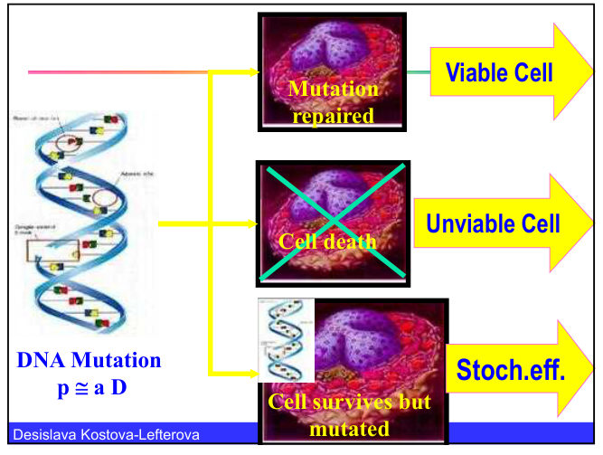
Diagram of DNA mutation after irradiation, showing three outcomes: a viable cell after the mutation is repaired, cell death, and a cell that survives but is mutated, giving a stochastic effect

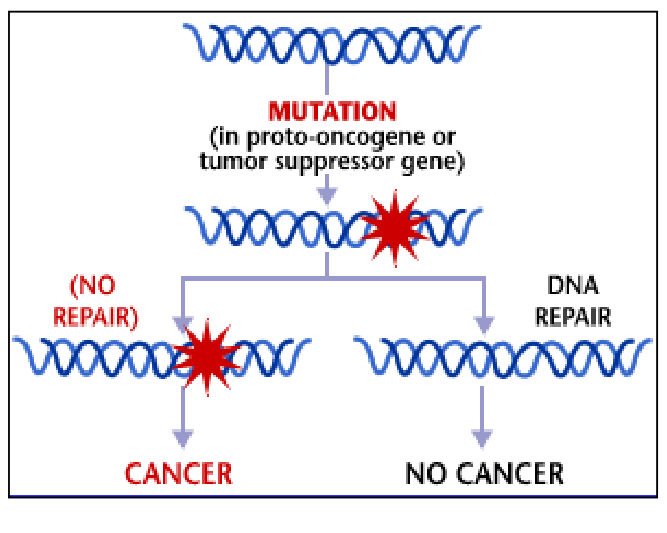
When radiation hits a cell nucleus, one of several outcomes follows. If the radiation causes no change, the cell is unaffected. If it causes a DNA mutation, the mutation may be repaired, leaving a viable cell. If the mutation is not repaired, the cell may die, or it may survive but carry the mutation, which is a stochastic effect. A mutation in a proto-oncogene or a tumour suppressor gene leads to cancer if it is not repaired, and to no cancer if DNA repair takes place.

Diagram showing a mutation in a proto-oncogene or tumour suppressor gene, followed by either no repair, which leads to cancer, or DNA repair, which leads to no cancer

The biological effect of radiation depends on the type of ionising radiation, the organs and tissues exposed, the energy transferred to them, and the duration of exposure.

Deterministic effects

A deterministic effect is one in which the severity depends on the radiation dose. Deterministic effects have a threshold dose: they appear only at high dose levels, and their severity increases with dose. They are predictable and preventable, which is why radiation protection aims to prevent them completely.

OrganEffectAcute exposure (Sv)Chronic exposure (Sv per year)
Eye lensCataract0.5more than 0.15
SkinErythema, dry desquamation3 to 5not stated
SkinNecrosis20not stated
TestesPermanent infertility3.5 to 6.02.0
OvaryInfertility2.5 to 6.0more than 0.2
Bone marrowHaematopoietic suppression0.5more than 0.4

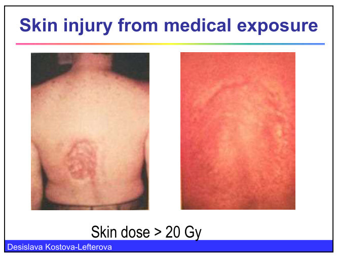
Skin injury from medical exposure is shown below. The skin dose in these cases was above 20 Gy.

Two photographs of skin injury from medical exposure on the back, the first showing a circular area of skin damage and the second a red area of skin, with the caption that the skin dose exceeded 20 Gy

Interventional specialists have developed radiation-induced cataracts from the high dose rates of scattered radiation from X-ray equipment above the couch. Deterministic effects can also cause burns, organ failure and death at high doses.

Stochastic effects

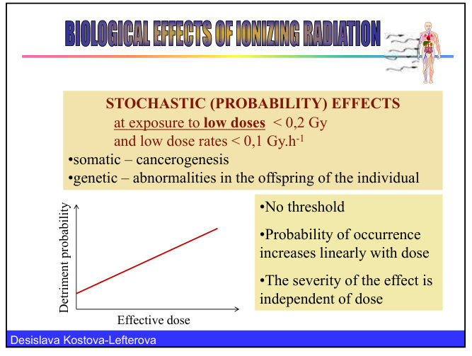
A stochastic effect is one in which the probability of occurrence increases with dose, but the severity does not depend on the dose. Stochastic effects have no threshold, and they appear at low doses, typically below 0.2 Gy at low dose rates below 0.1 Gy per hour.

  • Somatic effectscancer, which is called carcinogenesis.
  • Genetic effectsabnormalities in the offspring of the exposed individual. Radiation has not been identified as a cause of genetic effects in humans, although it does cause them in plants and animals.

Stochastic effects appear years after the exposure, which makes them hard to attribute. The evidence comes from large groups of people who received higher doses and were followed over time. Examples include the survivors of Hiroshima and Nagasaki, miners of uranium, patients treated with X-rays for non-malignant inflammatory conditions, patients treated with iodine-131 for non-malignant thyroid disease, and the populations affected by Chernobyl and Fukushima.

The dose-effect relation is extrapolated to low doses using the linear non-threshold model (LNT). It is the model used for radiation protection, but it should not be used to assess the health effects on an individual.

Graph of detriment probability against effective dose, a straight line with no threshold, showing that the probability rises linearly with dose and that the severity is independent of dose

The nominal probability coefficient for cancer risk is 5 per cent per sievert for adult workers, which is 5 fatal cancers per 100 000 people exposed to 1 mSv. For comparison, about 320 new cases of cancer per 100 000 people occur each year in Bulgaria.

Children are more sensitive than adults. The lecture gives a cancer risk 3 to 10 times higher for ages under 10, 2 times higher for ages 10 to 20 and 1.5 times higher for ages 20 to 30. Girls are about twice as sensitive as boys, and the lecture also gives a figure of up to 10 times the risk for adults at the youngest ages.

Stochastic and deterministic effects compared

FeatureDeterministic effectStochastic effect
Dependence on doseSeverity increases with doseProbability increases with dose, severity does not
ThresholdYes, high doseNone
Dose rangeHigh dosesLow doses
ExamplesCataract, skin burns, infertility, bone marrow suppressionCancer, genetic effects in offspring
PreventionPreventable by keeping doses below thresholdRisk reduced by keeping doses as low as reasonably achievable

All 57 Diagnostic Imaging topics · exam study support, not clinical guidance. SuperMed is not affiliated with the Medical University of Sofia.

Start free. No card needed.

Every account starts free, with Urology open in full and free topics in Cytology, Medical Biology, Anatomy, Biophysics, Medical Genetics, Pharmacology, Preclinics of Prosthetic Dental Medicine, Preclinics of Pediatric Dental Medicine, Dental Prophylaxis, Diagnostic Imaging, Dental Allergology, Dental Implantology, Physiotherapy, Cytology & Histology, Anatomy I and Medical Biology. Super opens the rest. Super is €14.99 a month.

Create a free account