Dental Implantology · Year 5 · Medical University of Sofia
01
Introduction in dental implantology. Brief historical overview. Classification of dental implants and terminology
Free notes for topic 01 of the Dental Implantology syllabus, open without an account. Written by a senior student against the syllabus question and checked line by line by a second student before publishing. How content is made
Updated
1. Introduction to dental implantology
note
Dental implantology focuses on replacing missing teeth with biocompatible substitutes that integrate with the bone. The substitute is typically made of titanium or zirconia. Implants restore function and aesthetics, and they act as a stable base for crowns, bridges or dentures. They also help preserve the bone structure of the jaw and improve the patient's quality of life.
Advantages of implants
note
Implants offer several advantages over other forms of tooth replacement.
Bone fuses with the implant, which gives long-term stability.
Functional restoration
Implants restore chewing (mastication) efficiency and speech, and they perform better than removable dentures.
2. Brief historical overview
note
Early developments
Early civilisations, including the Egyptians and the Mayans, experimented with tooth replacements made of shell or metal. Shell and ivory replacements have been found in Egyptian and Mayan skulls. These early replacements lacked long-term function or integration with bone.
During the 18th and 19th centuries, early attempts used gold and other alloys. These often failed because of infection and poor biocompatibility.
In the 1930s, subperiosteal frames were introduced. They sat on top of the bone, under the gum tissue. They provided some stability, but they often caused complications because they did not osseointegrate.
The osseointegration breakthrough
note
In 1952, a Swedish orthopaedic surgeon discovered osseointegration while studying bone healing in rabbits. Titanium chambers fused with the bone, and this led him to develop the first modern dental implants.
The titanium implant from this work was placed in 1965. It was revolutionary and laid the foundation for contemporary implantology. Osseointegration describes the direct attachment of bone to the implant without soft tissue forming between them, which gives long-term stability. The first titanium dental implants were placed successfully in the 1960s, and they showed long-term stability.
Modern advances
note
In the 1980s and 1990s, dental implants became widely accepted. Companies such as Nobelpharma (later Nobel Biocare) commercialised this research, and this era saw the development of proprietary implant systems. Improved surgical techniques and materials supported the wider adoption of implants during the 1980s.
The introduction of tapered screw implants improved primary stability, especially in softer bone, and reduced surgical complexity. The main reason is the wedge effect. A tapered implant has a conical or slightly tapered shape, so it compresses the surrounding bone as it is inserted. This creates mechanical engagement with the bone and increases the immediate primary stability of the implant. The effect is particularly important in areas of low bone density.
In the late 20th and early 21st centuries, 3D imaging, CAD/CAM technology and minimally invasive procedures further advanced the field. Today, dental implants are a standard and highly predictable solution for tooth replacement.
3. Classification of dental implants
note
Dental implants are classified by their design, material, structure and surface properties. Each classification offers benefits that depend on the clinical need.
Classification by design
note
Most implants are endosseous implants, which are inserted directly into the jawbone. Endosseous implants are subdivided by their shape.
Shape
Description
Screw-shaped
Threads increase the surface area, which enhances stability and osseointegration. Widely used in a range of bone conditions.
Cylinder
Press-fit implants designed for simpler placement. They were popular in the 1980s but have been phased out because of complications.
Blade
Used in narrow ridges. They were common before root-form designs became dominant.
Root-form
Shaped like a natural tooth root, which helps distribute load and gives better stability.
Tapered
Allow better bone condensation during insertion and greater initial stability, particularly in low-density bone such as the posterior maxilla.
Classification by material
note
Material
Properties
Titanium
The most commonly used material because of its high biocompatibility, strength and capacity for osseointegration. It forms a protective titanium oxide (TiO₂) layer that interacts positively with bone cells and encourages them to grow around and bond with the implant surface.
Zirconia
A non-metallic ceramic with aesthetic advantages, especially for patients with metal sensitivities and fragile gingival tissues. Zirconia implants osseointegrate well, although they may be slightly less versatile than titanium.
Classification by structure
note
Type
Features
Limitations
One-piece
The abutment and implant are fused. Ideal for limited space, and used in narrow ridges or in immediate loading when faster integration is wanted. Needs sufficient bone volume and optimal placement.
Requires precise surgical technique. The abutment is part of the implant, so it cannot be adjusted or replaced independently.
Two-piece
Has a separate abutment, which gives flexibility in restoration and loading. The implant is placed first. After a healing period, the abutment is attached in a second stage.
The healing period can last from 3 to 6 months, depending on the case. Micromovement can occur at the implant-abutment interface, which can affect the longevity of the restoration.
Surface treatment
note
Implant surfaces are modified to enhance osseointegration, particularly in low-density bone. Two groups of techniques are used.
Subtractive processes remove material from the surface to increase its roughness. The larger surface area gives bone more contact, which accelerates osseointegration.
Acid etching uses hydrochloric, sulfuric or hydrofluoric acid. It creates micropores that increase the surface area and give better bone anchorage. The mechanical interlocking between bone and implant improves, cellular attachment increases and bone forms faster around the implant.
Sandblasting uses large grit particles of aluminium oxide or titanium oxide. They are blasted at the implant surface at high pressure, which creates macroscopic surface roughness.
Additive processes add materials or substances to the implant surface to improve bone-to-implant interaction.
Plasma spraying coats the implant with bioactive titanium or hydroxyapatite using a high-temperature plasma spray. This creates a rough surface for osseointegration. Hydroxyapatite mimics the mineral content of bone, so it further improves biological fixation.
Anodisation forms a thick oxide layer electrochemically, which produces nanoporous structures. This increases surface hydrophilicity and improves the biological response by promoting protein adsorption and cell attachment. Anodised implants are reported to have superior bone-to-implant contact rates and faster healing times.
Zygomatic implants
note
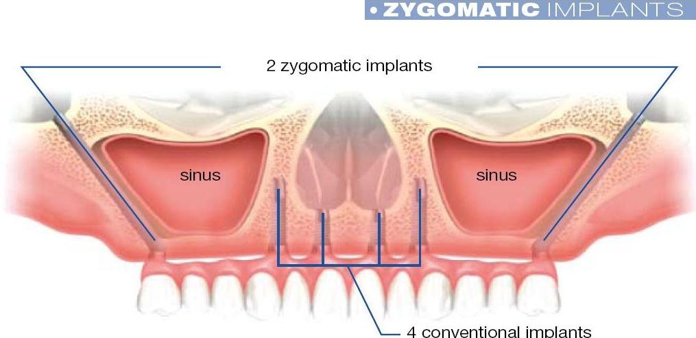
The figure below compares two zygomatic implants, which are anchored in the zygomatic bone and pass through the maxillary sinus, with four conventional implants in the anterior maxilla.
Maxilla with two zygomatic implants running from the lateral maxilla through each maxillary sinus, compared with four conventional implants placed in the anterior maxilla
4. Basic terminology
note
Components of an implant restoration
Several terms describe the parts of an implant restoration, from the visible crown down to the fixture in the bone.
Term
Meaning
Abutment
Connects the implant fixture to the prosthetic restoration, such as a crown, bridge or denture. Abutments can be standard (off-the-shelf) or custom-made to match the patient's anatomy. They are crucial for the right gingival emergence profile and mechanical retention of the prosthesis. The abutment is secured with screws or cement, depending on the implant system.
Abutment screw
Secures the abutment to the implant. It is engineered for precise torque control, to avoid loosening or overtightening, which can lead to mechanical failure such as screw loosening or fracture. The torque value matters for the longevity of the connection.
Implant fixture (body)
The part embedded in the jawbone. It integrates with the bone by osseointegration and provides the stability needed to support the prosthetic tooth or teeth. Titanium is the most common material because of its excellent biocompatibility and its ability to bond with bone.
Osseointegration
Living bone fuses directly with the titanium implant surface without any intervening fibrous tissue. This direct bone-to-implant contact ensures long-term stability and function.
Platform switching
The abutment diameter is smaller than the implant platform. This design helps preserve the peri-implant bone by reducing bone loss at the implant neck. It also improves soft tissue stability around the implant, which leads to better aesthetic outcomes.
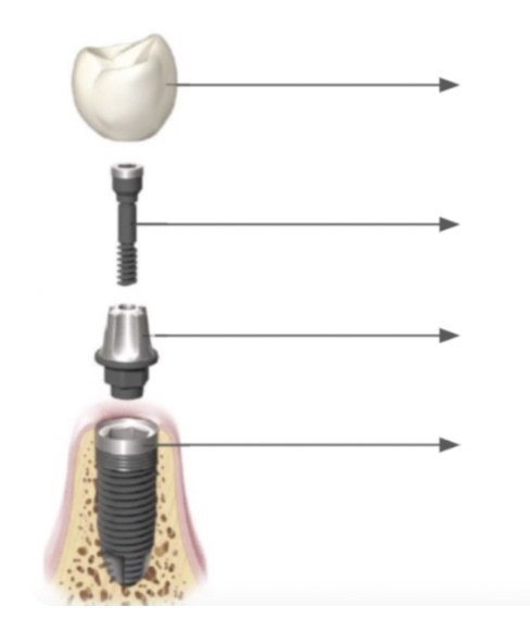
The exploded diagram below shows the components from the top down. The arrows are for naming the parts: the crown, the abutment screw, the abutment and the implant fixture.
Exploded view of an implant restoration from top to bottom: crown, abutment screw, abutment and implant fixture placed in the jaw, each with an arrow for labelling
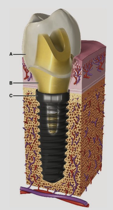
The cross-section below shows the same components in bone. Labels A, B and C mark the crown, the abutment at the gum margin and the implant at the crestal bone level.
Single implant restoration in bone, cross-section with the crown, a gold-coloured abutment and the implant fixture in the jaw, labelled A, B and C
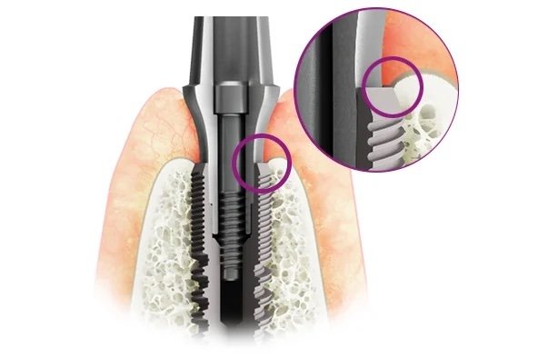
The narrower junction between the abutment and the implant neck is shown in the next figure. The circled inset magnifies the neck of the implant at the crestal bone level, where the threads sit in the bone and the bone surrounds the top of the fixture.
Implant with abutment seated in the jaw, with a circled magnified inset of the implant neck at crestal bone level showing the threads and surrounding bone
Implant structure and connection terms
Term
Meaning
Implant macrostructure
The overall shape and dimensions of the implant, designed to maximise initial stability and the distribution of force.
Implant microstructure
Surface modifications of the implant fixture that enhance osseointegration, such as acid etching, sandblasting or plasma spraying.
Internal hex connection
A hexagonal recess inside the implant fixture, into which a matching hex on the abutment fits. It helps the abutment seat precisely and provides rotational stability.
Conical hex connection
A tapered fit between the implant and the abutment. It gives excellent mechanical stability and minimises micromovement at the implant-abutment interface. It also reduces microbial infiltration, which leads to less peri-implant bone loss.
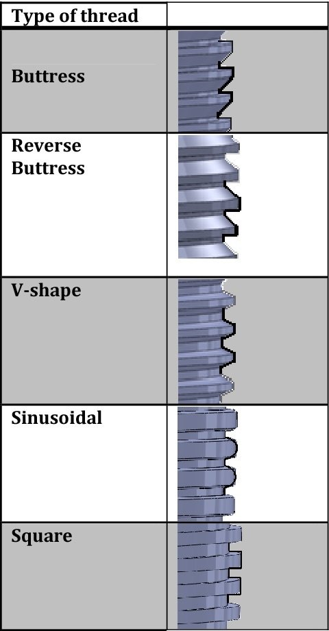
Buttress threads
A thread design on the implant fixture with a sloped surface on one side, which distributes occlusal forces effectively. They enhance stability, particularly in high-load situations.
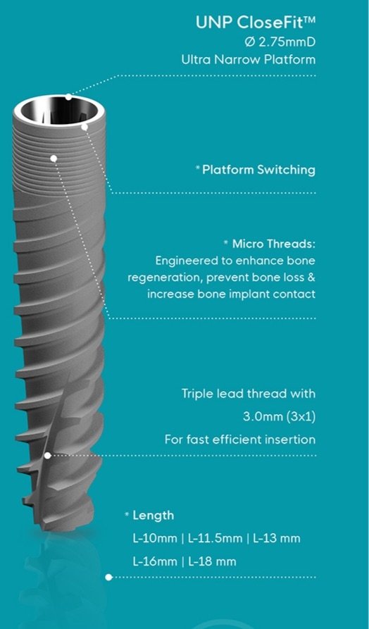
Microthreads
Fine threads at the collar of the implant. They improve the distribution of occlusal forces at the crestal bone level, help preserve the peri-implant bone and reduce stress concentration in the bone around the implant neck.
The internal hex connection is compared with the external hex connection below.
Internal hex and external hex implant-abutment connections, shown as end-on views, with the internal hex as a hexagonal recess inside the fixture
Thread profiles vary between implant designs. The figure below shows five of them: buttress, reverse buttress, V-shape, sinusoidal and square threads.
Thread profiles of implant fixtures: buttress, reverse buttress, V-shape, sinusoidal and square threads
The narrow-platform implant below illustrates platform switching, micro threads and the thread lead. It has a 2.75 mm diameter, a triple-lead thread with a 3.0 mm lead for fast, efficient insertion, and lengths from 10 mm to 18 mm.
Ultra-narrow platform implant, 2.75 mm in diameter, labelled for platform switching, micro threads, a triple-lead thread with 3.0 mm lead, and lengths from 10 mm to 18 mm
Implant quality scale
note
Implant success can be graded in four groups, from success to failure.
Group
Survival status
Clinical conditions
I
Success (optimum health)
No pain or tenderness on function. No mobility. Less than 2 mm radiographic bone loss from the initial surgery. No history of exudate.
II
Satisfactory survival
No pain on function. No mobility. 2 to 4 mm radiographic bone loss. No history of exudate.
III
Compromised survival
May have sensitivity on function. No mobility. Radiographic bone loss of more than 4 mm, which is less than half the implant body. Probing depth of more than 7 mm. May have a history of exudate.
IV
Failure (clinical or absolute failure)
Any of the following: pain on function, mobility, radiographic bone loss of more than half the implant length, uncontrolled exudate, or the implant is no longer in the mouth.
All 46 Dental Implantology topics · exam study support, not clinical guidance. SuperMed is not affiliated with the Medical University of Sofia.
Start free. No card needed.
Every account starts free, with Urology open in full and free topics in Cytology, Medical Biology, Anatomy, Biophysics, Medical Genetics, Pharmacology, Preclinics of Prosthetic Dental Medicine, Preclinics of Pediatric Dental Medicine, Dental Prophylaxis, Dental Allergology, Dental Implantology, Physiotherapy, Cytology & Histology, Anatomy I and Medical Biology. Super opens the rest. Super is €14.99 a month.