


Internal Medicine (Clinical) · Year 5 · Medical University of Sofia

01

Acute bronchitis, tracheobronchitis, bronchiolitis

Free notes for topic 01 of the Internal Medicine (Clinical) syllabus, open without an account. Written by a senior student against the syllabus question and checked line by line by a second student before publishing. How content is made

Updated

Summary

note

Acute bronchitis and tracheobronchitis are a common infection of the large airways, usually caused by a virus. The main symptom is a cough, often with sputum. The illness is self-limiting and usually settles within one to three weeks. It is diagnosed by excluding pneumonia, and it is managed mainly with supportive care, because antibiotics have little benefit when there is no other illness.

Bronchiolitis is an infection of the small airways, the bronchioles, which become inflamed and blocked. It mainly affects infants under two years of age, and respiratory syncytial virus (RSV) is its commonest cause. It causes cough, wheeze, crackles and increased work of breathing, and it can lower the oxygen level in the blood. Most cases are mild, but severe cases need close monitoring and sometimes hospital care. Treatment is supportive, with oxygen when saturations fall below 92%, and antibiotics only when a secondary bacterial infection is likely.
1. Definition

note

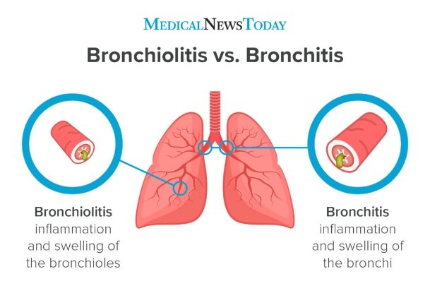
Acute bronchitis and bronchiolitis are both acute infections of the airways, but they involve different parts of the airway tree. In acute bronchitis the larger airways are inflamed. In bronchiolitis the smallest airways, the bronchioles deep in the lung, are inflamed and blocked. The diagram shows the difference.

Diagram comparing bronchiolitis with bronchitis: a lung drawing with two magnified airway cross-sections, one labelled bronchiolitis, inflammation and swelling of the bronchioles in the lower lung, and one labelled bronchitis, inflammation and swelling of the bronchi near the centre of the lung

Acute bronchitis: definition

note

Acute bronchitis is inflammation of the large airways of the lung, extending as far as the tertiary bronchi. It is a short-term illness that is self-limiting, usually lasting one to three weeks. Tracheobronchitis is the same process with the trachea also involved. The two terms are almost interchangeable.

Acute bronchitis is a diagnosis of exclusion. A patient with a cough, and without additional clinical or radiological evidence of pneumonia, is assumed to have acute bronchitis or tracheobronchitis.

Bronchiolitis: definition

note

Bronchiolitis is inflammation of the bronchioles, the smallest airways of the lung, which leads to their blockage. It is usually caused by a viral infection and usually occurs in children under two years of age. Epidemics occur in winter. It is the most common infection in the first years of life, and it accounts for about half of paediatric hospital admissions.

2. Classification

note

There is no single formal classification for either condition. The groupings below are the useful ways of sorting them in practice.

Acute bronchitis: classification

note

By site, bronchitis involves the bronchi. Tracheobronchitis also involves the trachea, and the two are managed in the same way.

By duration, acute bronchitis is short-term. Its cough is self-limiting over one to three weeks. A cough with sputum lasting more than three months points towards chronic bronchitis rather than an acute illness.

By cause, most cases are viral and bacterial causes are uncommon. The causes are set out in the aetiology section.

Bronchiolitis: classification

note

There is no formal classification. Two practical groupings are used.

By cause, RSV is the most common cause, and other respiratory viruses cause the remaining cases.

By severity, most cases are mild. Severe disease is recognised by the signs set out under clinical features.

3. Aetiology and risk factors

note

Each condition has its own causes. Acute bronchitis is usually viral, while bronchiolitis is caused most often by one virus, and the causes of each are set out below.

Acute bronchitis: causes

note

Most cases are viral. The viruses that cause it are influenza A and B, parainfluenza, respiratory syncytial virus, coronavirus, adenovirus and rhinovirus. Acute bronchitis can follow an upper respiratory tract infection, such as a cold, when the infection spreads down into the trachea and bronchi.

Bacterial causes are uncommon. They include Haemophilus influenzae, Moraxella catarrhalis, Mycoplasma pneumoniae and Streptococcus pneumoniae. Sputum analysis can identify the specific cause.

Smoking slows recovery, and stopping smoking helps the illness settle sooner.

Bronchiolitis: causes and risk factors

note

RSV is the most common cause of bronchiolitis. Most of the remaining cases are caused by other respiratory viruses, such as parainfluenza and influenza viruses. Most children are infected with RSV in the first years of life, and natural immunity is not fully protective, so infection can recur throughout life.

Prematurity, chronic lung disease, congenital heart disease and immunocompromise are risk factors for more severe disease.

4. Pathogenesis

note

The two conditions involve inflammation of the airways, but the site of the inflammation and the way it blocks airflow differ.

Acute bronchitis: mechanism

note

Irritation and damage to the airway lining cause inflammation, and neutrophils infiltrate the affected airway tissue. Substances released by the neutrophils promote hypersecretion of mucus into the airways. The infection also injures the ciliated lining of the airways, which slows the clearance of secretions.

Inflammation sensitises the cough reflex. The cough is then triggered more readily by changes in air temperature and by irritants such as cigarette smoke or perfumes.

Bronchiolitis: mechanism

note

RSV is an enveloped RNA virus that spreads by droplets. Its G protein helps the virus attach to the respiratory epithelium, and its F protein allows the virus to fuse with the host cell. Infection of the bronchioles causes inflammation and swelling, which narrows the airway.

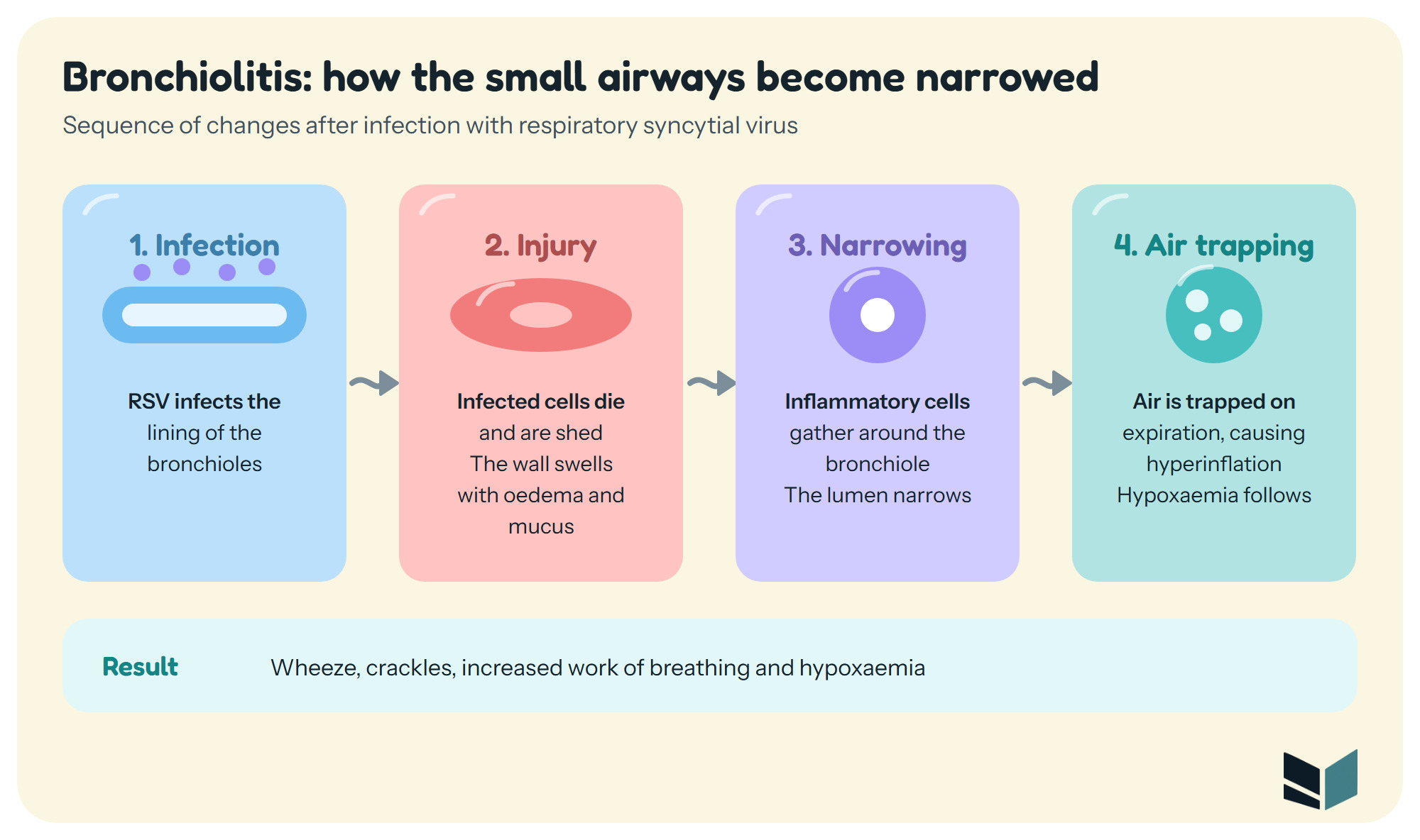
The infected epithelial cells are destroyed and shed, the wall swells with oedema and mucus, and inflammatory cells collect around the airway. Air is trapped on expiration, which causes hyperinflation, and ventilation and blood flow become poorly matched, which causes hypoxaemia.

Four-step sequence of bronchiolitis: 1 Infection, RSV infects the lining of the bronchioles; 2 Injury, infected cells die and are shed and the wall swells with oedema and mucus; 3 Narrowing, inflammatory cells gather around the bronchiole and the lumen narrows; 4 Air trapping, air is trapped on expiration causing hyperinflation, with wheeze, crackles and hypoxaemia as the result

5. Clinical features

note

The typical features of each condition are set out separately below, followed by a comparison of the two.

Acute bronchitis: symptoms and signs

note

The typical features are a productive cough with shortness of breath (dyspnoea) and wheezing. Sputum production and cough usually last less than three months. Chest pain, fever, fatigue or malaise may also occur.

The cough may be dry or productive, and it is often worse in the morning. When the trachea is involved, coughing causes raw pain behind the sternum. Acute bronchitis is one of the causes of haemoptysis. Chest tightness and wheeze, similar to asthma, can occur, and these are usually self-limiting.

Bronchiolitis: symptoms and signs

note

Bronchiolitis causes increased work of breathing and cough, and it can affect feeding. The symptoms are cough, wheeze and dyspnoea. Fever occurs in about 30% of cases, usually below 39 °C. Acute otitis media can also accompany the illness.

The symptoms are generally mild, but they can become severe and should be closely monitored. Signs of severe disease are:

  • poor feeding
  • significant decrease in activity
  • respiratory rate above 70 breaths per minute
  • severe chest wall recession (Hoover's sign)
  • bluish skin

In the severe form, tachycardia, hyperinflation of the lungs, a barrel chest and cyanosis may be present. The disease is characterised by wheezing and hypoxaemia.

Bronchiolitis and bronchitis compared

note

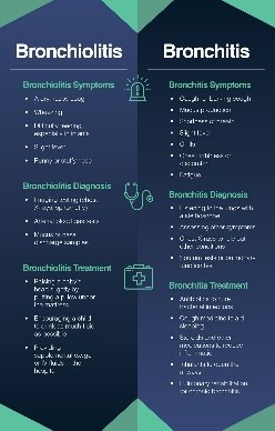
The two conditions are often compared side by side, as in the infographic below, which sets out the symptoms, diagnosis and treatment of each.

Infographic comparing bronchiolitis on the left and bronchitis on the right, each with panels for symptoms, diagnosis and treatment, with a stethoscope icon and a medical bag icon

6. Diagnosis

note

Diagnosis rests on the examination, with a chest X-ray and laboratory tests used to exclude other causes and to support the diagnosis.

Acute bronchitis: diagnosis

note

Diagnosis rests on the clinical picture, with pneumonia excluded. Physical examination may show a decreased intensity of breath sounds, wheezing, rhonchi and prolonged expiration. A dry or wet cough that persists is typical.

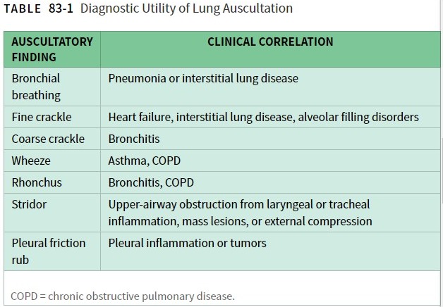
Auscultation findings help to place the sound in context. Coarse crackles are associated with bronchitis, rhonchi with bronchitis or COPD, and wheeze with asthma or COPD. Wheeze is heard on expiration and stridor on inspiration. Both reflect narrowing of the airways, but stridor indicates narrowing of the upper airway, while wheeze indicates narrowing of the lower airways. Stridor, a noise on inspiration, indicates partial obstruction of a major airway and needs urgent investigation. Fine crackles are heard when there is non-mucous fluid in the alveoli, and coarse crackles when there is mucous fluid in them.

Table of auscultatory findings and their clinical correlation: bronchial breathing with pneumonia or interstitial lung disease, fine crackle with heart failure, interstitial lung disease or alveolar filling disorders, coarse crackle with bronchitis, wheeze with asthma or COPD, rhonchus with bronchitis or COPD, stridor with upper airway obstruction, and pleural friction rub with pleural inflammation or tumours

The investigations are:

  • a chest X-ray, to exclude pneumonia
  • a sputum sample, which shows neutrophil granulocytes on microscopy, and sputum culture
  • blood tests, which may show a raised white cell count and a raised CRP

Where the illness is clearly uncomplicated, specific tests are often not needed.

Pneumonia should be suspected when two or more of the following are present:

FindingThreshold
Temperatureoutside 36 to 38 °C
Pulseabove 90 beats per minute
Respiratory rateabove 20 breaths per minute
White cell countoutside 4000 to 12000 cells/mm³

A cough lasting more than three months should prompt consideration of chronic bronchitis.

Bronchiolitis: diagnosis

note

Physical examination shows coughing, wheezing and dyspnoea, with crackles and wheezes on auscultation.

RSV testing is done on a nasopharyngeal aspirate, by direct immunofluorescence. A chest X-ray excludes pneumonia. Oxygen saturation is measured, because a persistent saturation below 92% is a reason for urgent referral.

7. Treatment

note

Treatment is supportive for both conditions. Antibiotics are used selectively, and the decision depends on the condition and the patient's risk.

Acute bronchitis: treatment

note

Most cases are self-limiting over a few weeks and do not need antibiotics. Treatment is usually symptomatic. Stopping smoking helps speed recovery.

Antibiotics have little benefit when there is no comorbidity. The following approach is used:

  • A delayed prescription may be considered, with advice, because symptoms can take three weeks to resolve.
  • Immediate antibiotics are considered for people over 80 years old with recent hospitalisation, oral steroids, diabetes and/or congestive heart failure.
  • Antibiotics are given to people over 65 years old who have two of the factors listed above.

When antibiotics are given, the options are:

  • amoxicillin 500 mg three times a day for five days
  • oxytetracycline 250 to 500 mg four times a day
  • doxycycline 200 mg on the first day, then 100 mg daily

Bronchiolitis: treatment

note

Treatment is supportive.

  • Oxygen is given if the saturation falls below 92%.
  • Paracetamol can be given to babies for fever.
  • Antibiotics are given only if there is good evidence of a secondary bacterial infection.
  • Nebulised hypertonic saline (3%) thins the mucus in the airways.
  • Bronchodilators are not really recommended for bronchiolitis, but a trial may be useful to help rule out asthma.

Refer immediately if the infant has:

  • apnoea, observed or reported
  • a seriously unwell appearance
  • severe respiratory distress, such as grunting or chest recession
  • a respiratory rate above 70 breaths per minute
  • central cyanosis
  • a persistent oxygen saturation below 92%

Infants admitted to hospital may need intravenous fluids or tube feeding if they cannot feed, and ventilatory support if respiratory failure develops.

8. Complications and prognosis

note

The complications and the outlook for each condition are set out below.

Acute bronchitis: complications and prognosis

note

Symptoms can take up to three weeks to resolve. A cough that lasts beyond three months needs reassessment for chronic bronchitis.

Pneumonia can develop as a complication of acute bronchitis. Viral infections of the airways can also trigger exacerbations of asthma or COPD.

Older people, and people with diabetes, heart failure or recent hospitalisation, are the groups in whom antibiotics are considered earlier. Most cases are self-limiting.

Bronchiolitis: complications and prognosis

note

Hypoxaemia can persist and needs oxygen. Apnoea is a warning sign that needs immediate referral. Pneumonia can complicate bronchiolitis, and acute otitis media is another recognised feature. Severe disease can progress to respiratory failure needing ventilation, and a secondary bacterial infection can develop.

About 2 to 3% of cases need hospital admission. Without active treatment, about 90% of cases resolve within three weeks. Most infants recover fully. Reinfection with RSV occurs throughout life.

9. Differential diagnosis

note

Both conditions must be distinguished from other causes of cough, wheeze and breathlessness, as set out below.

Acute bronchitis: differential diagnosis

note

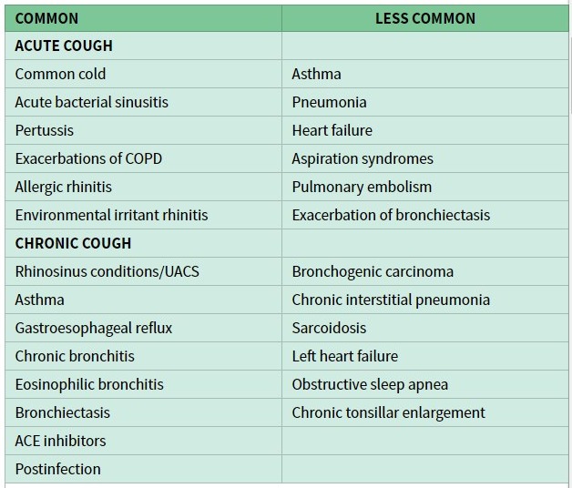
The table below sets out the common and less common causes of acute and chronic cough, which should be considered before a diagnosis of acute bronchitis is made.

Table of causes of cough: common causes of acute cough are the common cold, acute bacterial sinusitis, pertussis, exacerbations of COPD, allergic rhinitis and environmental irritant rhinitis; less common causes are asthma, pneumonia, heart failure, aspiration syndromes, pulmonary embolism and exacerbation of bronchiectasis; common causes of chronic cough are rhinosinusitis, asthma, gastro-oesophageal reflux, chronic bronchitis, eosinophilic bronchitis, bronchiectasis, ACE inhibitors and postinfection; less common causes of chronic cough are bronchogenic carcinoma, chronic interstitial pneumonia, sarcoidosis, left heart failure, obstructive sleep apnoea and chronic tonsillar enlargement

The cause of the cough can be placed by where it starts:

  • The upper airway: post-nasal drip, which follows a history of chronic rhinitis. Laryngitis or a laryngeal tumour alters the voice and gives a harsh or painful cough. Whooping cough or croup gives paroxysms of cough, often with stridor.
  • The trachea: tracheitis, which causes raw retrosternal pain with coughing.
  • The bronchi: acute bronchitis and COPD give a dry or productive cough, worse in the morning. Asthma usually gives a dry cough, worse at night. Eosinophilic bronchitis resembles asthma but without airway hyper-reactivity. Lung cancer gives a persistent cough, often with haemoptysis.
  • Drugs: ACE inhibitors cause a dry cough that can take up to six months to resolve after the drug is stopped.
  • Reflux and aspiration: gastro-oesophageal reflux disease, with a history of acid reflux, heartburn or hiatus hernia, and aspiration.
  • Infection: Bordetella pertussis infection in adults can cause a cough lasting up to three months.

Pneumonia is suspected when the findings listed under diagnosis are present.

Bronchiolitis: differential diagnosis

note

The differential diagnosis includes:

  • acute bronchitis, sometimes called a cold, which is the main comparison
  • bronchial asthma
  • congestive cardiac failure
  • inhaled foreign body
  • pertussis
  • bronchopneumonia, in which the child may be febrile and toxic
  • cystic fibrosis

Lower respiratory tract sounds, such as wheeze and crackles, point towards bronchiolitis when they are heard on auscultation. Wheeze in general is most often caused by asthma, COPD, pulmonary oedema and bronchiolitis. A trial of a bronchodilator may help rule out asthma.

10. Prevention

note

Prevention is considered for each condition. For bronchiolitis it centres on RSV, and for acute bronchitis it centres on avoiding smoking.

Bronchiolitis: prevention

note

RSV prevention in infants has one of two strategies:

  • maternal vaccination in pregnancy at 32 to 36 weeks' gestation, using one approved recombinant protein subunit vaccine
  • nirsevimab, a long-acting monoclonal antibody against RSV, given to infants up to 8 months old whose mothers have not been vaccinated, or whose mothers are at risk of not responding to vaccination

Nirsevimab is also recommended for certain infants entering their second RSV season who are at high risk of severe RSV. These include infants with immunocompromise, chronic lung disease of prematurity, severe cystic fibrosis or severe congenital heart disease. Nirsevimab is preferred to palivizumab, which was recommended before nirsevimab was licensed, because of its efficacy and because it protects for a full RSV season.

Acute bronchitis: prevention

note

Not smoking reduces the chance of repeated airway infections, and it also helps recovery when an infection occurs.

All 136 Internal Medicine (Clinical) topics · exam study support, not clinical guidance. SuperMed is not affiliated with the Medical University of Sofia.

Start free. No card needed.

Every account starts free, with Urology open in full and free topics in Cytology, Medical Biology, Anatomy, Biophysics, Medical Genetics, Pharmacology, Internal Medicine (Clinical), Preclinics of Prosthetic Dental Medicine, Preclinics of Pediatric Dental Medicine, Dental Prophylaxis, Diagnostic Imaging, Dental Allergology, Dental Implantology, Physiotherapy, Cytology & Histology, Anatomy I and Medical Biology. Super opens the rest. Super is €14.99 a month.

Create a free account