Preclinics of Pediatric Dental Medicine · Year 3 · Medical University of Sofia
01
Prenatal human development - periods, stages, and processes of development after fertilization
Free notes for topic 01 of the Preclinics of Pediatric Dental Medicine syllabus, open without an account. Written by a senior student against the syllabus question and checked line by line by a second student before publishing. How content is made
Updated
Prenatal human development: the short version
note
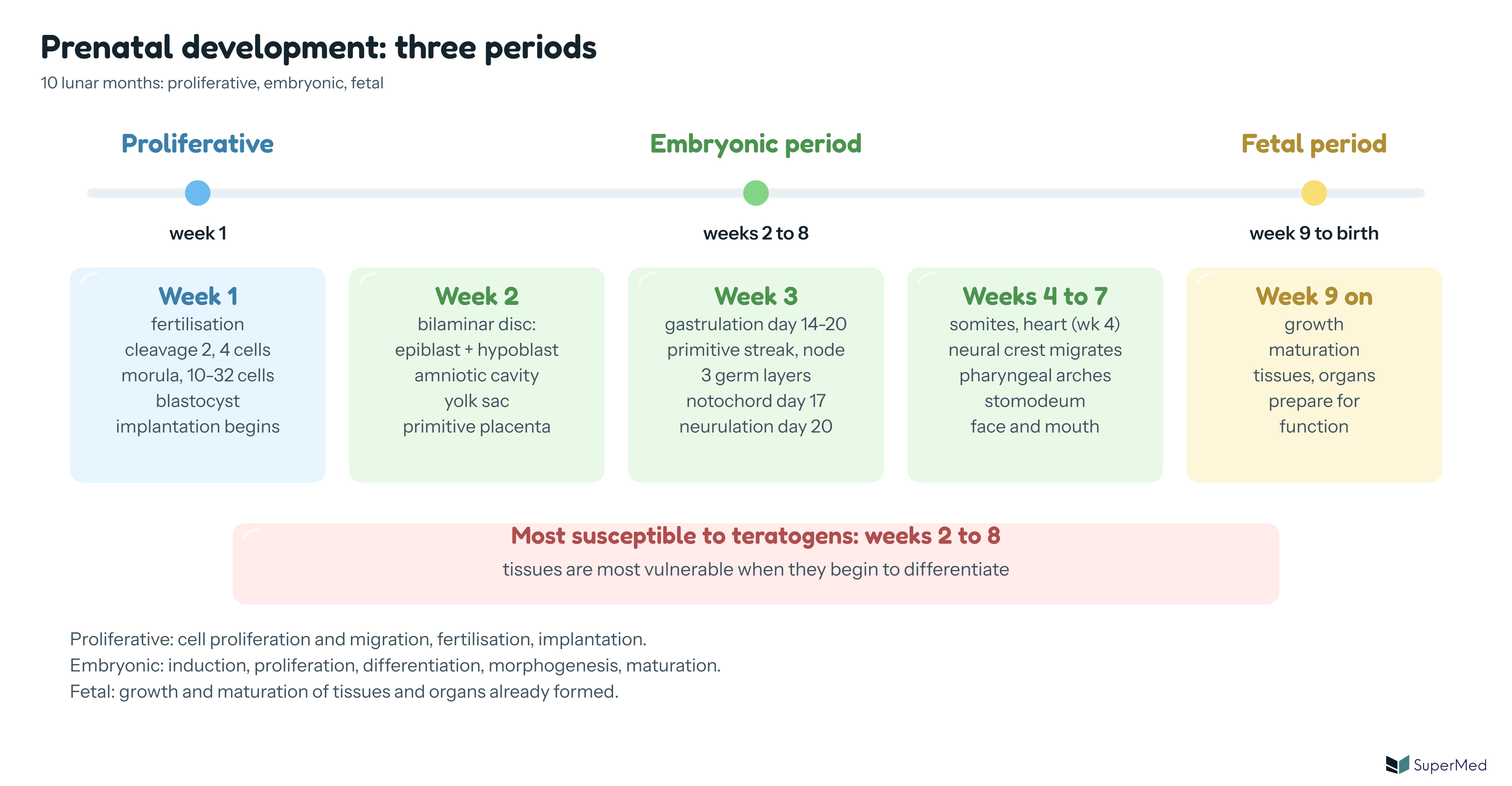
Prenatal development lasts 10 lunar months and runs through three periods. The first week is the proliferative period, weeks 2 to 8 are the embryonic period, and from week 9 to birth is the fetal period.
The points to hold on to:
Fertilisation = union of egg and sperm, in the upper third of the uterine (fallopian) tube; the result is one large cell, the zygote (about 140 μm).
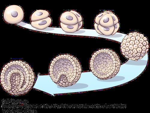
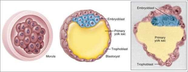
The zygote cleaves: 2 cells, 4 cells, then the morula (10 to 32 cells), which reaches the uterus at the end of the first week and becomes the blastocyst (inner cell mass plus trophoblast).
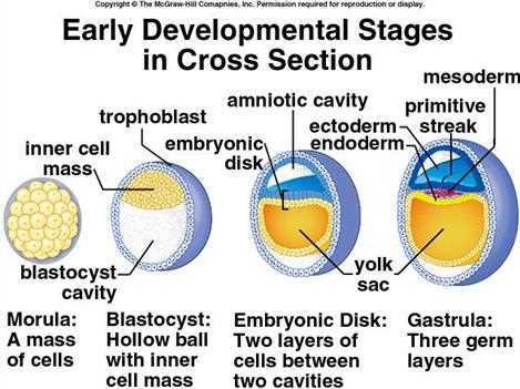
In the second week the inner cell mass becomes the bilaminar disc (epiblast above, hypoblast below) with an amniotic cavity above and the yolk sac below.
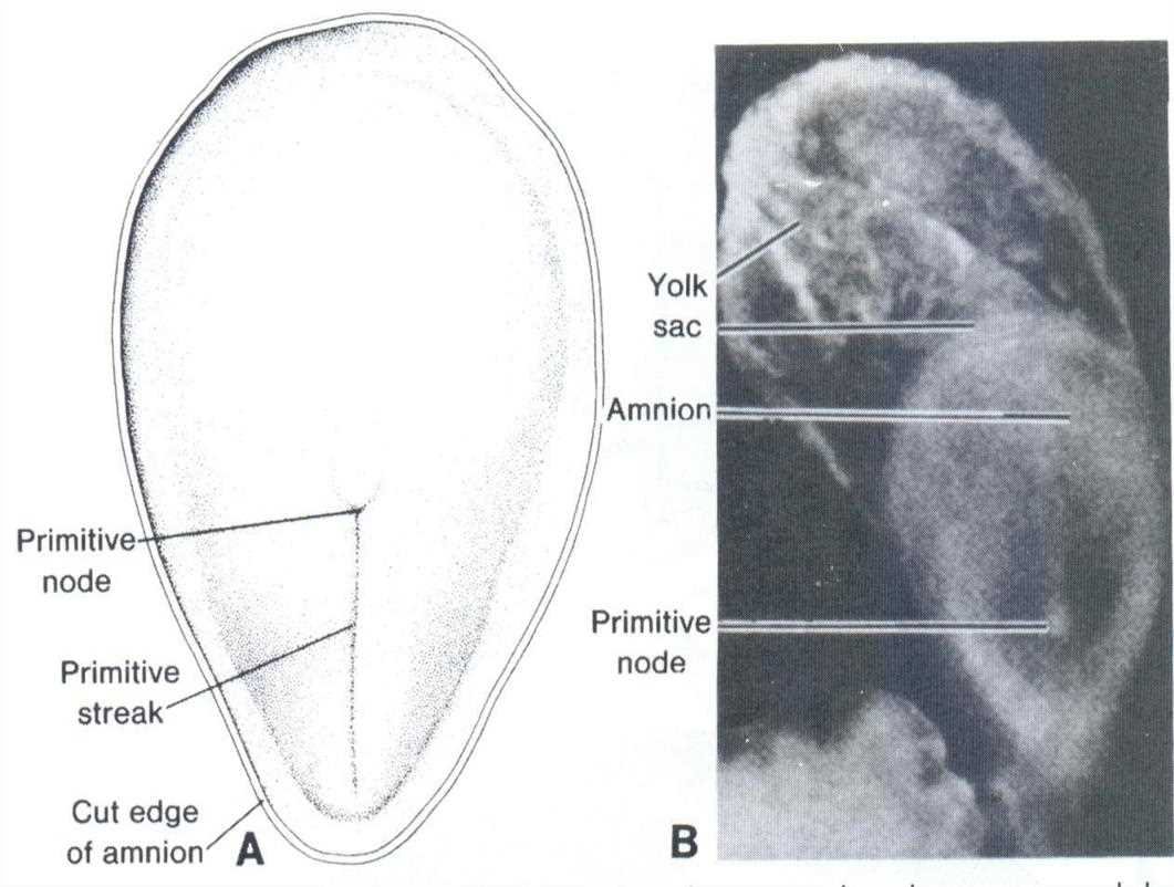
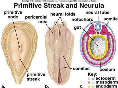
Gastrulation (days 14 to 20) turns the bilaminar disc into a trilaminar disc: ectoderm, mesoderm, endoderm. The primitive streak and primitive node drive it.
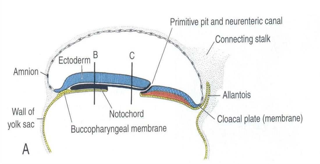
Late in the third week three events follow: the notochord forms (day 17), the pharyngeal (oropharyngeal) membrane marks the beginning of the primary mouth, and the neural plate forms. Neurulation (about day 20) closes the neural plate into the neural tube.
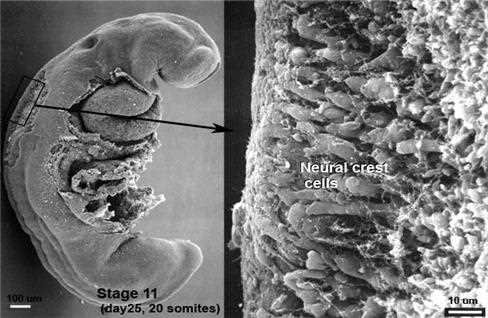
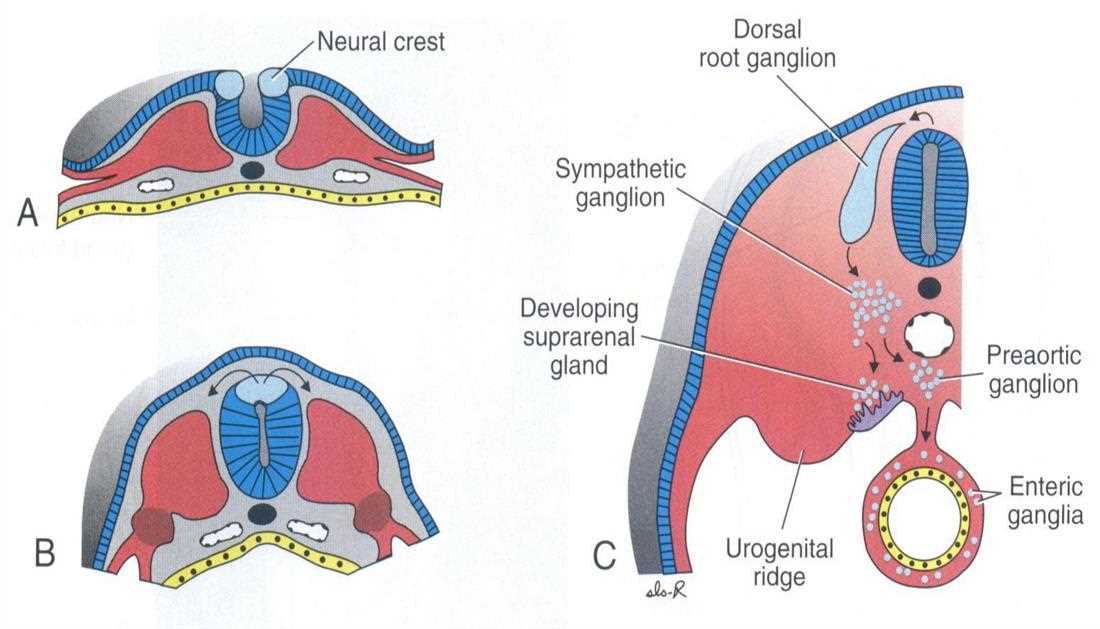
The neural crest cells separate from the neuroectoderm and migrate. In the head they form the ectomesenchyme, the "fourth germ layer"; every tissue of the tooth except enamel, and its supporting apparatus, comes from them.
The face and oral structures form between the 4th and 7th weeks. This is the time of greatest susceptibility to teratogens (weeks 2 to 8).
Schematic: prenatal development in three periods (proliferative week 1, embryonic weeks 2 to 8, fetal week 9 to birth) with the main events of weeks 1 to 4-7 and the window of teratogen sensitivity
1. Periods of prenatal development
note
Prenatal development is the process of growth and development within the womb, during which the zygote becomes an embryo, then a fetus, and finally a baby. The prenatal period lasts 10 lunar months (40 weeks). Development passes through three phases (periods):
Period
When
What happens
Proliferative (pre-implantation)
after fertilisation, spans the first week
fertilisation and implantation; largely cellular proliferation and migration; formation of the blastocyst and the embryonic disc
Embryonic
2nd to 8th week
induction, proliferation, differentiation, morphogenesis and maturation; differentiation of all major internal and external organs (= morphogenesis)
Fetal
9th week to the end of pregnancy
active growth and maturation of the tissues and organs already formed
The proliferative period is also called the preimplantation period. Its events are fertilisation, implantation and the embryonic disc, together with the formation of the blastocyst.
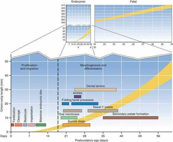
Sequences of prenatal development from Ten Cate: embryonic and fetal stages above; below, the embryonic part expanded against crown-rump length (mm) and days, showing proliferation and migration, then morphogenesis and differentiation, with the timing of fertilisation, cleavage, blastocyte, implantation, bilaminar disc, oral membrane, somite stage, arches, folding facial processes, dental lamina, nasal and primary palate, secondary palate formation
The three phases in one line: 1st week = proliferative stage (cellular proliferation, migration and differentiation); 4th week = differentiation stage (morphogenesis of internal and external organs); 8th week onward = fetal stage (growth and maturation of the fetus), then birth.
Exam tip: the proliferative period is the first week (fertilisation to implantation). Learn it as: proliferative = week 1, embryonic = weeks 2 to 8, fetal = week 9 to birth.
Day-by-day schedule
A day-by-day outline (from the student notes, for the days the lecture does not date) fits the lecture's periods:
Days
Events
Day 1
fertilisation, forming the zygote
Days 2-3
cleavage; the zygote divides and forms a morula (a solid ball of cells)
Days 4-5
the morula becomes a blastocyst with an inner cell mass and an outer trophoblast layer
Days 6-7
the blastocyst implants into the uterine wall
Days 8-14
the bilaminar disc forms (epiblast and hypoblast)
Day 17
the notochord develops, setting the stage for neurulation (gastrulation is dated days 14 to 20 and neurulation about day 20 in the lecture, see below)
Week 4
pharyngeal arches form and contribute to facial structures
2. Fertilisation
note
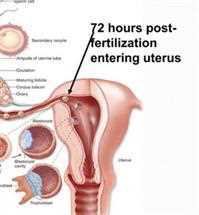
Fertilisation is the union of egg and sperm (fusion of a sperm cell and an egg cell, the oocyte). The origin of the tissues begins with it, when the sperm contacts the egg in the distal part of the uterine tube. It happens in the upper third of the oviduct (fallopian tube), in its widest part, the ampulla, which lies close to the ovary.
The ovum is a big round cell. The sperm is small and highly mobile.
After ovulation the ovum (0.15 mm in diameter) is picked up by the fimbriae of the tube and moved along by the cilia and the peristaltic movement of the tube.
Under the influence of oestrogen the cervix secretes a flow of alkaline mucus. Of the sperm deposited in the vagina only thousands, capacitated sperm, enter the uterine tube; only 300 to 500 reach the ovum. The rest are destroyed by the acid medium of the vagina. It takes about 1 hour for sperm to reach the site.
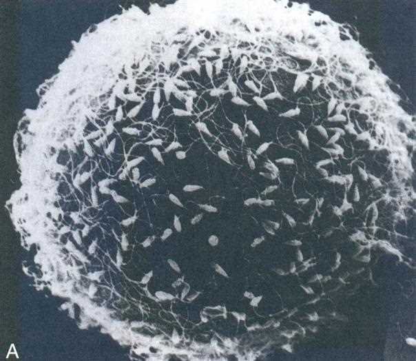
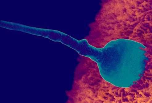
Scanning electron micrograph of many sperm on the surface of the egg with its surrounding cells (A)Coloured scanning electron micrograph of a sperm head attached to the egg surface
The three phases of fertilisation
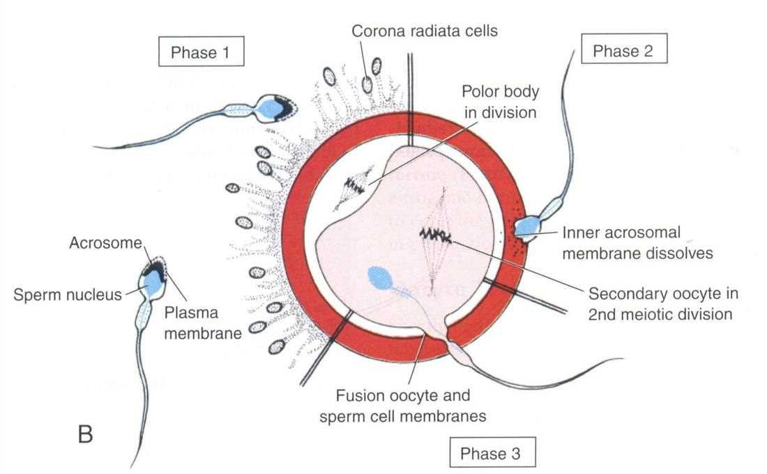
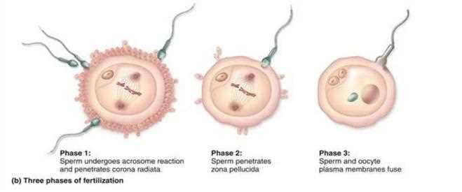
Phase 1: the sperm passes through the corona radiata cells by the effect of the enzyme hyaluronidase secreted from the sperm, and by the movement of its tail. In the acrosome reaction the acrosome releases its contents.
Phase 2: the inner acrosomal membrane dissolves and the sperm penetrates the zona pellucida by acrosin.
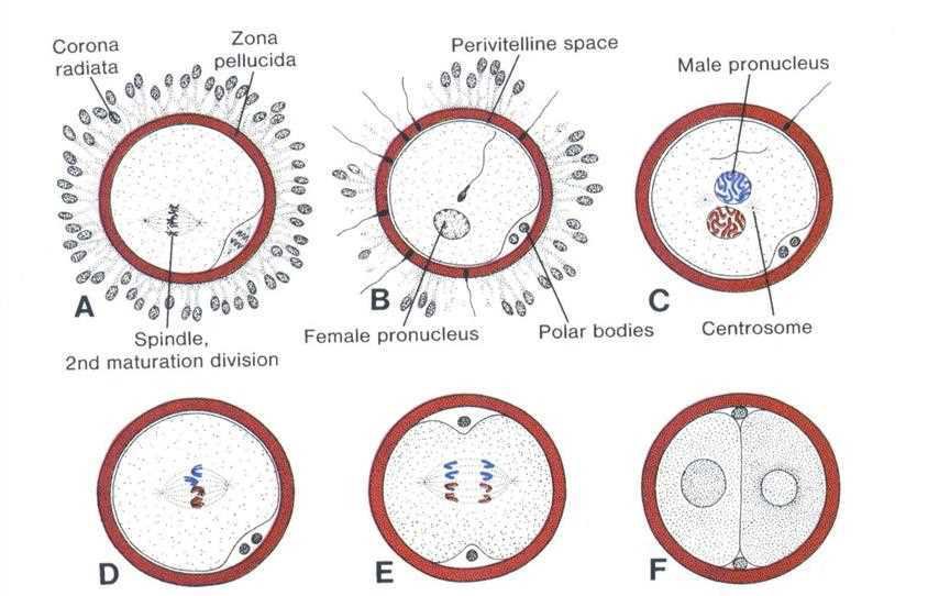
Phase 3: the plasma membranes of oocyte and sperm fuse. The secondary oocyte (arrested at metaphase II) immediately restarts and completes its second meiotic division, and a male and a female pronucleus form.
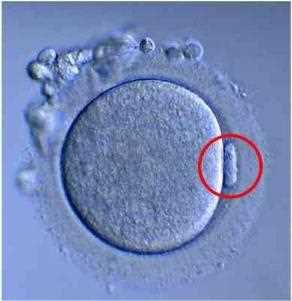
Polar bodies, seen in division in the figure, are cast off by the oocyte during the maturation divisions.
Three phases of fertilisation: corona radiata cells, polar body in division, acrosome, sperm nucleus, plasma membrane, inner acrosomal membrane dissolves, secondary oocyte in second meiotic division, fusion of oocyte and sperm cell membranes (phase 1, phase 2, phase 3)Human oocyte with the polar body circled in red, inside its zona pellucida and corona radiataThree phases of fertilisation drawn on the oocyte: phase 1 sperm undergoes acrosome reaction and penetrates corona radiata, phase 2 sperm penetrates zona pellucida, phase 3 sperm and oocyte plasma membranes fuseUterus and uterine tube: the most common site of conception is the ampulla, the widest part of the tube close to the ovary; 72 hours after fertilisation the embryo enters the uterus
3. The zygote
note
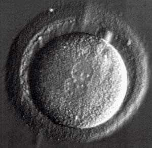
After fertilisation, the single large cell with a complete (diploid) set of 46 chromosomes is called the zygote. The fertilised egg grows and is termed the zygote; it is a large single cell, 140 μm in diameter. In the centre of the zygote the male and the female genetic material (DNA) can be seen as two nucleoli (the male and female pronuclei).
Human zygote: a large round cell inside the zona pellucidaStages from fertilisation to the first division, A to F: corona radiata, zona pellucida, spindle of the 2nd maturation division, perivitelline space, female pronucleus, male pronucleus, polar bodies, centrosome
4. Cleavage and the morula
note
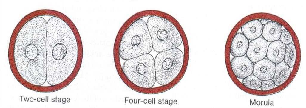
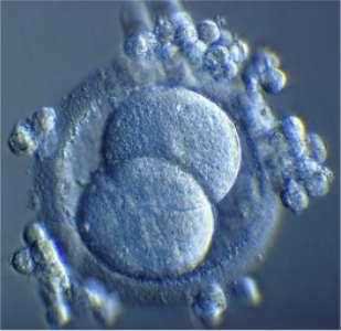
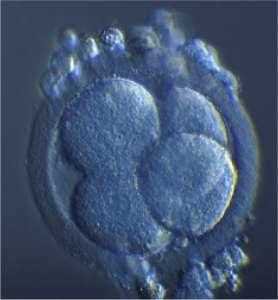
The continuous process of cell division follows fertilisation. The zygote divides in the primary (first) division: rapid cell division forming two new cells. The next division gives the four-cell stage.
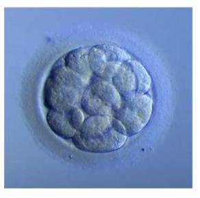
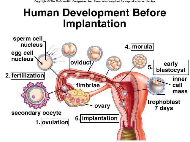
The cell mass produces a ball of cells, the morula, in the uterine tube. It has 10 to 32 cells. The morula grows and begins its migration medially towards the uterus at the end of the first week. Once in the uterine cavity the morula floats freely for the next 2 days, covered by endometrial fluid and mucus.
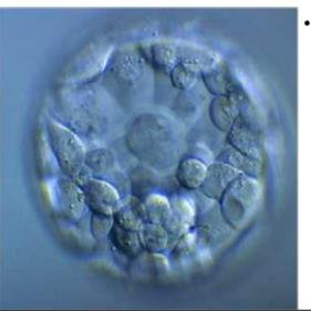
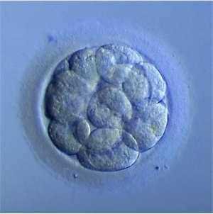
Two-cell stage, four-cell stage and morula drawn side by sideTwo-cell stage: two large blastomeres inside the zona pellucidaPhotograph of the cleaving embryo at the three to four-cell stage, blastomeres in the zona pellucidaPhotograph of an early blastocyst, a ring of cells around a cavity with a cell clump (student notes)Photograph of a morula, a rounded ball of cellsPractical exam slide 2, the morula: a ball of 10 to 32 cells in the zona pellucidaCleavage path from the first cell divisions through the morula to the blastocyst stagesTen Cate figure: morula, the blastocyst with embryoblast, primary yolk sac and trophoblast, and a section of the blastocyst
On the slide: the morula is a solid ball of 10 to 32 round cells inside the zona pellucida, with no cavity yet. Slide 3 (below) has a cavity and an eccentric clump of cells.
5. The blastocyst
note
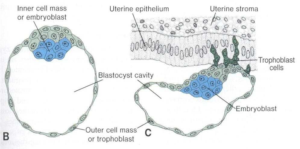
Shortly after the morula enters the uterus from the fallopian tube it becomes the blastocyst through cellular differentiation and cavitation. As the blastocyst swells it becomes hollow and develops a small inner cell mass. The morula's cells differentiate into two types:
an inner cell mass (embryoblast), growing on the interior of the blastocoel;
trophoblast cells, growing on the exterior.
In the student notes the blastocyst is formed by the trophoblasts pumping sodium ions so that water enters by osmosis. The fluid is absorbed through the canaliculi of the zona pellucida; the morula starts to accumulate fluid and forms a cavity between its cells. Once the cavity appears it is called a blastocyst. The zona pellucida becomes stretched, thinned and gradually disappears shortly before implantation.
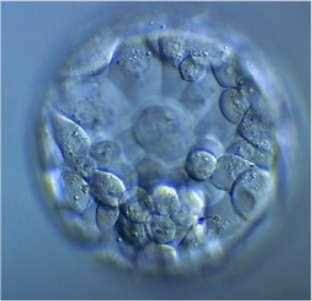
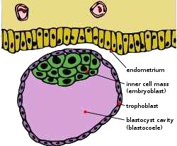
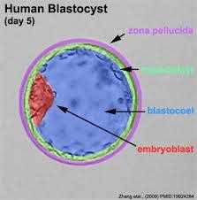
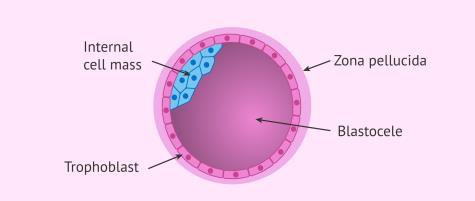
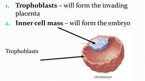
The blastocyst has an outer layer of cells (trophoblast, which becomes the placenta), an inner cell mass (embryoblast, which forms the embryo) and a fluid-filled cavity, the blastocoel. The formation of the blastula (blastocyst, a hollow ball of cells) marks the beginning of morphogenesis, the shaping of the embryo, and of the migration of dividing cells to specific locations.
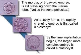
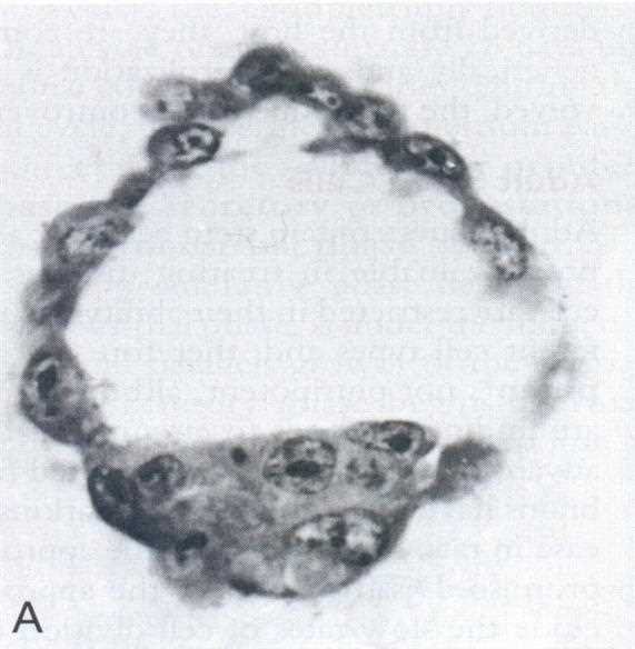
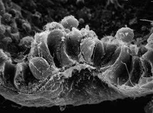
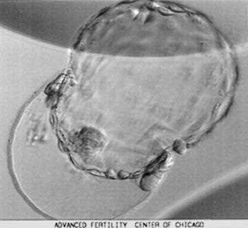
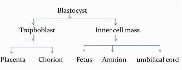
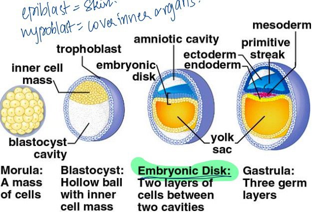
Practical exam slide 3, the blastocyst: a hollow ball with fluid-filled cavity and a small inner cell massSchematic of the blastocyst: endometrium, inner cell mass (embryoblast), trophoblast, blastocyst cavity (blastocoele)Human blastocyst, day 5: zona pellucida, trophoblast, blastocoel and embryoblast labelled (Zhang et al.)Blastocyst scheme: internal cell mass, zona pellucida, trophoblast, blastoceleMorula of 3 days, still travelling down the uterine tube (zona pellucida), then the cavity forms and the embryo is called a blastocyst; at implantation the larger, more complex embryo is still called a blastocystBlack and white section through a blastocyst: a thin trophoblast wall and a clump of inner cells (A)Human development before implantation: ovulation, fertilisation, oviduct, fimbriae, ovary, secondary oocyte, morula, early blastocyst, inner cell mass, trophoblast at 7 days, implantationFormation of the inner cell mass: blastocyst with inner cell mass (embryoblast), blastocyst cavity and outer cell mass (trophoblast); on the right the blastocyst at the uterine epithelium and stroma with trophoblast cellsScanning electron micrograph of the cells of the inner cell mass of the blastocystEarly developmental stages in cross section: morula (a mass of cells), blastocyst (hollow ball with inner cell mass), embryonic disk (two layers of cells between two cavities), gastrula (three germ layers)Blastocyst photographed in an IVF laboratory, a hollow ball with a cell clump inside the zona pellucidaTrophoblasts will form the invading placenta; inner cell mass will form the embryo (late blastocyst)Flow chart: the blastocyst divides into trophoblast (placenta, chorion) and inner cell mass (fetus, amnion, umbilical cord)
6. Implantation
note
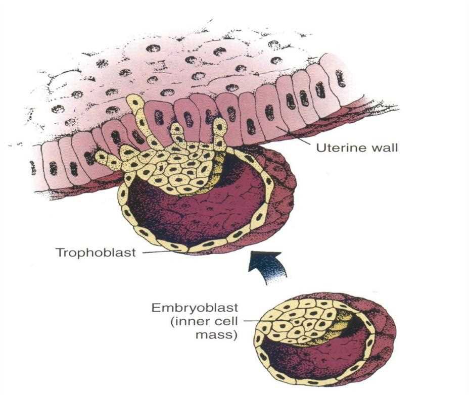
Implantation is the process in which the blastocyst digests the uterine endometrium, permitting deeper penetration. It begins about 6 to 7 days after fertilisation (about the 20th day of a regular menstrual cycle). The blastocyst attaches to and burrows into the thickened uterine lining (the endometrium) to establish a connection with the maternal blood supply. That connection is essential for the exchange of nutrients, waste and gases between mother and embryo.
As the zona pellucida disappears the trophoblast cells become more adhesive to the endometrium.
The trophoblast cells invade the stromal cells lying between the secretory endometrial glands, by histolytic action of the blastocyst, bringing about deeper penetration into the decidua.
The trophoblast cells facilitate this adhesion by penetrating the endometrium with protrusions of the cytoskeleton of their cells.
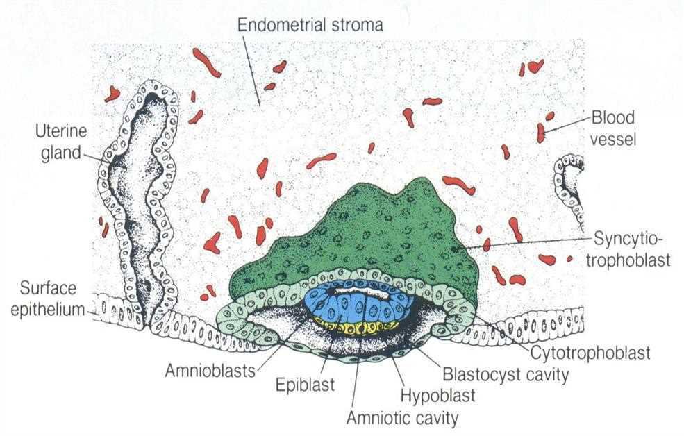
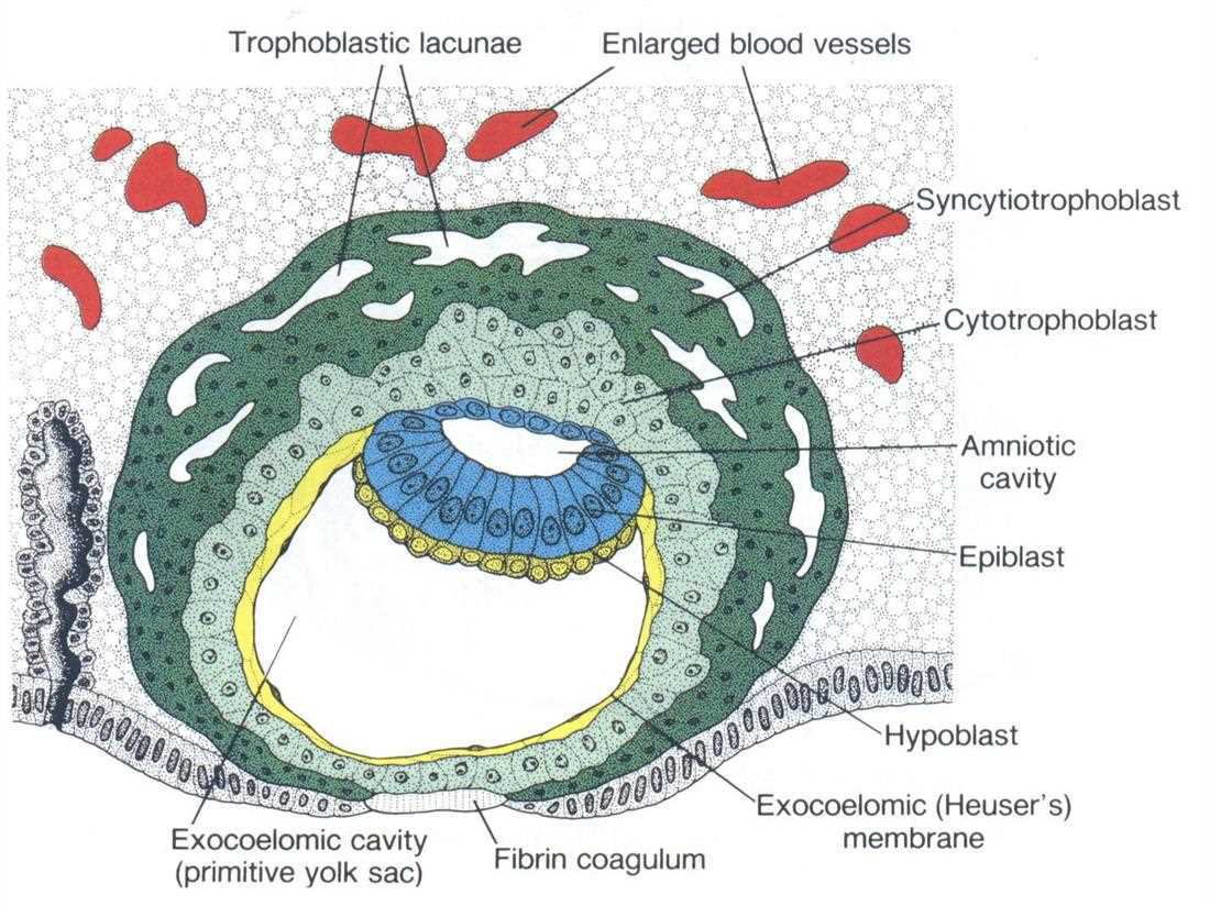
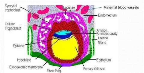
The trophoblast cells differentiate into syncytiotrophoblast (where hCG, human chorionic gonadotropin, is produced) and cytotrophoblast. The syncytiotrophoblast ruptures maternal capillaries and establishes a route of passive exchange between mother and embryo (lacunae filled with maternal blood).
The trophoblast also begins to differentiate to form a primitive placenta; the embryo connects to the developing placenta through a stalk.
hCG
hCG (human chorionic gonadotropin) is produced by the trophoblast starting on day 6. It causes the endometrium of the uterus to grow and proliferate and prevents the menstrual cycle from occurring. This is why a woman misses her periods when pregnant.
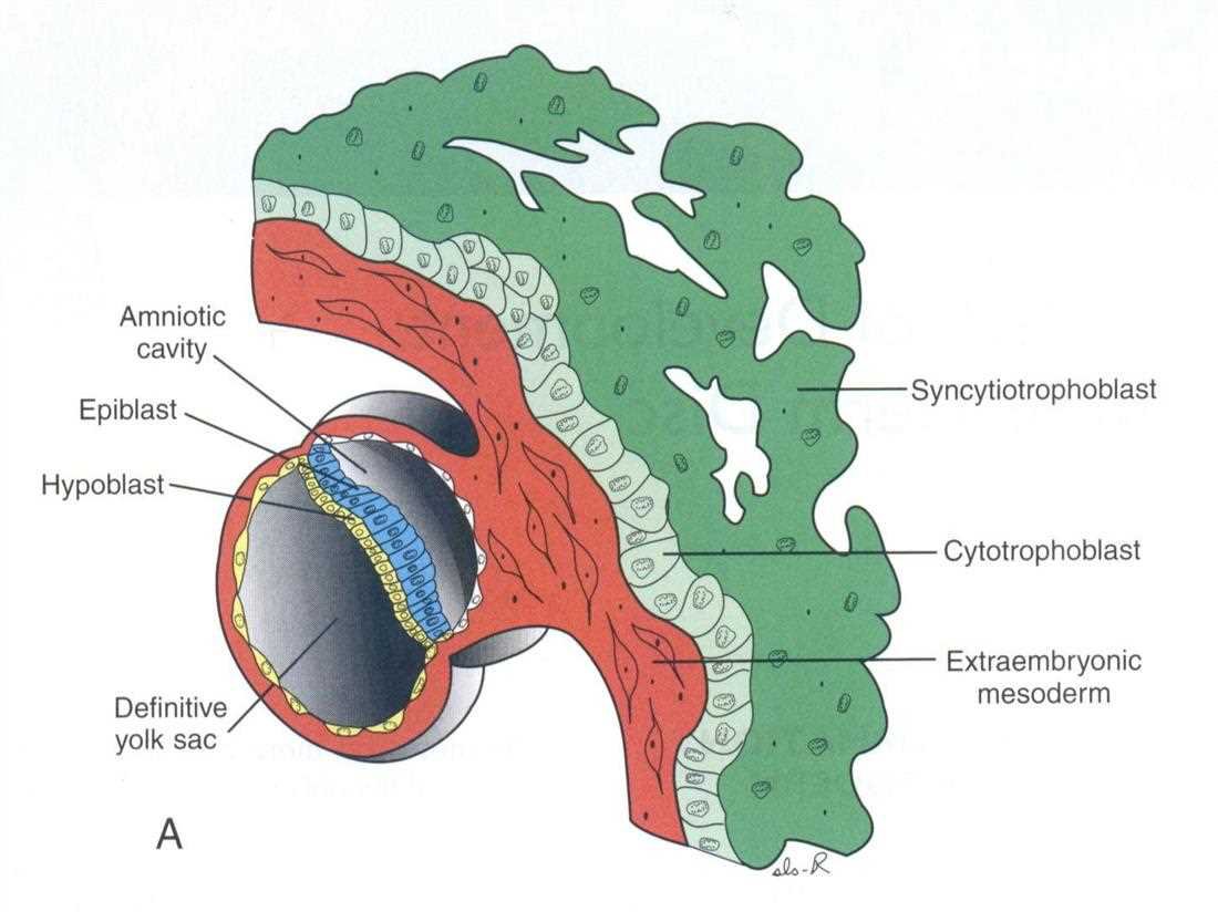
Coloured scanning electron micrograph of the implanting blastocyst, red, embedded in the uterine surfaceBlastocyst implanting into the uterine wall: trophoblast; embryoblast (inner cell mass) belowImplanted blastocyst with endometrial stroma, uterine gland, surface epithelium, syncytiotrophoblast, cytotrophoblast, amnioblasts, epiblast, hypoblast, blastocyst cavity, amniotic cavity, blood vesselImplanted bilaminar embryo: trophoblastic lacunae, enlarged blood vessels, syncytiotrophoblast, cytotrophoblast, amniotic cavity, epiblast, hypoblast, exocoelomic cavity (primitive yolk sac), exocoelomic (Heuser's) membrane, fibrin coagulumLater stage of implantation: amniotic cavity, epiblast, hypoblast, definitive yolk sac, syncytiotrophoblast, cytotrophoblast, extraembryonic mesodermStudent notes version of the implanted embryo: syncytial trophoblast, cellular trophoblast, epiblast, hypoblast, exocoelomic membrane, fibrin plug, lacunae, maternal blood vessels, endometrium, amnion, amniotic cavity, uterine gland, epithelium, primary yolk sac
7. The second week: the embryonic disc
note
In the second week the blastocyst increases in size by proliferation. A blastocyst cavity forms between the inner cell mass and the trophoblast, and differentiation of the inner cell mass begins.
Formation of the embryonic disc
Two small cavities develop on either side of the inner cell mass. They reach each other in the centre, where a small disk is formed: the embryonic disk.
The embryonic disk becomes the embryo, composed of the common walls of the two adjacent sacs.
One sac is lined with ectodermal cells, which will form the outer body covering (the epithelium). The other sac is lined with endodermal cells.
The embryonic disc is also called the embryoblast (lecture term).
The bilaminar disc
The result is a bilaminar embryonic disc of two cell layers:
Layer
Position
Fate
Epiblast
upper
will give ectoderm, mesoderm and endoderm
Hypoblast
lower
extraembryonic endoderm
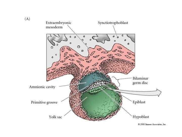
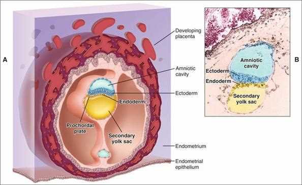
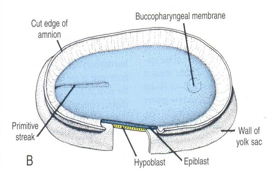
Above the disc is an upper amniotic cavity, below it is the lower yolk sac (a primitive haematopoietic organ for the embryo and fetus). The trophoblast also begins to differentiate to form a primitive placenta, and the embryo connects to the developing placenta through a stalk.
Exam tip: bilaminar = two layers (epiblast + hypoblast), trilaminar = three layers (ectoderm, mesoderm, endoderm). Do not mix up the second and third week.
Practical exam slide 4, the bilaminar germ disc: extraembryonic mesoderm, syncytiotrophoblast, amniotic cavity, primitive groove, yolk sac, bilaminar germ disc, epiblast, hypoblastTen Cate: human blastocyst in schematic and histologic section, with amniotic cavity within the ectoderm layer, secondary yolk sac and a well established bilaminar embryoStudent notes: morula, blastocyst with inner cell mass and blastocyst cavity, embryonic disk with epiblast, hypoblast, amniotic cavity, ectoderm, endoderm, yolk sac, and gastrula with three germ layers: mesoderm, primitive streak
8. Gastrulation: the third week
note
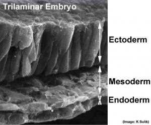
Gastrulation is the process by which the bilaminar embryonic disc is converted into a trilaminar one. It lasts from the 14th to the 20th day and establishes the three germ layers by stratification. A single-layered blastula is reorganised into a multilayered gastrula.
Towards day 14 the primitive ectoderm thickens, forming the primitive plaque.
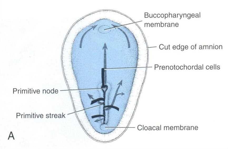
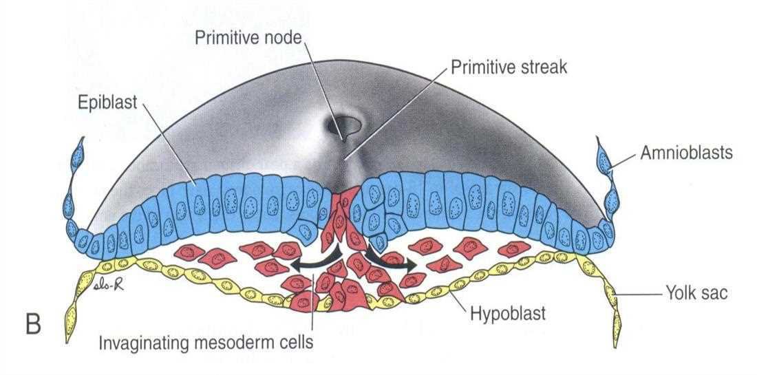
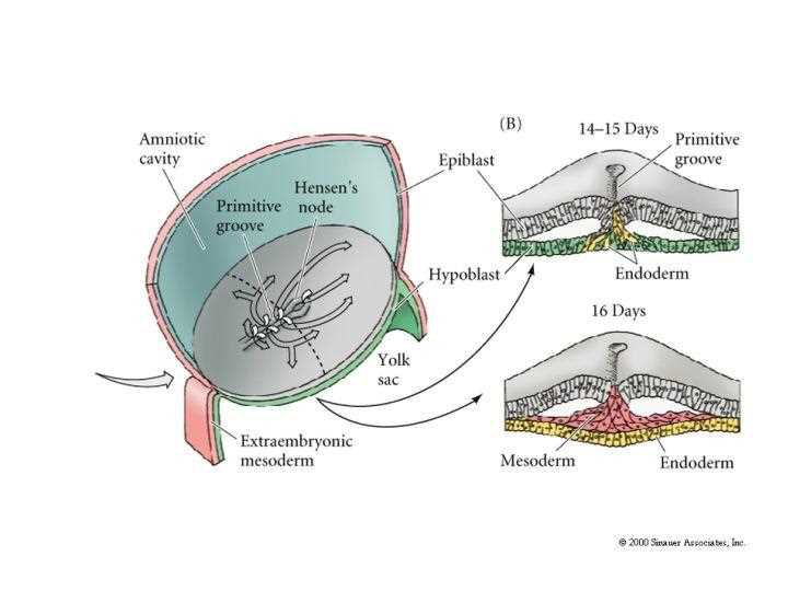
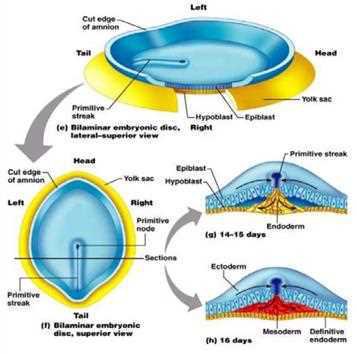
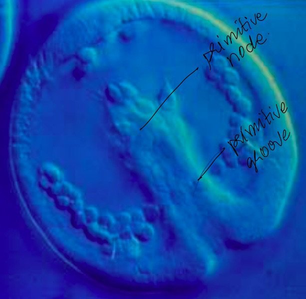
A primitive groove forms in it (by invagination), and at its cranial edge the primitive node (Hensen's node) forms. Together the groove, node and pit are the primitive streak.
The cells of the primitive groove and node acquire mitotic activity. They divide and penetrate between the ectoderm and the endoderm.
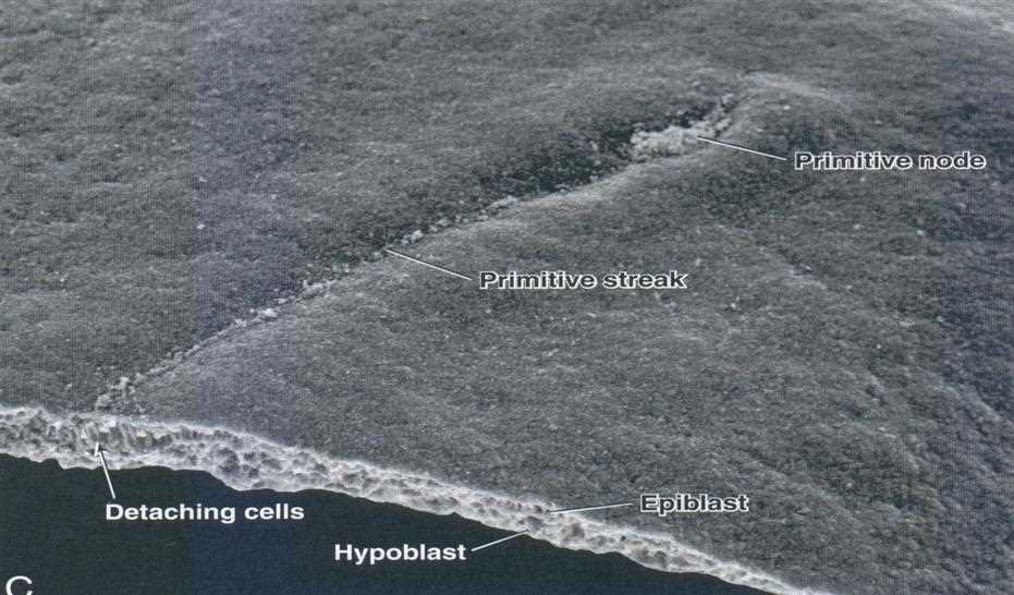
Formation of the primitive streak within the embryonic disc is critical. The epiblast cells migrate through the primitive streak towards the hypoblast, which eventually creates three tissue layers: ectoderm, mesoderm, endoderm.
The beginning of mesoderm formation is the invagination of the ectoderm under the primitive streak: the inserted cells divide ectoderm and endoderm.
Reading the figure of cell movements
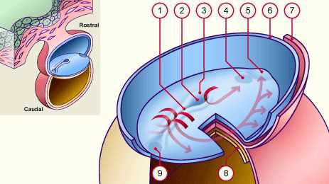
The numbered scheme of the embryonic disc seen dorsally has these labels: 1 primitive groove, 2 primitive pit, 3 primitive node, 4 oropharyngeal membrane, 5 cardiac plate, 6 sectional edge of the amniotic membrane, 7 mesoderm, 8 endoderm, 9 future cloacal membrane; 1+2+3 together form the primitive streak. The red arrows show schematically the migration directions of the epiblast cells to their points of final destination.
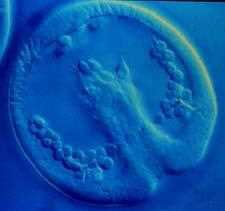
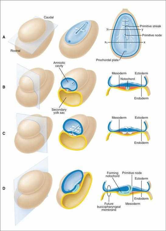
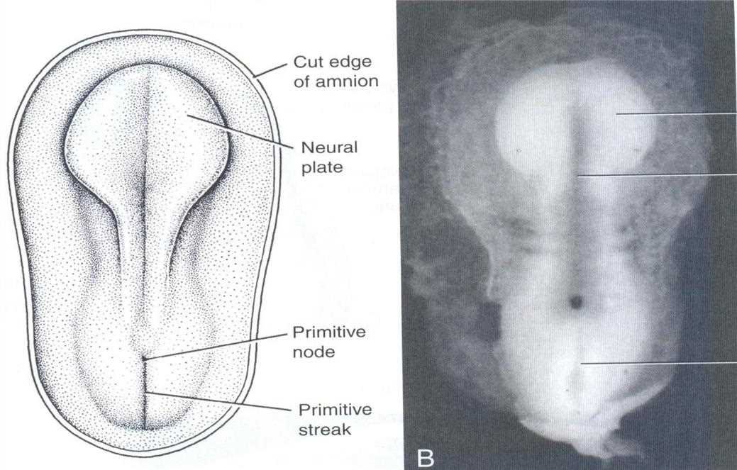
Gastrulation: embryonic disc seen from above in a blue-tinted contrast micrograph with the primitive node and primitive streak on the midlineA: drawing of the disc with primitive node, primitive streak, cut edge of amnion; B: photograph of the embryo with yolk sac, amnion and primitive node labelledPrimitive streak, cut edge of amnion, wall of yolk sac, hypoblast, epiblast and buccopharyngeal membrane (B)Dorsal view: buccopharyngeal membrane, cut edge of amnion, prenotochordal cells, primitive node, primitive streak, cloacal membrane, with arrows for cell migration (A)Cross section through the primitive streak: epiblast, primitive node, primitive streak, amnioblasts, invaginating mesoderm cells, hypoblast, yolk sacScanning electron micrograph of the primitive node and primitive streak: detaching cells, epiblast and hypoblastNumbered scheme 1 to 9 of the cell movements during gastrulation, rostral and caudal ends, with red arrowsTrilaminar embryo in a scanning electron micrograph: ectoderm, mesoderm, endodermGastrulation scheme: amniotic cavity, Hensen's node, primitive groove, epiblast, yolk sac, extraembryonic mesoderm; sections at 14 to 15 days (primitive groove, epiblast, hypoblast, endoderm) and at 16 days (ectoderm, mesoderm, definitive endoderm)Student notes: bilaminar embryonic disc, lateral-superior and superior views with primitive streak, primitive node, epiblast, hypoblast, yolk sac, cut edge of amnion, and sections at 14 to 15 days and 16 daysTen Cate: gastrulation, conversion of the bilaminar to a trilaminar embryo, with the plane of section, the three-dimensional view and the two-dimensional view; ectodermal cells converge on the primitive streak and primitive node and migrate between ectoderm and endodermStudent notes: primitive node and primitive groove labelled on a photograph of the disc
9. Mesoderm formation and the fate of the germ layers
note
The mesoderm has a twofold origin:
the majority is derived from the proliferation (inward movement) of the ectoderm through the primitive streak;
a smaller part is formed later, from the migrating cells of the neural crests (the ectomesenchyme).
The germ layers are the ectoderm, the mesoderm and the endoderm. The initial process starts with the primitive streak and the primitive node.
The third week of development: the bilaminar embryonic disk converts into a trilaminar disk of ectoderm, mesoderm and endoderm (gastrulation). The primitive streak is critical.
Fate of the germ layers
Germ layer
Main derivatives
Ectoderm
epidermis, hair, nails, glands of the skin; brain and spinal cord (neuroectoderm); neural crest: sensory nerve cells and some nervous structures, pigment cells, portions of the skeleton, blood vessels of head and neck; sensory organs (eyes, ears, parts of the nose); tooth enamel
Mesoderm
skeleton (bones, cartilage); muscles (skeletal, smooth, cardiac); circulatory system (heart, blood vessels, blood cells); kidneys and ureters (excretory); gonads and associated ducts; connective tissues: dermis of the skin, tendons, ligaments
Endoderm
digestive system (lining of the gastrointestinal tract, liver, pancreas); respiratory system (lining of lungs and trachea); urinary system (lining of the bladder); endocrine glands (parts of the thyroid and parathyroid glands)
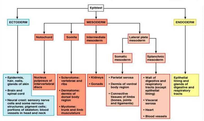
The mesoderm in detail
Part of the mesoderm
Derivatives
Notochord
nucleus pulposus of the intervertebral discs
Somite
sclerotome: vertebrae and ribs; dermatome: dermis of the dorsal body region; myotome: trunk and limb musculature
Intermediate mesoderm
kidneys, gonads
Lateral plate, somatic mesoderm
parietal serosa; dermis of the ventral body region; connective tissues of the limbs (bones, joints, ligaments)
Lateral plate, splanchnic mesoderm
wall of the digestive and respiratory tracts (except the epithelial lining); visceral serosa; heart; blood vessels
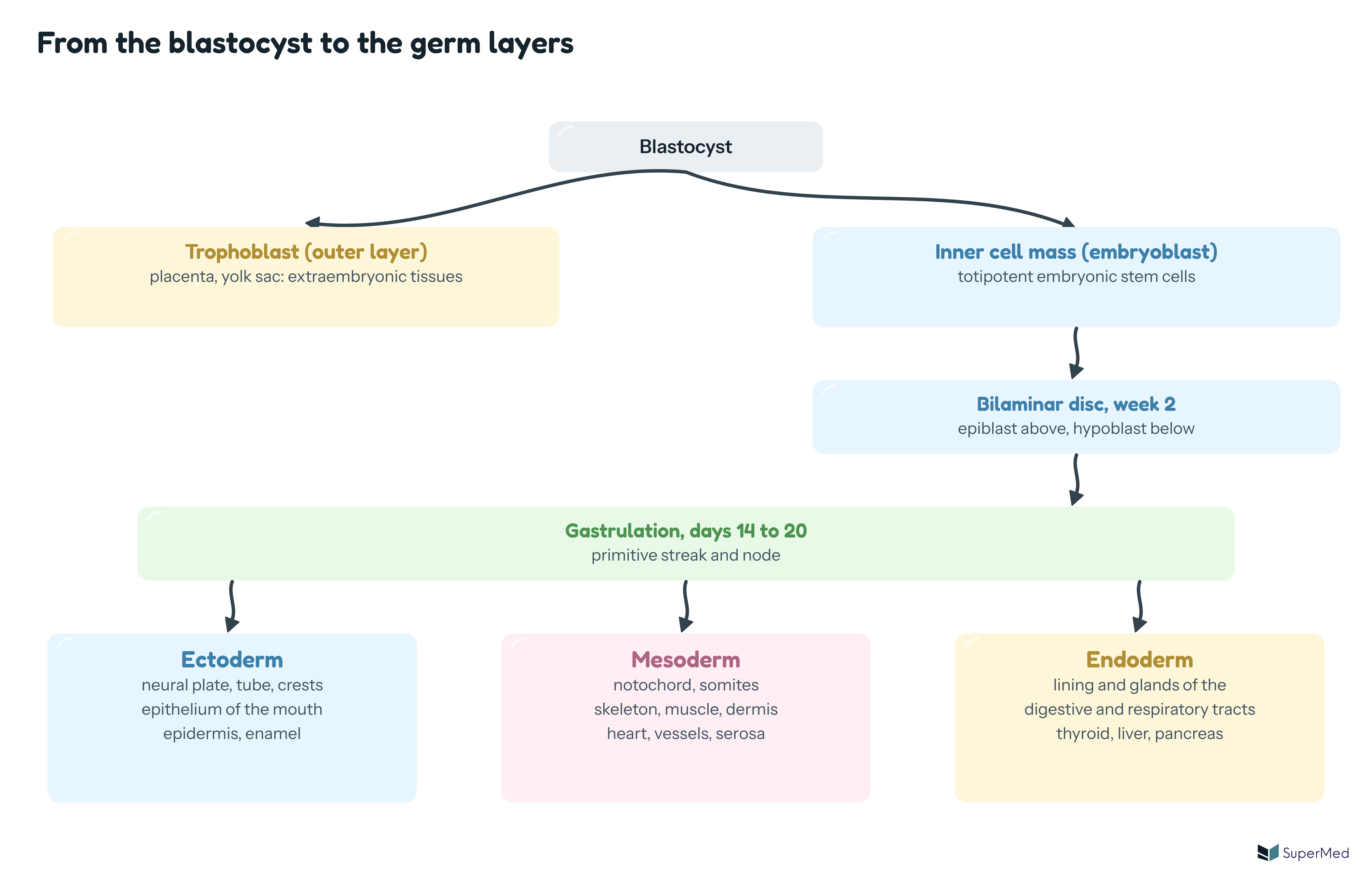
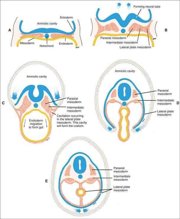
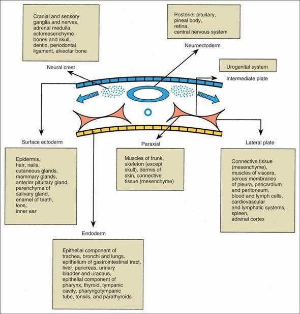
Schematic: the blastocyst divides into trophoblast (placenta, yolk sac) and inner cell mass; the inner cell mass becomes the bilaminar disc, gastrulation follows, and the three germ layers ectoderm, mesoderm and endoderm are formed with their main derivativesFlow chart from the epiblast: ectoderm (epidermis, hair, nails, glands of skin, brain, spinal cord, neural crest derivatives), mesoderm (notochord, somite, intermediate mesoderm, lateral plate mesoderm split into somatic and splanchnic), endoderm (epithelial lining and glands of digestive and respiratory tracts)Ten Cate: cross-sectional profiles, the mesoderm between ectoderm and endoderm differentiating into paraxial, intermediate and lateral plate mesoderm, with the amniotic cavity enclosing the embryo and the intraembryonic coelom forming in the lateral plateTen Cate: derivatives of the germ layers and cranial neural crest. Neural crest gives cranial and sensory ganglia and nerves, adrenal medulla, ectomesenchyme, bones and skull, dentin, periodontal ligament and alveolar bone; surface ectoderm gives epidermis, hair, nails, cutaneous glands, mammary glands, anterior pituitary, parenchyma of the salivary gland, enamel of teeth, lens and inner ear; neuroectoderm gives posterior pituitary, pineal body, retina and CNS; paraxial mesoderm gives muscles of trunk, skeleton except skull and dermis; lateral plate gives connective tissue, serous membranes, cardiovascular and lymphatic systems, spleen, adrenal cortex; intermediate plate gives the urogenital system; endoderm gives the epithelial components of the respiratory and gastrointestinal tracts, liver, pancreas, bladder, pharynx, thyroid, tympanic cavity, tonsils and parathyroids
10. Events of the late third and early fourth week: the fateful moment
note
Through complex movements the embryo develops from a simple bilaminar disk into different forms. Three fateful events occur:
Notochord formation: the organiser of the whole embryo.
Formation of the pharyngeal (oropharyngeal, buccopharyngeal) membrane: the beginning of the primary mouth.
Formation of the neural plate: an incentive for the formation of tissues, organs and organ systems.
The notochord
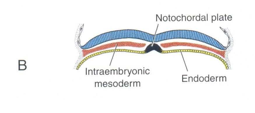
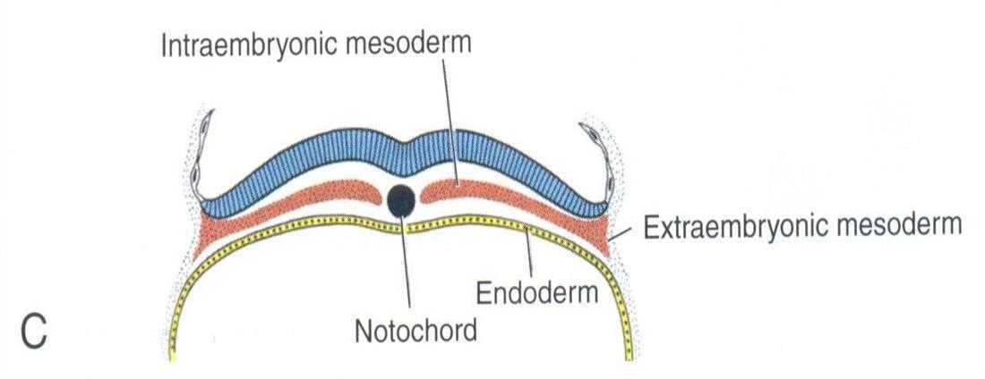
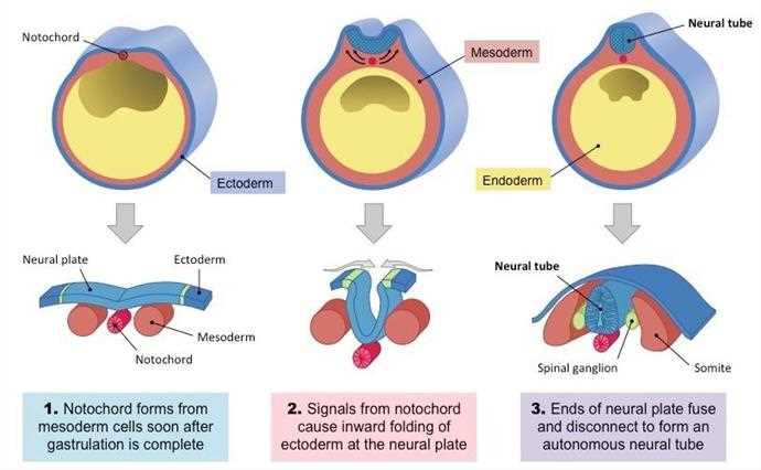
Towards day 17 of the primitive plaque a new proliferation of cells forms in depth: the notochord. In front of the primitive streak lies the primitive node, mesodermal cells that will form the notochord. The notochord activates (induces) the overlying ectoderm to form the neural plate. Portions of the mesoderm that do not form the notochord divide into sections called somites.
By the end of the third week the trilaminar embryonic disc has a definite orientation. During the fourth week the embryo begins to form a tubular structure.
Cross sections
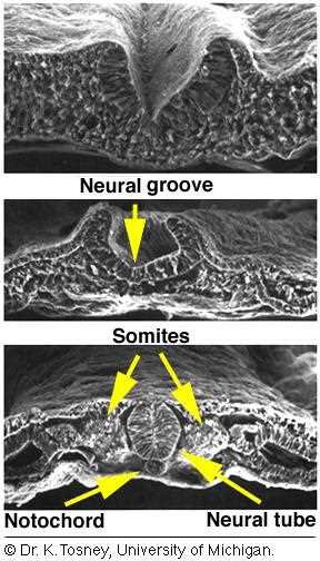
In cross section the notochordal plate lies in the midline between the intraembryonic mesoderm on both sides, beneath the ectoderm and above the endoderm; the notochord then separates and the neural groove, neural tube, somites and notochord appear in order.
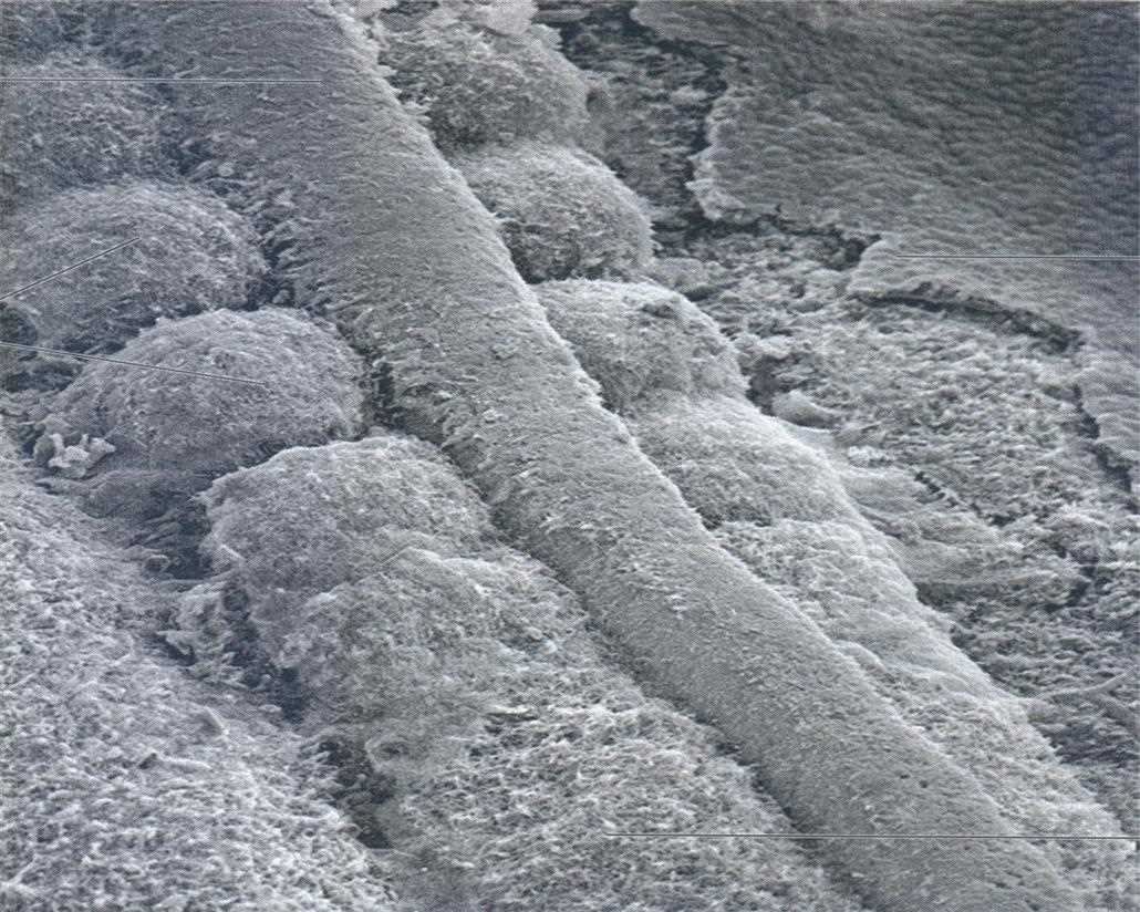
Sagittal view: primitive pit and neuroenteric canal, connecting stalk, amnion, ectoderm, notochord, buccopharyngeal membrane, wall of yolk sac, allantois, cloacal plate (membrane) (A)Cross section: notochordal plate, intraembryonic mesoderm and endoderm (B)Cross section: intraembryonic mesoderm, extraembryonic mesoderm, endoderm and the notochord (C)Primitive streak and neurula: primitive node, pericardial area, neural folds, neural tube, notochord, somite, gut, coelom, primitive streak, somites, with the key ectoderm, mesoderm, endoderm (a, b, c)Scanning electron micrograph of the notochord as a ridge between the somitesThree steps: 1 the notochord forms from mesoderm cells soon after gastrulation is complete; 2 signals from the notochord cause inward folding of the ectoderm at the neural plate; 3 ends of the neural plate fuse and disconnect to form an autonomous neural tube
11. Somites and the sections of the embryo
note
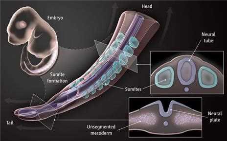
During the fourth week portions of the mesoderm that do not form the notochord divide into sections called somites. They are specific body regions and structures. There are 38 somite pairs; they give rise to most of the skeletal structures of the head, neck and trunk.
From the student notes: somites are segmented blocks of mesodermal tissue that form on both sides of the neural tube. They are first observed during the third week and are one of the earliest visible signs of body segmentation.
Formation: the notochord induces the overlying mesoderm to differentiate into paraxial mesoderm, which becomes segmented into somites (somitogenesis).
Order: the somites form in a cranio-caudal (head-to-tail) sequence along the length of the embryo.
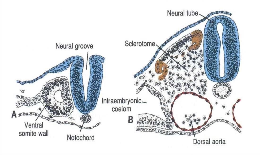
Derivatives: the sclerotome (medial part of each somite) forms the axial skeleton, vertebrae and ribs; the dermatome (lateral part) forms the dermis of the skin; the myotome (intermediate part) forms skeletal muscles of the trunk, limbs and diaphragm.
Each pair of somites is a segment that contributes to a specific region of the body.
Abnormalities in somitogenesis can lead to vertebral defects, scoliosis and other musculoskeletal abnormalities.
Somite formation: embryo, head, neural tube, somites, unsegmented mesoderm, neural plate, tailCross section of the neural groove and neural tube with ventral somite wall, notochord, sclerotome, intraembryonic coelom, dorsal aorta (A and B)
12. The embryonic period and its sensitivity
note
The embryonic period runs from the second to the eighth week. The mass of cells becomes an embryo, the different tissues develop, and the organs and organ systems are organised. The heart is formed in the fourth week. The face and the oral structures are formed between the 4th and 7th week.
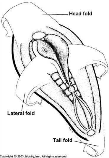
Folding of the embryo
The embryo is folded by the head fold, the lateral folds and the tail fold. The development of a head fold is critical to the formation of the primitive oral cavity. Details are given with the 4th week in topic I.02.
Folding embryo: head fold, lateral fold and tail fold
Clinical comment: teratogens
Environmental teratogens may affect the development of normal cells, tissues, organs or organ systems. The smaller and less complex the development, the less extensive the problem created. Development is also related to timing. Tissues are most susceptible to defective development when they begin to differentiate in the embryonic period (2 to 8 weeks).
Epithelial-mesenchymal interactions
Epithelial-mesenchymal interactions are the necessary interactions of an epithelium and the underlying mesenchyme that determine the terminally differentiated tissue. They matter for the face and the teeth.
Formation of the neural tube and the fate of the germ layers
The key events of the first weeks: the series of events leading to the formation of the three-layered (triploblastic) embryo during the first 3 weeks of development involves cell proliferation and migration. During the next 3 to 4 weeks the major tissues and organs differentiate from the triploblastic embryo. The key events are the differentiation of the nervous system and neural crest tissues from the ectoderm, the differentiation of the mesoderm, and the folding of the embryo.
13. Neurulation and the neural tube
note
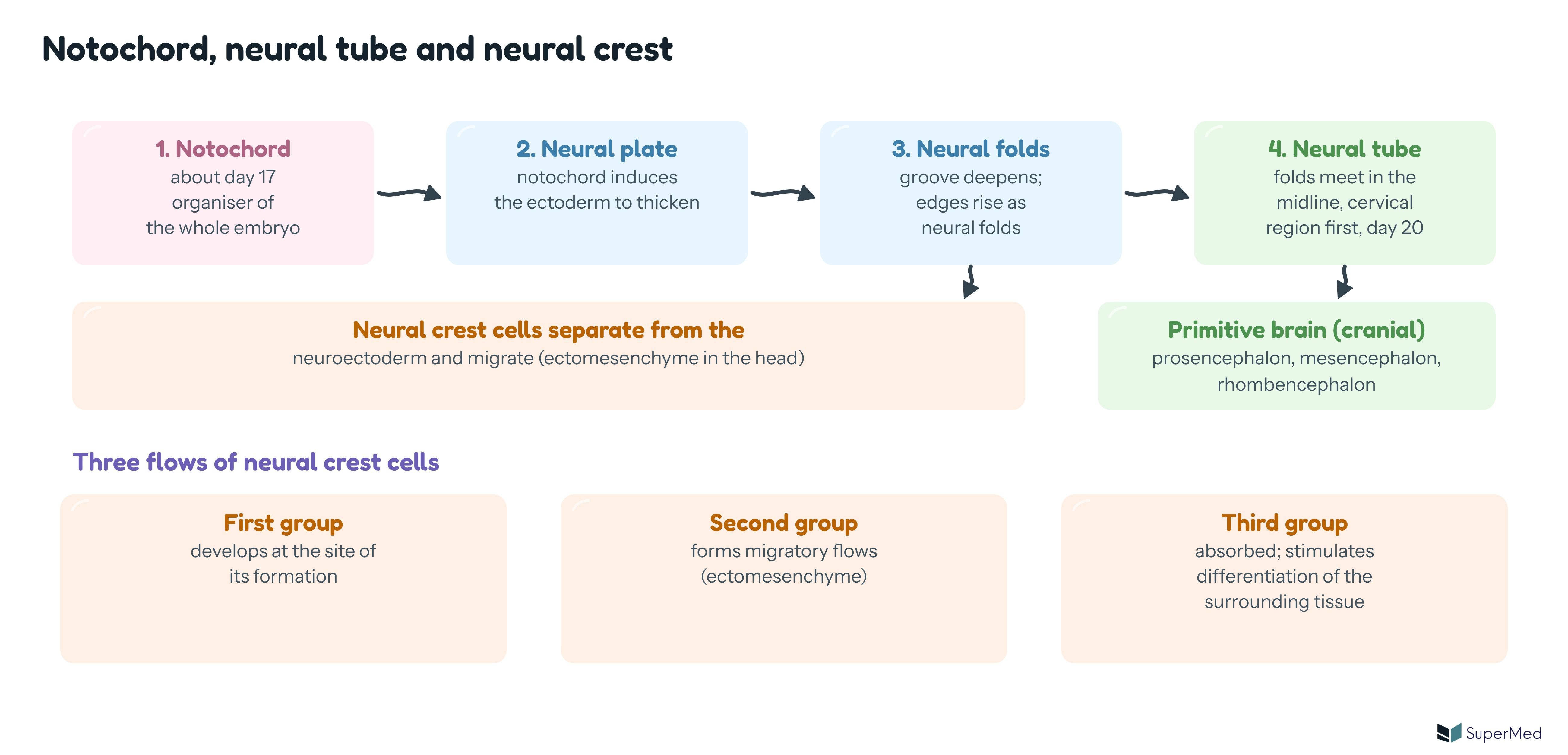
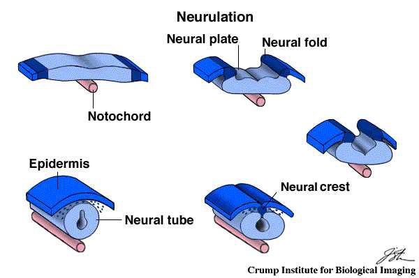
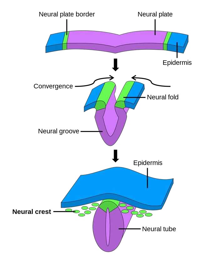
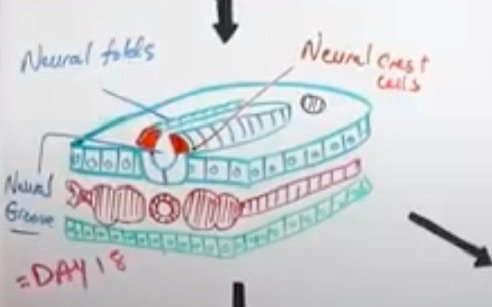
The cranial part of the embryo grows first. In the ventral direction the pharyngeal membrane is formed. Distally over the notochord starts an ectodermal thickening, the neural plate. The neural folds appear during the third prenatal week: the lateral edges of the neural plate begin to elevate as folds, arising dorsally. These folds represent the first change in shape of the embryo's body from the flat sheet of cells.
At approximately 20 days the gastrula enters the next phase, neurulation. The neural folds reach the midline; the neural tube closes first in the cervical region and then closes both anteriorly and posteriorly.
Elements of neurulation
The elements are the neural plate, the neural tube and the neural crests. The steps:
A neural plate forms in the cranial end (neuroectoderm: ectodermal cells above the notochord thicken and elongate).
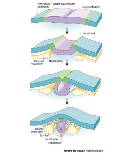
At the end of the third week the lateral edges of the plate become elevated and move together to form the neural folds. The space created is the neural groove.
The neural folds fuse and the plate becomes the neural tube. Fusion begins at the future cranial end and proceeds towards the caudal end.
During closure the cells at the crest of the neural folds detach, forming the neural crest cells.
When the neural tube has fused completely, neurulation is complete and the tube is separated from the surface ectoderm.
The neural groove deepens and is surrounded by the two neural folds, and the neural crest cells develop from these folds. The neural tube gives rise to the central nervous system (brain and spinal cord). It has a hollow lumen.
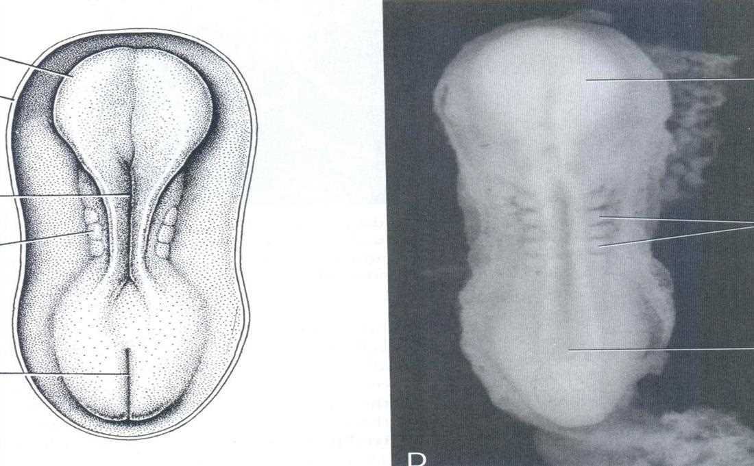
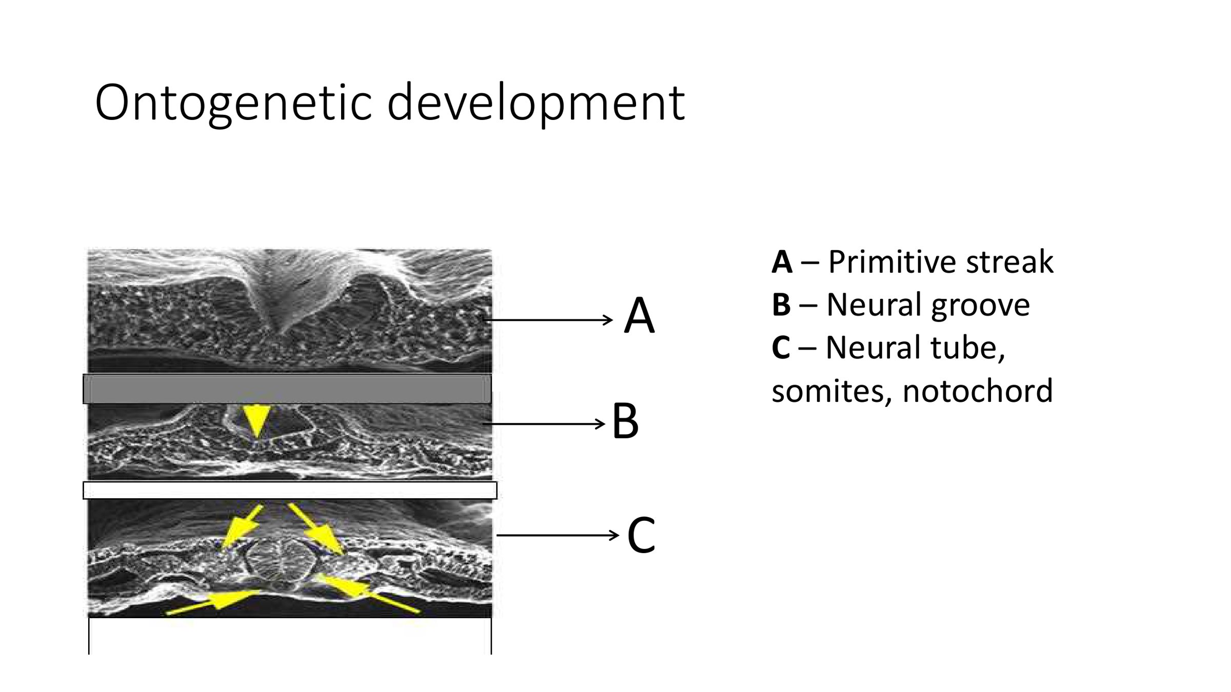
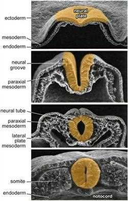
Schematic: the notochord induces the neural plate; the plate invaginates to the groove with raised folds; the folds meet to form the neural tube, and neural crest cells separate and migrate (three flows)Dorsal view of the embryo: neural plate, primitive node, primitive streak, cut edge of amnion (A) and embryo photograph (B)Dorsal view of a later embryo with the neural folds, somites and primitive streak (C, D)Neurulation: neural plate, neural fold, notochord, epidermis, neural crest, neural tube (Crump Institute)Neural plate border, neural plate, convergence of the neural folds, neural groove, epidermis, neural crest and neural tube in three stepsNeurulation in three SEM sections: neural groove, somites, notochord and neural tube (yellow arrows)Practical exam slide 9, Ontogenetic development: A primitive streak, B neural groove, C neural tube, somites and notochordTen Cate: scanning electron micrographs of the formation and closure of the neural fold elevations; ectoderm, neural plate, neural groove, neural tube, paraxial mesoderm, lateral plate mesoderm, somite, notochord, endodermNeurulation with non-neural ectoderm, neural plate border, neuroectoderm, neural fold, paraxial mesoderm, neural plate, neural crest cells, somite, neural tube, notochord
Primitive brain
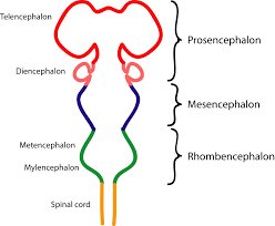
In the cranial part the neural tube builds the primitive brain. At this stage it consists of the prosencephalon, the mesencephalon and the rhombencephalon. The bilateral neural crests stimulate the maxillofacial area. In the head region the anterior part of the tube expands as forebrain, midbrain and hindbrain; the hindbrain forms 8 bulges, the rhombomeres.
In the student notes: the cranial (anterior) portion expands to form the prosencephalon (forebrain), the middle portion the mesencephalon (midbrain) and the caudal portion the rhombencephalon (hindbrain). These primary vesicles differentiate into secondary vesicles: the prosencephalon gives the telencephalon and diencephalon, the mesencephalon remains relatively unchanged, and the rhombencephalon gives the metencephalon and myelencephalon.
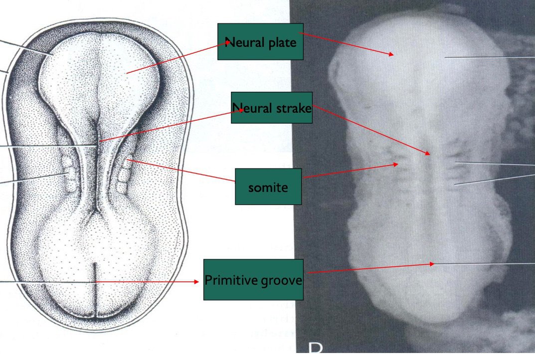
Dorsal view of the embryo with neural plate, neural streak (groove), somite and primitive groove labelled on a drawing and a photographBrain vesicles: telencephalon, diencephalon (prosencephalon); mesencephalon; metencephalon, myelencephalon (rhombencephalon); spinal cord
14. The neural crest and the ectomesenchyme
note
As the neural tube forms, a group of cells separates from the neuroectoderm. These neural crest cells have the capacity to migrate and differentiate extensively within the developing embryo. They are the basis for nerve structures. The neural crests are bilaterally paired strips of cells arising in the ectoderm at the margins of the neural tube.
The neural crest in the head
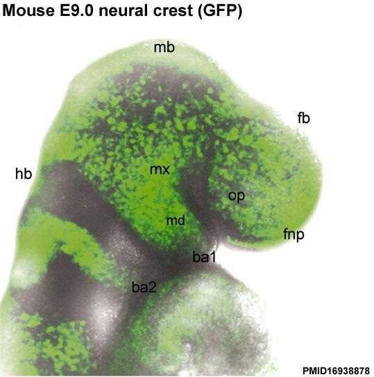
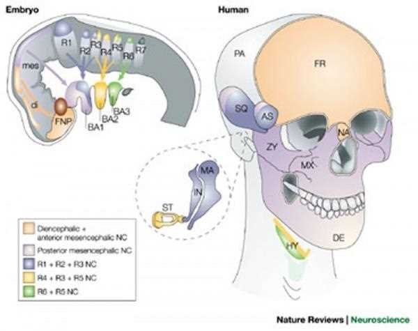
Neural crest cells in the head region have an important role. They differentiate to form most of the connective tissue of the head. Embryonic connective tissue elsewhere is derived from mesoderm and is known as mesenchyme; in the head it is known as ectomesenchyme, reflecting its origin from neuroectoderm. In the head region neural crest cells migrate into the pharyngeal arches and form ectomesenchyme, contributing tissues that in the body region are typically derived from mesoderm.
Neural crest cells are a migratory cell population that gives rise to the majority of the cartilage, bone, connective tissue and sensory ganglia in the head. Many different systems (neural, skin, teeth, head, face, heart, adrenal glands, gastrointestinal tract) have a contribution from neural crest cells.
Exam tip: All tissues of the tooth (except enamel) and its supporting apparatus are derived directly from neural crest cells. The proper migration of the neural crest cells is essential for the development of the face and the teeth.
Three flows
The neural crest cells divide rapidly. Three cell flows with different biological functions form:
the first group develops at the site of its formation;
the second group forms migratory flows;
the third group is intended to be absorbed and stimulates differentiation of the surrounding tissue.
Ectomesenchyme
The migration flows of the neural crest are called ectomesenchyme. They represent the fourth germ layer. They are another source (besides the primitive mesoderm) for mesenchymal tissue.
Migration pathway
Ectomesenchymal cells transmit active products into the intercellular spaces: fibronectin and glycosaminoglycans. The migration pathway of the neural crest cells from the neural folds is achieved by these products. Their broadcasting stops when the cells reach their destination.
From the student notes: when the crest cells migrate they differentiate into sensory neurons, autonomic neurons, glial cells, pigment cells (melanocytes), parts of the skull and face and other tissues. Dysfunction of the neural crest can lead to a variety of congenital conditions known as neurocristopathies.
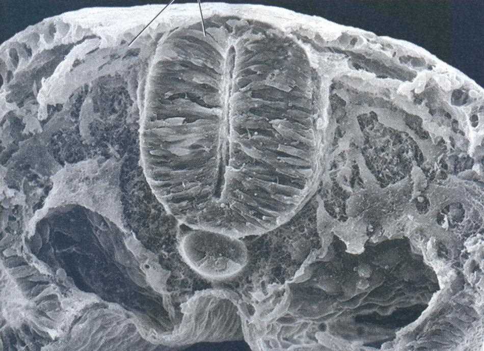
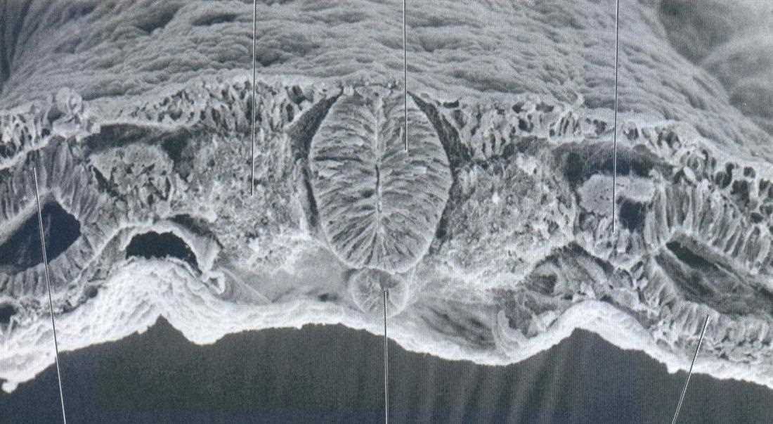
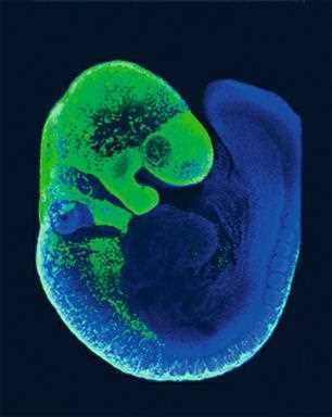
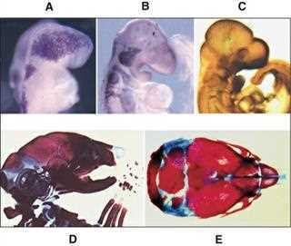
Embryo (stage 11, day 25, 20 somites) with the neural crest cells enlarged in the cranial regionMouse embryo at E9.0 with neural crest cells shown in green: mb midbrain, fb forebrain, hb hindbrain, op optic, fnp frontonasal process, mx maxillary, md mandibular, ba1 and ba2 branchial archesScanning electron micrograph, transverse section: neural tube and neural crests (lecture labels: neural tube, neural crests)Scanning electron micrograph, transverse section of the embryo (lecture labels: somites, neural tube, notochord, paraxial mesoderm, somatic mesoderm)Neural crest in sections A and B and derivatives in C: dorsal root ganglion, sympathetic ganglion, developing suprarenal gland, preaortic ganglion, enteric ganglia, urogenital ridgeThe embryo with colonisation of the head and pharyngeal arches by diencephalic, anterior and posterior mesencephalic and rhombencephalic neural crest cells, colour coded, with the skull regions they form on the human headTen Cate: migration of neural crest cells throughout the embryo traced in a Pax3-GFP transgenic mouse model (green)Ten Cate: migration and differentiation of cranial neural crest cells (A migrating NCC, B and C neuronal differentiation, D skeletal differentiation, E neurocranium, bone red and cartilage blue)Hand-drawn student diagram: neural folds, neural crest cells, neural groove, day 18
15. Parts of the embryonic tube and the embryo at 22 to 28 days
note
The embryo in the fourth week is a tube with three parts:
Part
Forms
Pars cranialis (the fastest growing)
the head
Pars umbilicalis
the torso with its organs
Pars caudalis
the limbs
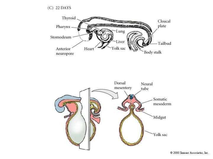
The cranial part is the largest. At about day 22 the embryo carries the stomodeum, pharynx, thyroid, lung, heart, liver, yolk sac, body stalk, tailbud, cloacal plate and the anterior neuropore.
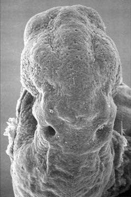
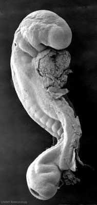
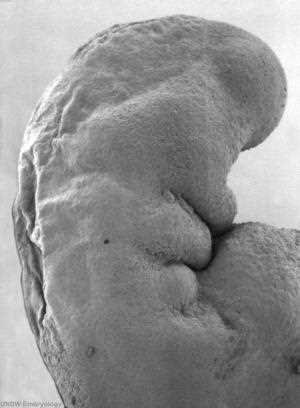
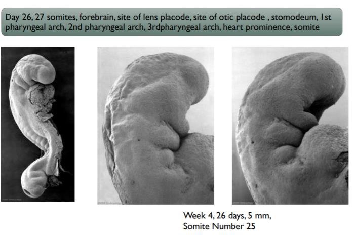
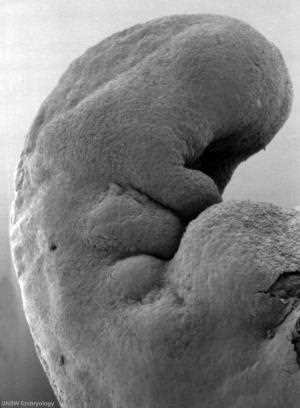
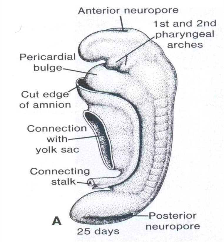
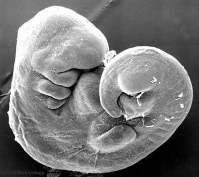
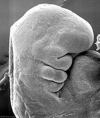
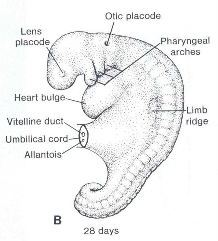
The embryo at day 26 to 27 shows: 27 somites, forebrain, site of the lens placode, site of the otic placode, stomodeum, 1st, 2nd and 3rd pharyngeal arch, heart prominence and somites. At week 4, day 26, the embryo is 5 mm long and has 25 somites.
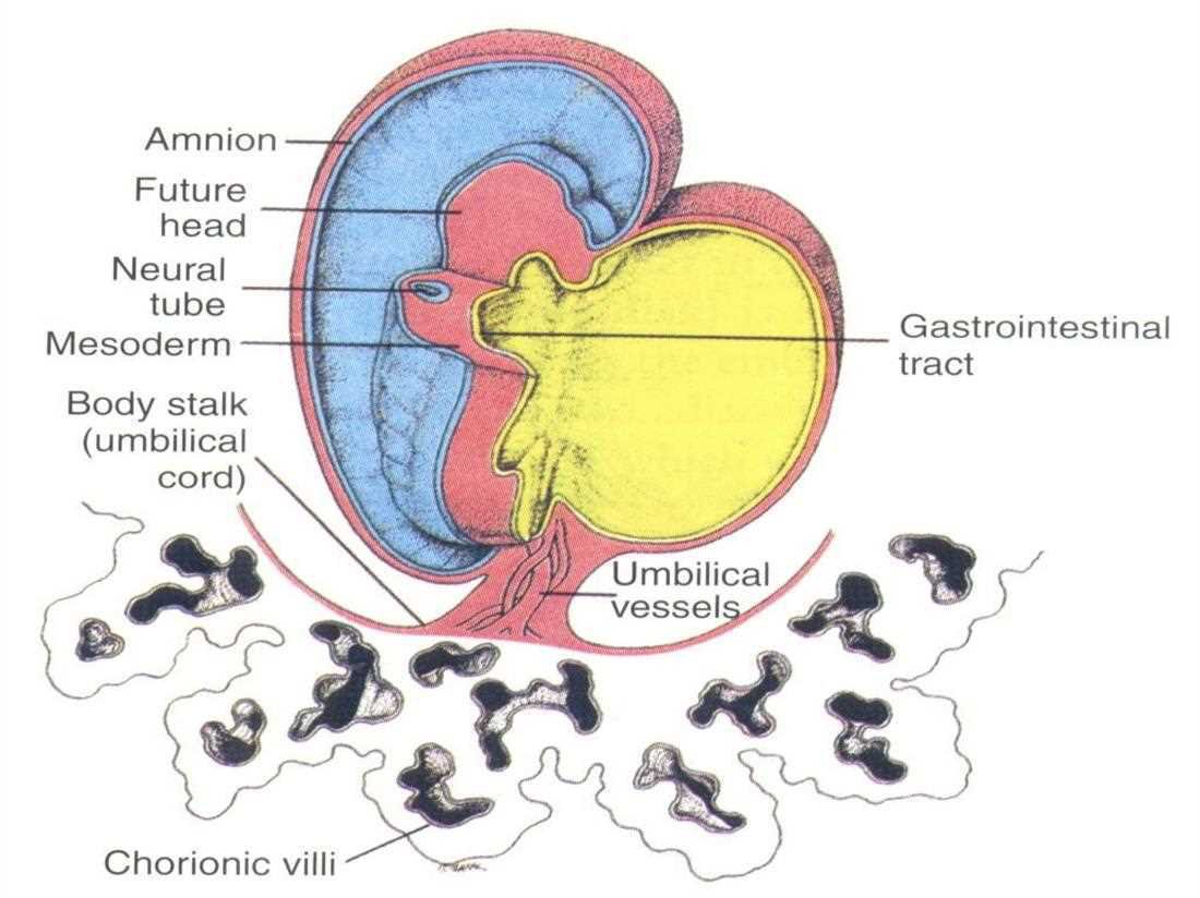
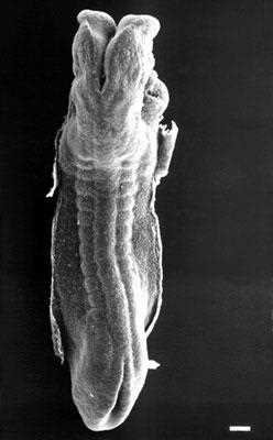
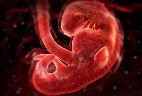
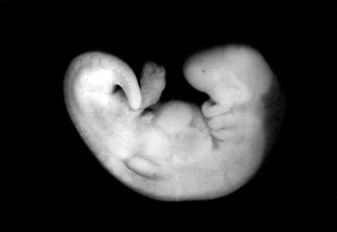
C: embryo at 22 days (thyroid, pharynx, lung, stomodeum, anterior neuropore, heart, liver, yolk sac, cloacal plate, tailbud, body stalk) and below a cross section: dorsal mesentery, neural tube, somatic mesoderm, midgut, yolk sacEmbryo with amnion, future head, neural tube, mesoderm, body stalk (umbilical cord), umbilical vessels, chorionic villi and the gastrointestinal tractScanning electron micrograph, dorsal view of the embryo: open neural folds cranially, somites along the sidesScanning electron micrograph of the cranial end of the embryo seen from the frontWhole embryo at week 4, day 26, 5 mm, somite number 25, from the side (SEM)Head of the 26-day embryo seen from the side: forebrain bulge, pharyngeal arches and heart prominence (SEM)Student notes: day 26 to 27 embryo with 27 somites, forebrain, sites of the lens and otic placodes, stomodeum, 1st to 3rd pharyngeal arches, heart prominence; SEMs of week 4, 26 days, 5 mm, somite number 25Head of the 26-day embryo, second view, with the pharyngeal arches and the heart prominenceEmbryo at 25 days: anterior neuropore, 1st and 2nd pharyngeal arches, pericardial bulge, cut edge of amnion, connection with the yolk sac, connecting stalk, posterior neuroporeScanning electron micrograph of the embryo head and pharyngeal arches with the heartCloser scanning electron micrograph of the head, pharyngeal arches and heartEmbryo at 28 days: lens placode, otic placode, pharyngeal arches, heart bulge, vitelline duct, umbilical cord, allantois, limb ridgeThree-dimensional drawing of a human embryo in the fourth week with its curved bodyHistology practical: human embryo in the stage of the three parts, with pars cranialis, pars umbilicalis and pars caudalis
16. The fetal period and the morphogenetic mechanisms
note
The fetal period continues until birth. Both values are found in the lectures for its start: the 8th week (p173) and the 9th week (p3). During this period growth continues, the already formed tissues and organs differentiate, and the fetus is prepared for function. All major structures are already formed but they continue to grow and develop.
As all major organs are formed, the fetus is not as sensitive to damage from environmental exposure as the embryo was. Instead, toxic exposure often causes physiological abnormalities or minor congenital malformation. The major organs continue to develop and mature, and the fetus becomes increasingly capable of independent survival outside the womb as it nears the end of the pregnancy.
Morphogenetic mechanisms for the formation of the three-dimensional embryo
A summary of what has happened until now, in five steps:
Developing mesoderm converts the bilaminar embryonic disk to a trilaminar disk; this involves cell proliferation and migration.
Formation of the neural plate and primary brain ventricles.
Formation of the neural tube and neural crests; formation of migratory flows, stimulating differentiation.
Multiple stimulation between the neural crests and the ectoderm.
Expression of different biophysical impacts associated with morphogenetic changes.
Every account starts free, with free topics in Cytology, Medical Biology, Anatomy, Biophysics, Pharmacology and Preclinics of Pediatric Dental Medicine. Super opens the rest. Super is €15 a month.