


Preclinics of Prosthetic Dental Medicine · Years 1-2 · Medical University of Sofia

01

Prosthetic Dental Medicine - subject, orofacial complex, maxillofacial region and masticatory system

Free notes for topic 01 of the Preclinics of Prosthetic Dental Medicine syllabus, open without an account. Written by a senior student against the syllabus question and checked line by line by a second student before publishing. How content is made

Updated

The short version

note

  • Prosthetic Dental Medicine (PDM) explores, prevents and treats permanent alterations in the dental arches and the maxillofacial region which violate mastication, speech and aesthetics.
  • Permanent alterations come from tooth loss, tooth decay or fracture, congenital defects such as cleft palate, and acquired jaw and facial defects, usually after cancer surgery.
  • A prosthesis (from the Greek prothesis, an artificial device for replacing part of the body) restores, replaces, consolidates or protects organs and tissues of the masticatory system.
  • There are two basic types of prosthetic restoration. Fixed restorations are permanently fixed to natural teeth and cannot be removed by the patient. Removable restorations can be removed, and should be removed by the patient, for cleaning and disinfection.
  • The masticatory system is made of five sections: supporting, motor, secretory, neuro-trophic and soft (covering) tissues.
  • The orofacial complex is the broadest term. The maxillofacial region is the area from the forehead to the chin, and the masticatory system is the functional system that lets you chew and move your jaw.
  • The masticatory system carries out five functions: mastication, speech, facial expression, swallowing and aesthetics.
  • The textbook treats the shape of human dentition as the expression of its phylogenetic evolution.
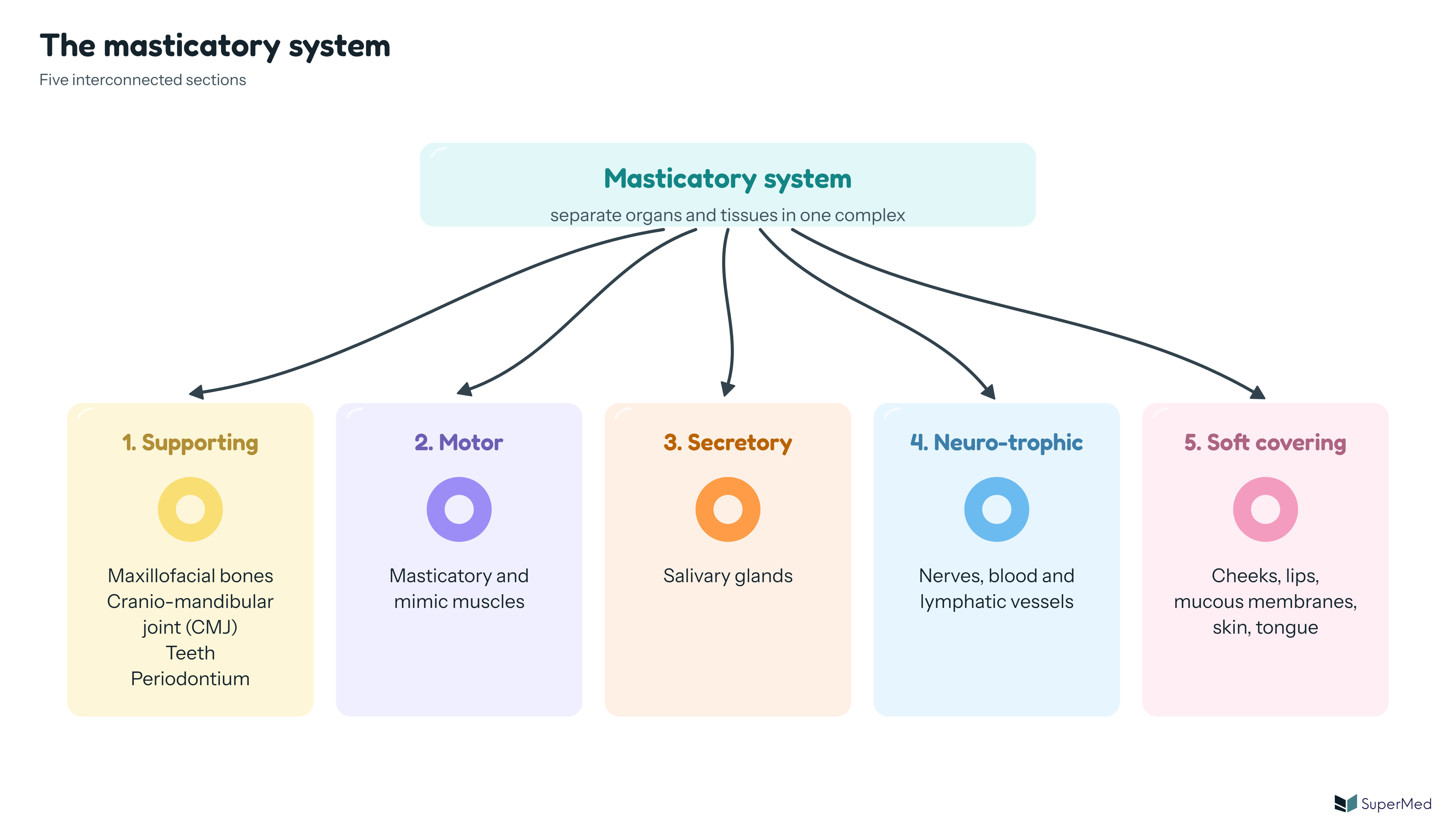
Schematic: the masticatory system with five sections: 1. Supporting (maxillofacial bones, cranio-mandibular joint, teeth, periodontium), 2. Motor (masticatory and mimic muscles), 3. Secretory (salivary glands), 4. Neuro-trophic (nerves, blood and lymphatic vessels) and 5. Soft covering (cheeks, lips, mucous membranes, skin, tongue)

1. Prosthetic Dental Medicine: subject and scope

note

Prosthetic Dental Medicine (PDM) is the branch of dental medicine concerned with diagnosing, preventing and treating anatomical and functional changes in the dentition and the maxillofacial region. Its aims include restoring mastication, speech, aesthetics and overall orofacial harmony.

The textbook gives the object of PDM in the same terms: the exploration, prevention and treatment of permanent alterations in the dental arches and the maxillofacial region, which violate mastication, speech and aesthetics.

PDM focuses on permanent alterations in the oral and facial structures. The permanent alterations it can explore are:

  • loss of hard tissue as a result of tooth decay
  • tooth fracture from trauma
  • single or multiple tooth loss
  • congenital jaw deformities (cleft palate is an example)
  • acquired jaw and facial defects, usually as a result of cancer and the surgical treatment that follows (trauma is also an example)
  • completely edentulous dental arches

Defects in the maxillofacial region, such as congenital clefts, trauma or surgical defects, need complex prosthetic rehabilitation.

2. Prosthetic restorations: types and purpose

note

The restorative devices of prosthetic dental medicine are special individual constructions. The textbook calls them prosthetic constructions, prostheses or simply dentures. The word prosthesis is Greek and means an artificial device for replacing part of the body.

Prostheses are used to:

  • replace or restore organs and tissues of the masticatory system which have been destroyed, lost or are not aesthetic
  • consolidate teeth and tissues after injury
  • prevent further pathological changes or destruction in oral structures
  • protect and stabilise the surrounding tissues

There are two basic types of prosthetic restoration.

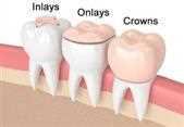
Two inlays, onlays and crowns, shown on a row of teeth from left to right: an inlay, an onlay and a full crown
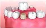
Fixed bridge: a pontic filling a gap between two prepared teeth, with a crown on each abutment
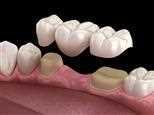
Fixed bridge over a row of teeth: crowns joined as one unit, with the pontic filling the gap

Fixed prosthetic restorations

note

Fixed prosthetic restorations are permanently fixed to natural teeth by means of special glue (cement). The patient cannot remove them.

Examples given by the textbook: inlay, onlay, partial crown, complete crown, fixed bridge and splint.

Fixed restorations can be cemented onto implants according to some accounts, while the textbook says natural teeth.

Removable prosthetic restorations

note

Removable prosthetic restorations can be removed, and should be removed by the patient for cleaning and disinfection.

Examples given by the textbook: acrylic resin partial dentures, one-piece cast framework partial dentures, partial dentures with precision attachments, complete (full) dentures, removable surgical prostheses and facial prostheses.

The textbook notes that although fixed restorations are always preferred by patients and clinicians, the best choice is sometimes a removable denture rather than a fixed restoration.

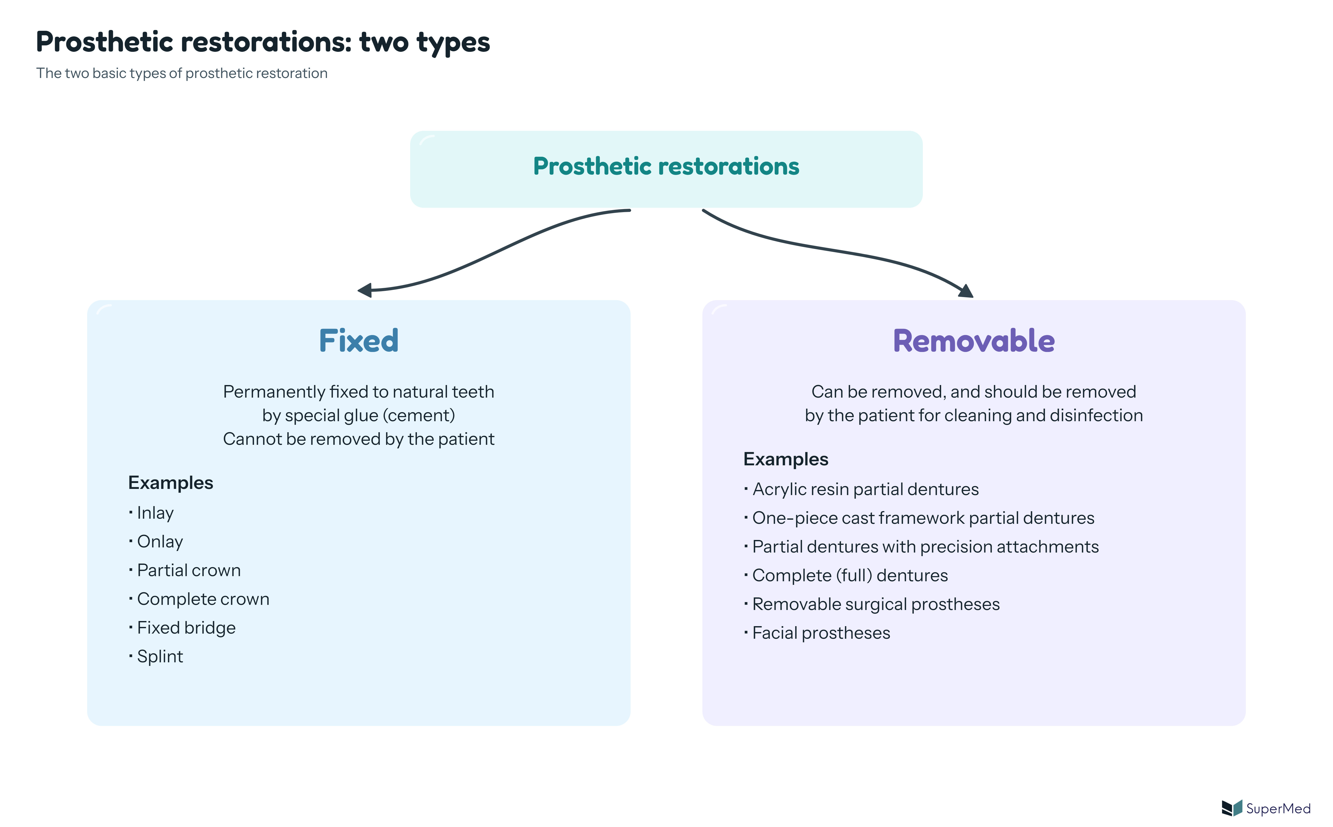
Schematic: prosthetic restorations split into two types. Fixed: permanently fixed to natural teeth by special glue (cement), examples inlay, onlay, partial crown, complete crown, fixed bridge, splint. Removable: can be removed and should be removed by the patient for cleaning and disinfection, examples acrylic resin partial dentures, one-piece cast framework partial dentures, partial dentures with precision attachments, complete dentures, removable surgical prostheses, facial prostheses
FeatureFixed restorationRemovable restoration
Attached toNatural teeth, permanently, by special cementRemoved from the mouth by the patient
Who removes itNot the patientThe patient, for cleaning and disinfection
ExamplesInlay, onlay, partial crown, complete crown, fixed bridge, splintAcrylic resin partial dentures, one-piece cast framework partial dentures, partial dentures with precision attachments, complete dentures, removable surgical prostheses, facial prostheses

3. The orofacial complex

note

The orofacial complex refers to the functional and anatomical structures that contribute to chewing, swallowing, speaking, breathing and facial expression. It is described as an organ system of its own, which works in an integrated manner to provide the functions of mastication, swallowing, breathing and speech, and of smell, taste and vision.

It includes both hard and soft tissues:

  • hard tissues: maxilla, mandible, cranio-mandibular joint (CMJ), teeth
  • soft tissues: tongue, lips, cheeks, skin, mucosa
  • muscles: masticatory muscles and the muscles of facial expression (mimic muscles)
  • secretory glands: major and minor salivary glands
  • neurovascular supply: sensory and motor nerves, arterial and venous systems, and lymphatics

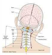
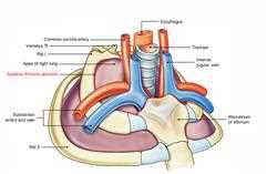
The orofacial complex is associated with the brain, the neck, the cervical spine and the thoracic inlet.

Head and neck: skull, brain, brain stem, cervical spine vertebrae C1 to C7 and spinal cord, labelled
Thoracic inlet: oesophagus, trachea, manubrium of the sternum, subclavian arteries and veins, internal jugular vein and the apex of the right lung, labelled

4. The maxillofacial region

note

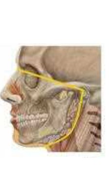
The maxillofacial region is the anatomical area from the forehead to the chin. It includes:

  • craniofacial bones: maxilla, mandible, zygomatic, nasal and frontal bones
  • soft tissues: facial skin, lips, muscles and mucosa
  • joints: primarily the cranio-mandibular joint (CMJ), also called the TMJ

The maxillofacial region is important for biomechanical function (mastication and jaw movement), facial aesthetics and speech articulation.

The maxillofacial region is the bones and soft tissues of the face and jaws, which is the anatomical focus. The masticatory system is the functional focus (see section 8).

Lateral view of the skull and jaws, with the maxillofacial outline traced in yellow around the maxilla and mandible

5. The masticatory system and its five sections

note

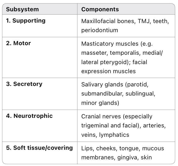
The masticatory system is composed of separate organs and tissues in the maxillofacial region, united in a complex that is responsible for the masticatory function of chewing. A completely formed masticatory system is made up of five basic sections.

SectionComponents (textbook)Detail
SupportingMaxillofacial bones, cranio-mandibular joint (CMJ), teeth, periodontiumMaxilla, mandible, TMJ, teeth, periodontium
MotorMasticatory and mimic musclesMasticatory muscles (masseter, temporalis, medial and lateral pterygoid) and facial expression muscles
SecretorySalivary glandsParotid, submandibular, sublingual and minor salivary glands
Neuro-trophic (nutritional)Nerves, blood and lymphatic vesselsCranial nerves (especially trigeminal and facial), arteries, veins and lymphatics
Soft (covering) tissuesCheeks, lips, mucous membranes, skin, tongueLips, cheeks, tongue, mucous membranes, gingiva and skin
Table of the five subsystems of the masticatory system: 1. Supporting (maxillofacial bones, TMJ, teeth, periodontium), 2. Motor (masticatory muscles and facial expression muscles), 3. Secretory (salivary glands), 4. Neurotrophic (cranial nerves, arteries, veins, lymphatics), 5. Soft tissue or covering (lips, cheeks, tongue, mucous membranes, gingiva, skin)

The motor section includes the mimic muscles, such as occipitofrontalis, temporoparietalis, nasal, buccinator, zygomatic and mentalis. The muscles themselves are described in the topic on masticatory and mimic muscles.

6. Functions of the masticatory system

note

The masticatory system is made of five interconnected subsystems, and together they do the following jobs.

FunctionWhat takes part
Mastication (chewing)Coordinated activity of the teeth, muscles, TMJ and saliva
Speech articulationPrecise positioning and movement of the tongue, lips and teeth
Facial expressionMuscles innervated by the facial nerve (cranial nerve VII)
Swallowing (deglutition)Coordinated by the tongue, soft palate and pharyngeal muscles
AestheticsProper alignment of the dental arches and facial support, which prevents an aged or sunken appearance

The textbook names the same three areas that PDM treats when they are violated: mastication, speech and aesthetics.

7. Phylogenetic development of the maxillofacial region

note

This section covers the phylogenetic background that the textbook gives for the masticatory system and the maxillofacial region. The ontogeny and eruption theories that follow in the textbook belong to other topics.

Phylogenetics is the study of evolutionary relationships among groups of organisms (species and populations). It is discovered through molecular sequencing data and morphological data matrices. The textbook derives the term from the Greek words phyle and phylon, denoting tribe and birth. In the past, phylogenetic relationships were reconstructed from phenotypes, often anatomical characteristics. Today molecular data, including protein and DNA sequences, are used to build phylogenetic trees. Evolution takes place over long periods that cannot be observed directly, so biologists reconstruct phylogenies by inferring the evolutionary relationships among present-day organisms. In biological systematics as a whole, phylogenetic analyses have become essential in researching the evolutionary tree of life.

Physical anthropology is one of the most important divisions of phylogeny. It is the science of humans in their biological aspect: origin, anatomy and physiology of the different nationalities. Other branches are palaeontology, anthropometry and forensic anthropology. Anthropometry measures the physical characteristics of people.

Palaeontology is the study of fossils to determine the evolution of organisms and their interactions with each other and with their environments (palaeoecology). As a historical science, it explains causes rather than conducting experiments to observe effects. Body fossils and trace fossils are the main types of evidence about ancient life. Geochemical evidence helps to study the evolution of life before there were organisms large enough to leave fossils.

Palaeodentistry is the palaeontological study of teeth. Its objects of investigation are:

  • teeth anatomy
  • skeleton variations
  • the TMJ and cheek bone alterations
  • craniometry

Its specific observations include the Carabelli and George cusps, spacing and diastemata, groove depths, the eminence of dental crowns, and dental caries and erosions in different types of humans.

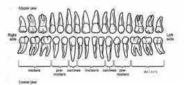
Upper and lower dentition chart, seen from the front, with right and left sides labelled and the groups of molars, premolars, canines and incisors marked

Phylogenetic terms and classification

note

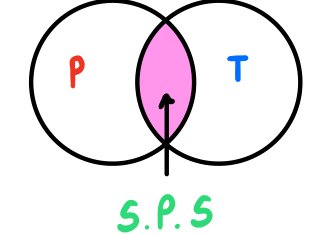
Taxonomy is the classification, identification and naming of organisms. It is richly informed by phylogenetics, but remains methodologically distinct. The two fields overlap in phylogenetic systematics, which is one method used to create cladograms and to delimit taxa (clades). A group whose members all descend from one common ancestor is called monophyletic.

Venn diagram: two overlapping circles labelled P (phylogenetics) and T (taxonomy). The overlap, labelled S.P.S., is the science of phylogenetic systematics, which creates cladograms

Phylogenesis (phylum meaning order or class, and genesis meaning origin) involves:

  • morphological characteristics of organisms, sorted into categories and periods (morphogenesis)
  • the development and evolutionary history of species in taxonomic groups
  • the origin and history of the evolution of species

The evolutionary connections between organisms are shown graphically in phylogenetic trees. Evolution is a branching process, in which populations change over time and may split into separate branches, hybridise together or end in extinction. Classification uses features such as the mouth (stoma), movable cartilaginous jaws, the tongue and the number of teeth (homodont and polyphyodont dentitions).

Palaeogenesis gives information about the evolution of human organs. For example, in amphibians the first mouth formation is found; in reptiles the palate, bone and glands appear first; and in crocodiles cement and periodontium can be seen for the first time.

Comparative anatomy, as part of primatology, investigates the different types of human: Australopithecus from Africa, Paranthropus, Neanderthals, Homo erectus and Homo sapiens. Primates have incongruent temporomandibular joints, which differ in the angle of the articular heads. The shape of human dentition reflects all of its functions. Human dento-maxillary anatomy is the expression of phylogenetic evolution.

The study of comparative anatomy, dental phylogenetics and anthropology allows the dentist to understand the present form and function of human dentition. The study of human dentition has interested not only dentists but also palaeontologists, anthropologists and geneticists, who focus on dental anatomy and the phylogenesis of human dentition as the result of an evolutionary process.

Evolutionary timeline of the masticatory system

note

The textbook gives the following approximate timeline. The dates are as the textbook gives them.

PeriodDevelopment given in the textbook
Before about 3.5 billion yearsStromatolites appear
Cambrian period onwardsEvolution of fish, amphibians, reptiles, birds and mammals, over a long period
Before about 65 million yearsEvolution of primates begins
Before about 4.5 million yearsReal human evolution begins, when bipedal primates appear; this period is characterised by the evolution of the human brain
Modern human epochsPleistocene, Upper Palaeolithic and Holocene
Before about 45,000 yearsThe late glacial period
Before about 11,000 yearsCultural development: industry, agriculture, science and mathematics, and the beginning of contemporary urbanisation

8. Key facts and comparison tables

note

Three terms are used in this topic, and they describe the same body from different angles.

TermScopeFocus
Orofacial complexStructures that contribute to chewing, swallowing, speaking, breathing and facial expressionBroadest term: everything involved in facial and oral function
Maxillofacial regionBones and soft tissues of the face and jaws, from the forehead to the chinAnatomical focus
Masticatory systemSeparate organs and tissues united to chew and move the jawFunctional focus

Key facts to remember:

  • PDM treats permanent alterations, not temporary ones.
  • Fixed restorations are permanently fixed to natural teeth and cannot be removed by the patient.
  • Removable restorations are removed by the patient for cleaning and disinfection.
  • The masticatory system has five sections: supporting, motor, secretory, neuro-trophic and soft (covering) tissues.
  • Mastication, speech and aesthetics are the functions that PDM restores.

All 75 Preclinics of Prosthetic Dental Medicine topics · exam study support, not clinical guidance. SuperMed is not affiliated with the Medical University of Sofia.

Start free. No card needed.

Every account starts free, with free topics in Cytology, Medical Biology, Anatomy, Biophysics, Medical Genetics, Pharmacology, Preclinics of Prosthetic Dental Medicine and Preclinics of Pediatric Dental Medicine. Super opens the rest. Super is €15 a month.

Create a free account