Bone as an organ - types of bones, bone structure, development and growth. Periosteum
Free notes for topic 01 of the Anatomy syllabus, open without an account. Written by a senior student against the syllabus question and checked line by line by a second student before publishing. How content is made
Updated
What this topic covers
note
Bone as a living organ, not just a piece of mineral: how it is classified by shape, how it is built at the organ, tissue and cell level, and how it forms and grows. It covers the four (plus two minor) shape-based classes of bone, the macroscopic structure of a long bone, the trajectory and lamellar architecture of compact and spongy bone, the osteon, the chemical composition of bone and the three bone cell types, bone marrow, the two routes of ossification (intramembranous and endochondral), postnatal growth at the epiphyseal plate, and the biological properties of bone including osteoporosis and fracture. The periosteum and endosteum, the membranes that cover and line every bone, are treated as their own note.
1. Bone as an organ
note
The locomotor apparatus has a passive part, the skeleton, and an active part, the skeletal muscles. The passive part is made of bones (the subject of osteology, osteologia) and the joints that connect them (arthrology, arthrologia); the active part is the skeletal muscles (myology, myologia).
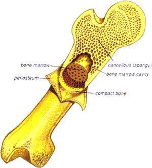
The adult human skeleton contains 206 bones. Each individual bone is built from two kinds of bone tissue: compact bone (substantia compacta, also called cortical bone, substantia corticalis), a dense outer shell, and cancellous or spongy bone (substantia spongiosa), a lattice of bone trabeculae filling the interior. Every bone surface is covered: by periosteum where it is not part of a joint, or by articular cartilage where it takes part in a joint, and the internal surfaces are lined by endosteum.
Femur cut in longitudinal section, labelled: periosteum, compact bone, cancellous (spongy) bone, bone marrow and bone marrow cavity
Bone tissue does more than form the rigid frame of the body: it supports the fleshy soft tissues, provides protected cavities for vital organs, and harbours the bone marrow. Bones are vascular and innervated: a bone is usually supplied by one artery, which enters the marrow directly (the nutrient artery, travelling in the nutrient canal, is shown in the long bone diagrams below).
Periosteum and endosteum
note
The periosteum is the dense connective-tissue membrane that covers the outer surface of a bone everywhere except at its articular surfaces, which are covered by articular cartilage instead, and at the attachment sites of tendons and ligaments. It has two layers: an outer fibrous layer of dense irregular connective tissue carrying the blood vessels and nerves that supply the bone, and an inner, more cellular osteogenic (cambium) layer containing osteoprogenitor cells and osteoblasts. The osteogenic layer is thin and largely inactive in the mature skeleton but becomes active again in bone repair after a fracture, and it is the layer responsible for a bone's growth in width. The periosteum is anchored firmly to the underlying bone by bundles of collagen fibres, Sharpey's fibres, that run into the outer compact bone, and it is through the periosteum that the nutrient artery reaches the nutrient canal on its way into the medullary cavity.
The endosteum is a much thinner, single-layered membrane that lines all of the internal surfaces of a bone: the medullary cavity, the surfaces of the trabeculae of spongy bone, and the walls of the Haversian and Volkmann's canals. It contains osteoprogenitor cells, osteoblasts and osteoclasts, and, like the periosteum's deep layer, is active in bone growth, remodelling and repair.
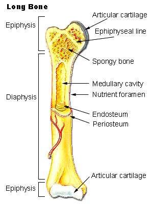
Long bone in longitudinal section, labelled: articular cartilage, epiphyseal line, spongy bone, medullary cavity, nutrient foramen, endosteum and periosteum
2. Types of bones
note
Bones are classified by their shape and proportions into four main groups, plus two smaller groups defined by a structural feature rather than shape:
long (trabecular) bones, ossa longa - most bones of the limbs
short bones, ossa brevia - the bones of the wrist and ankle
flat bones, ossa plana - most of the bones of the skull, and the sternum
irregular bones, ossa irregularia - the bones of the spine, the hip bones, and the bones of the cranial base and facial skull
pneumatic (hollow) bones, ossa pneumatica - the bones of the facial skull
sesamoid bones, ossa sesamoidea - small bones embedded within tendons, such as the patella
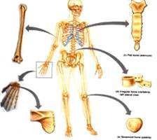
Four examples of bone shape: a long bone, the skull (flat and irregular bones), a vertebra (irregular bone) and the scapula (flat bone), arranged around a skeleton
Memory aids: bone shapes
note
Picture a house being classified by function, not by what it is made of: long bones are the load-bearing pillars of the limbs; short bones are the small bricks packed into the wrist and ankle; flat bones are the broad walls and roof (skull vault, sternum, ribs); irregular bones are the odd-shaped fittings that do not fit any other category (vertebrae, hip bone, cranial base); pneumatic bones are the hollow rooms with air inside (facial skull); and sesamoid bones are pebbles set into a rope, embedded within a tendon (the patella in the quadriceps tendon). This is SuperMed's own image, built directly from the six categories in the note above.
Long bones
note
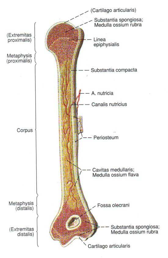
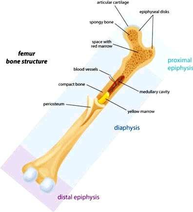
Long bones, ossa longa, form most of the bones of the limbs. A long bone has a shaft, the diaphysis, built of thick compact bone and enclosing the medullary cavity, which holds bone marrow (red in children, yellow in adults). At each end is a rounded epiphysis (proximal and distal), built of spongy bone with only a thin shell of compact bone. Between diaphysis and epiphysis lies the metaphysis, the region of the bone's growth plate (the epiphyseal plate).
The distinction between an epiphysis and an apophysis is one of function rather than appearance: an epiphysis lies at a joint surface and, through its own growth plate, contributes to the bone's longitudinal growth, whereas an apophysis is a separate ossification centre that grows onto the shaft as a projection (such as a trochanter or tuberosity) without taking part in a joint; it serves as an attachment site for muscles, tendons or ligaments rather than as a load-bearing joint surface.
Long bone (femur) in longitudinal section with Latin labels: extremitas proximalis and distalis, metaphysis, linea epiphysialis, substantia compacta, substantia spongiosa, corpus, periosteum, cavitas medullaris with medulla ossium flava, a. nutricia and canalis nutricius, fossa olecrani, cartilago articularisFemur in longitudinal section showing the proximal and distal epiphysis, the epiphyseal disks, spongy bone, compact bone, the diaphysis, the medullary cavity with yellow marrow, periosteum and the blood vessels supplying the bone
Short bones
note
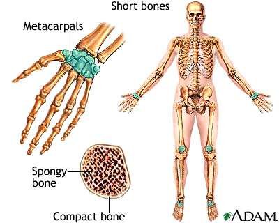
Short bones, ossa brevia, are found in the wrist and ankle (the carpal and tarsal bones) and in the vertebral bodies. They have a thin shell of cortical (compact) bone around a core of spongy bone, which contains red bone marrow.
Short bones of the hand: the metacarpals, shown on a whole skeleton and in cross-section with spongy bone and compact bone labelled
Flat bones
note
Flat bones, ossa plana, make up the skull vault, the sternum, the ribs, the scapula and the hip bone. They are built as a sandwich of two layers of compact bone with a layer of spongy bone (containing red bone marrow) between them. In the bones of the skull vault (calvaria), this internal spongy layer is given its own name, the diploe, and its thickness varies from place to place.
Irregular, pneumatic and sesamoid bones
note
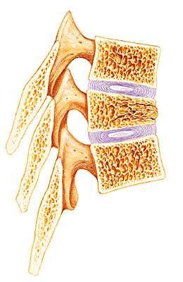
Irregular bones, ossa irregularia, include the vertebrae, the hip bones, and the bones of the facial skull and the cranial base; like short bones, they have spongy bone (with red marrow) surrounded by only a thin shell of compact bone.
Three lumbar vertebrae in longitudinal section, showing the spongy bone of the vertebral bodies and the intervertebral discs between them
Pneumatic (hollow) bones, ossa pneumatica, are bones of the facial skull that contain an air-filled cavity. Sesamoid bones, ossa sesamoidea, are small bones that develop within the substance of a tendon, the patella being the largest example.
3. Structure of mature bone
note
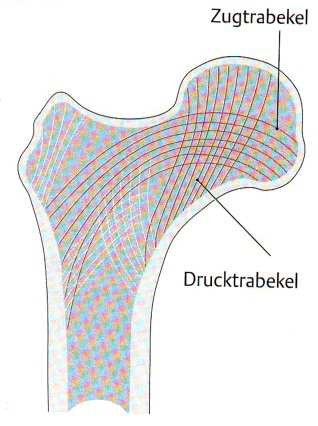
At the level of the whole organ, spongy bone shows a trajectory structure: its trabeculae are not arranged at random but follow the lines along which the bone is mechanically loaded, giving the greatest strength for the least amount of bone tissue. In the neck of the femur, for example, one system of trabeculae follows the lines of tension (Zugtrabekel) and another follows the lines of compression (Drucktrabekel), crossing one another to resist bending.
Femoral neck and head in section, showing tension trabeculae (Zugtrabekel) and pressure/compression trabeculae (Drucktrabekel) crossing one another
This architecture is not fixed for life: mechanical loading alters it. Increased use causes work-induced hypertrophy of bone, while disuse causes inactivity atrophy; disease processes such as degenerative joint disorders (arthroses) also alter the normal trajectory pattern.
Cutaway of a long bone end showing the trabecular (cancellous) bone of the epiphysis continuous with the compact bone of the shaft, with the marrow spaces and blood vessels running through it
Lamellar structure and the osteon (Haversian system)
note
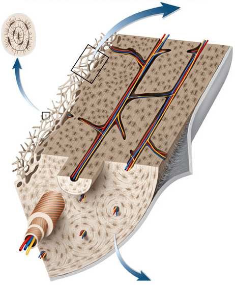
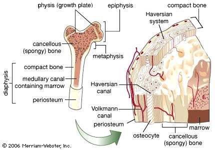
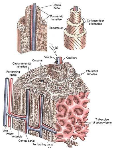
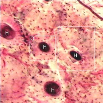
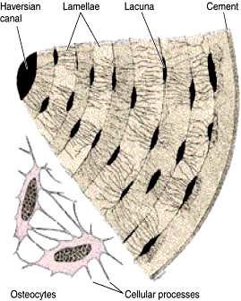
At the level of the tissue, mature compact bone is lamellar bone: it is built up from thin sheets, or lamellae, of matrix. These lamellae are organised into repeating structural units called osteons, or Haversian systems. Each osteon has 5 to 20 concentric (primary) lamellae wrapped around a central Haversian canal, which carries blood vessels and nerves. Osteons are linked to one another, and to the bone's surface, by transverse Volkmann's canals. Around the outside of the osteons lie circumferential lamellae (secondary lamellae), running around the whole circumference of the bone just under the periosteum and endosteum, and between neighbouring osteons lie the interstitial lamellae, the remnants of older osteons that have been partly remodelled away.
Diagram of compact bone from the whole bone down to the microscopic level: periosteum, compact bone, cancellous (spongy) bone, metaphysis, diaphysis, epiphysis, growth plate (physis), and, in the cutaway, the Haversian system with its Haversian canal, Volkmann's canal, osteocytes and marrowDetailed diagram of an osteon in compact bone: the central canal, concentric lamellae with alternating collagen fibre orientation, circumferential lamellae, interstitial lamellae, perforating (Volkmann's) canals, and the vein, artery and arteriole running with a venule and capillary into an osteon, with trabeculae of spongy bone alongsideGround section of compact bone under the microscope, with several Haversian canals (marked H) each surrounded by concentric lamellae and osteocyte lacunae
4. Bone as a tissue: chemical composition
note
By weight, fresh bone is roughly 50% water, 16% lipids, 12% protein (mostly collagen) and 22% inorganic constituents (bone salts, chiefly calcium in the form of hydroxyapatite).
Looked at as a tissue, the bone (extracellular) matrix itself is made of an organic and an inorganic part. The organic part (about 35% of the matrix) gives bone its elasticity: it is 95% type I collagen, with the remainder made of glycosaminoglycans - keratan sulfate, chondroitin sulfate and hyaluronic acid. The inorganic part (about 65% of the matrix) gives bone its hardness and rigidity: it is made of crystalline mineral salts, mostly crystals of hydroxyapatite, comprising calcium phosphate (85%), calcium carbonate (6-10%), magnesium phosphate (up to 1.5%) and traces of calcium fluoride.
Bone cells: osteoblasts, osteocytes, osteoclasts
note
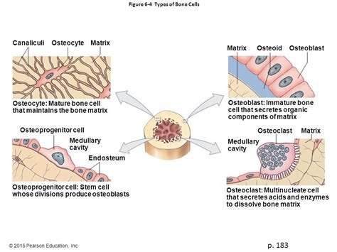
Bone is a specialised connective tissue: a calcified extracellular matrix populated by three cell types.
Cell
Origin and location
Function
Osteoblast, osteoblastus
Cuboidal to columnar cells on the outer (forming) surface of bone
Synthesises the organic components of the matrix: type I collagen, proteoglycans and several glycoproteins. Connected to neighbouring osteoblasts by gap junctions, through which nutrients pass from cell to cell. As it becomes surrounded by its own secretions it turns into an osteocyte ("blast > cyte")
Osteocyte, osteocytus
Lies within a small chamber, a lacuna, inside the mineralised matrix
Maintains the surrounding bony matrix; its death is followed by resorption of that matrix. Stays in contact with neighbouring osteocytes and with blood capillaries through gap junctions and thin channels called canaliculi, since nutrients and waste cannot diffuse through the calcified matrix itself
Osteoclast, osteoclastus
Giant, multinucleated cell of macrophage origin
Pumps out collagenase and other enzymes, creating a locally acidic environment that dissolves hydroxyapatite and digests collagen, resorbing bone
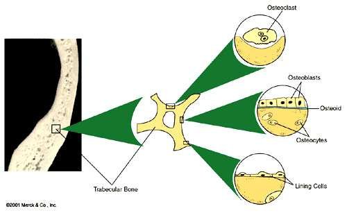
Diagram of bone cell lineage: osteoprogenitor cell (a stem cell), osteoblast (immature bone cell secreting organic matrix), osteocyte (mature bone cell maintaining the matrix, in canaliculi and matrix), and osteoclast (multinucleate cell dissolving bone matrix, arising near the medullary cavity and endosteum)Diagram tracing an osteoclast, osteoid, and lining cells back to a common trabecular bone surface, with osteoblasts laying down osteoid that matures into osteocytes embedded in the matrixOsteocytes embedded within the lamellae of bone matrix, each with fine cellular processes running through canaliculi to contact neighbouring osteocytes
Memory aids: bone cells
note
A common rule of thumb for the three cell names: osteoBLAST = Builds new matrix (a blast cell is a builder cell, as in "blast furnace" work); osteoCLAST = Crushes/Clears matrix away (sounds like "iconoclast", something that breaks things down); osteoCYTE = the mature cytizen just sitting quietly in its lacuna, maintaining what is already there. The source itself gives the lineage as "blast > cyte" - blasts that finish building become the resident cytes.
5. Bone marrow
note
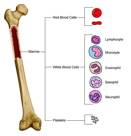
Bone marrow, medulla ossium, exists in two forms. Red bone marrow, medulla ossium rubra, is found in the epiphyses of long bones and in the spongy bone of short and flat bones - the vertebrae, sternum, ribs, pelvic bones and others - and totals roughly 1500 g in the adult. It is the site of haemopoiesis, producing red blood cells, the several types of white blood cell (lymphocytes, monocytes, eosinophils, basophils and neutrophils) and platelets, and it also contributes to the body's biological defence. Yellow (fatty) bone marrow, medulla ossium flava, fills the hollow interior of the middle portion (the diaphysis) of long bones, and consists mostly of fat cells once haemopoietic activity there has stopped.
Femur with the marrow cavity highlighted, and a tree diagram showing marrow giving rise to red blood cells, the white blood cell types (lymphocyte, monocyte, eosinophil, basophil, neutrophil) and platelets
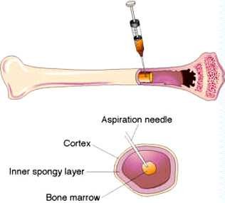
A sample of marrow can be obtained by bone marrow aspiration or biopsy, passing a needle through the compact bone (cortex) and the inner spongy layer to reach the marrow space, as shown below.
Diagram of a bone marrow aspiration needle passing through the cortex and inner spongy layer of bone into the bone marrow
Memory aids: bone marrow
note
A simple rule of thumb for the two marrow types: red marrow is really active, doing haemopoiesis, and is found where blood cell demand stays high throughout life (epiphyses of long bones, and the spongy bone of the vertebrae, sternum, ribs and pelvis). Yellow marrow is yellow because it is mostly fat, and it fills the shaft (diaphysis) of adult long bones once haemopoiesis there has stopped - "yellow" and "shaft" both bring to mind the long, hollow middle of a long bone.
6. Histogenesis of bone (osteogenesis)
note
Bone tissue arises from mesenchyme by a process called osteogenesis (histogenesis of bone), which follows one of two routes: intramembranous ossification (osteogenesis membranacea), in which bone forms directly within a sheet of connective tissue, and intracartilaginous or endochondral ossification (osteogenesis chondralis), in which bone replaces a pre-existing cartilage model. Endochondral ossification itself proceeds in two ways: a perichondral mechanism, which lays down a bone collar (ring) around the shaft of the cartilage model, and an endochondral mechanism proper, which establishes centres of ossification within the cartilage itself.
Intramembranous ossification
note
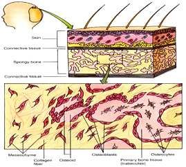
Intramembranous ossification occurs in only a few bones, for example the parietal bone. Bone is formed directly onto fibrous connective tissue, with no intermediate cartilage stage, in four steps: an ossification centre develops within the mesenchyme; calcification occurs as mineral is deposited; trabeculae form in the interior of the developing bone; and finally the surrounding mesenchyme is replaced by periosteum, with a thin layer of compact bone forming just beneath it.
Cross-section through developing membranous bone: mesenchyme differentiating into collagen fibre, osteoid and osteoblasts, which mature into osteocytes within the primary bone tissue (trabeculae)
Endochondral (intracartilaginous) ossification
note
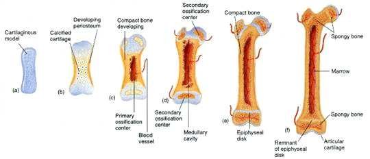
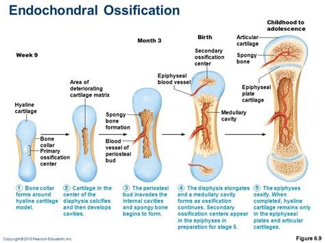
Endochondral ossification takes place within a piece of hyaline cartilage shaped like the bone that will replace it, and proceeds through a primary and then a secondary ossification centre.
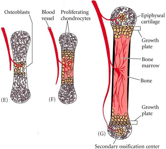
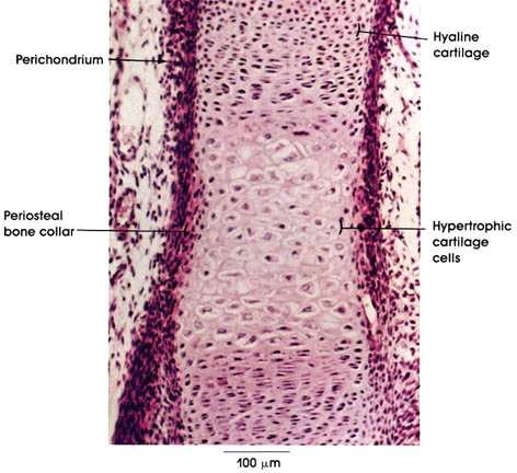
At the primary ossification centre, bone tissue first appears, through osteoblast activity, as a collar surrounding the cartilage model. This bone collar blocks the diffusion of nutrients to the cartilage underneath, so the cartilage begins to degrade: its chondrocytes produce alkaline phosphatase and swell, compressing the surrounding matrix and driving its calcification. The death of these chondrocytes leaves behind a porous, calcified scaffold, onto which osteoblasts then adhere and lay down layers of primary bone, surrounding the remnants of the cartilaginous matrix.
At the secondary ossification centre, bone begins to enlarge and its cells arrange themselves into rows. As the matrix increases in quantity the cells become more widely spread out, calcareous material is deposited between the rows of cells, and the matrix becomes progressively more calcified.
Three stages (E, F, G) of endochondral ossification in a long bone: osteoblasts and a blood vessel invading the cartilage model at (E) and (F), followed by (G) a fully formed bone with epiphyseal cartilage, growth plates at both ends, bone marrow and a secondary ossification centreMicroscopic section through a developing long bone end, labelled: perichondrium, hyaline cartilage, periosteal bone collar, and hypertrophic cartilage cellsFive stages of endochondral ossification: (a) cartilaginous model, (b) calcified cartilage with a periosteum forming, (c) primary ossification centre with a blood vessel, (d) secondary ossification centre and medullary cavity, (e) epiphyseal disks either side of the growing shaft, (f) mature bone with an articular cartilage remnant and spongy boneEndochondral ossification from week 9 of embryonic life to childhood: hyaline cartilage model developing an area of deteriorating cartilage matrix and a bone collar at the primary ossification centre, then spongy bone formation and a medullary cavity, and, by birth, secondary ossification centres with their own epiphyseal blood vessels, leaving hyaline cartilage only at the articular cartilage and the epiphyseal plate cartilage in childhood
7. Development and growth of individual bones
note
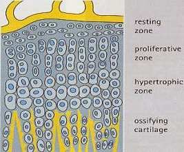
After birth, a long bone continues to lengthen at its growth (epiphyseal) plate, cartilago epiphysialis, the layer of cartilage in the metaphysis between diaphysis and epiphysis. Moving from the epiphysis towards the diaphysis, the plate has four growth zones: a resting zone, a proliferative zone (where chondrocytes divide and add new cartilage), a hypertrophic zone (where the chondrocytes enlarge) and a zone of ossifying cartilage, where the cartilage is finally replaced by bone.
Four zones of the epiphyseal growth plate in cross-section, from the epiphysis to the diaphysis: resting zone, proliferative zone, hypertrophic zone and ossifying cartilage
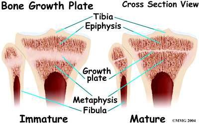
Growth at the plate is under hormonal control. It is stimulated by STH (somatotropic hormone, growth hormone), calcitonin and vitamin D3; a deficiency of vitamin D3 in children causes rickets (rachitis). It is inhibited by corticosteroids. Once growth is complete, the plate itself ossifies and is replaced by bone, leaving only a thin epiphyseal line.
Cross-section through an immature and a mature epiphysis of the tibia and fibula, comparing the thick growth plate of the immature bone with the thin, closed growth plate of the mature bone
Memory aids: growth-plate zones
note
Resting Proliferate Hypertrophy Ossify - "Rabbits Play Hide-and-seek Outside" (SuperMed's own) gives the four growth-plate zones in the order you cross them moving away from the epiphysis and towards the diaphysis: the cells rest, then multiply (proliferate), then swell up (hypertrophy), and finally the cartilage around them turns to bone (ossifies). A companion rule of thumb: the zone nearest the epiphysis is the quietest (resting) and the zone nearest the diaphysis is the one actually becoming bone (ossifying) - growth "travels" from quiet to solid.
8. Biological properties of bones
note
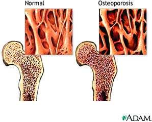
Bone tissue can lose mass, a process called bone atrophy or osteoporosis, of several kinds: immobilization osteoporosis, age-related (senile) osteoporosis, professional osteoporosis (from excessive physical activity), hormonal (menopause-related) osteoporosis, and gravity-induced osteoporosis (from the absence of normal mechanical loading, as in spaceflight). The opposite process, bone hypertrophy, also occurs, for example with increased mechanical use.
Comparison of normal and osteoporotic bone at the femoral head: the osteoporotic bone shows markedly thinned, sparser trabeculae
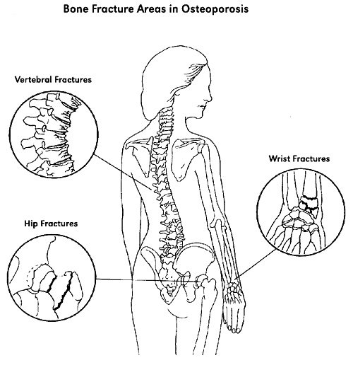
Osteoporosis particularly weakens certain sites, so that fractures there are characteristic of the disease: the vertebral bodies, the hip and the wrist.
Typical sites of osteoporotic fracture: vertebral (compression) fractures of the spine, hip fractures, and wrist fractures
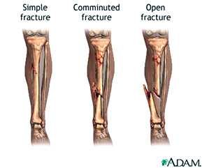
Mechanically, bone combines hardness and rigidity, which can give way to fracture under sufficient force, with strength and elasticity, which allow it to resist and absorb load without breaking. Fractures are classified by their pattern, for example as simple, comminuted (broken into several fragments) or open (with a wound communicating with the fracture site).
Three types of fracture of the tibia and fibula: a simple fracture, a comminuted fracture, and an open fracture with a break in the overlying skin
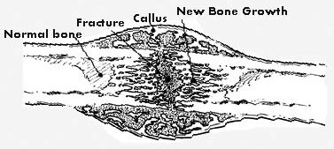
A fractured bone heals through a callus: new, initially disorganised bone that bridges the fracture site before being remodelled into new, organised bone.
Fracture of a long bone shaft, showing the callus bridging the fracture and new bone growth extending from it, compared with normal bone
Finally, bone is the body's principal store of calcium, holding around 97% of total body calcium.
Memory aids: osteoporosis
note
The five causes of osteoporosis in this note spell "I SHooP" (SuperMed's own): Immobilization, Senile (age-related), Hormonal (menopause-related), professional (from excessive physical activity), and gravity-induced (in weightlessp-type conditions such as spaceflight) - a bone loses density if it is not used enough (immobilization, gravity-induced weightlessness), used too much (professional), or its hormones or age work against it (hormonal, senile).
For fracture sites in osteoporosis, a simple count anchors the three classic locations: spine, hip, wrist - the three places a fall or everyday strain most often breaks osteoporotic bone, all sites rich in the spongy bone that osteoporosis thins fastest.
Memory aids and mnemonics
note
The note's best memory aids gathered in one place:
Bone shapes - long bones are pillars, short bones are bricks, flat bones are walls, irregular bones are odd fittings, pneumatic bones are hollow rooms, sesamoid bones are pebbles in a rope (a tendon) (SuperMed's own).
Bone cells - "blast > cyte": osteoBLAST builds, osteoCLAST crushes/clears, osteoCYTE just sits and maintains.
Marrow colour - red is really active (haemopoiesis, in axial spongy bone and long-bone epiphyses); yellow is full of fat (in the shaft of adult long bones).
Growth-plate zone order (epiphysis to diaphysis) - Resting, Proliferative, Hypertrophic, Ossifying: "Rabbits Play Hide-and-seek Outside" (SuperMed's own).
Osteoporotic fracture sites - spine, hip, wrist: a simple count of three to anchor.
The most important things to know
note
The skeleton has 206 bones, built of compact (cortical) bone and cancellous (spongy) bone, covered by periosteum or articular cartilage and lined internally by endosteum.
Bones are classified by shape: long (ossa longa), short (ossa brevia), flat (ossa plana), irregular (ossa irregularia), plus pneumatic (ossa pneumatica) and sesamoid (ossa sesamoidea) bones.
A long bone has a diaphysis (shaft, thick compact bone, medullary cavity), two epiphyses (spongy bone with a thin compact shell), and a metaphysis (the growth plate region). An apophysis is a separate, non-articular growth centre serving as a muscle attachment.
The periosteum is a two-layered membrane (outer fibrous, inner osteogenic) anchored by Sharpey's fibres; the endosteum is a thin membrane lining all internal bone surfaces. Both contain osteoprogenitor cells active in growth and repair.
At the organ level, spongy bone trabeculae form a trajectory structure following the lines of mechanical stress (tension and compression trabeculae); loading causes hypertrophy, disuse causes atrophy.
At the tissue level, compact bone is lamellar, organised into osteons (Haversian systems): a central Haversian canal, 5-20 concentric lamellae, circumferential and interstitial lamellae, linked by Volkmann's canals.
Bone matrix is 35% organic (mostly type I collagen, giving elasticity) and 65% inorganic (hydroxyapatite crystals, giving hardness and rigidity).
Three cell types: osteoblasts (build matrix, become osteocytes), osteocytes (maintain matrix, in lacunae, linked by canaliculi), osteoclasts (macrophage-derived, resorb bone).
Red bone marrow (epiphyses, spongy bone of axial bones) does haemopoiesis; yellow marrow (diaphysis of long bones) is fatty.
Osteogenesis proceeds by intramembranous ossification (bone forms directly in connective tissue, e.g. parietal bone) or endochondral ossification (bone replaces a cartilage model, via primary then secondary ossification centres).
Postnatal growth occurs at the epiphyseal plate (resting, proliferative, hypertrophic, ossifying cartilage zones); stimulated by growth hormone, calcitonin and vitamin D3, inhibited by corticosteroids; vitamin D3 deficiency causes rickets.
Biological properties: bone atrophy (osteoporosis - immobilization, senile, professional, hormonal, gravity-induced) and hypertrophy; mechanical hardness/rigidity (fracture risk) versus strength/elasticity; bone stores 97% of body calcium.
All 261 Anatomy topics · exam study support, not clinical guidance. SuperMed is not affiliated with the Medical University of Sofia.
Start free. No card needed.
Every account starts free, with free topics in Cytology, Medical Biology, Anatomy, Biophysics, Pharmacology and Preclinics of Pediatric Dental Medicine. Super opens the rest. Super is €15 a month.