


Cytology · Year 1 · Medical University of Sofia

01

Microscopy. Types of microscopes: principles and application

Free notes for topic 01 of the Cytology syllabus, open without an account. Written by a senior student against the syllabus question and checked line by line by a second student before publishing. How content is made

Updated

The short version

note

A microscope is an optical instrument that uses lenses to produce magnified images of objects too small to be seen by the unaided eye. Cytology, histology and embryology all exist because such an instrument exists.

Three things are worth carrying into the rest of the course. The light microscope cannot resolve anything closer together than 0.2 μm, which is why ribosomes and membranes are invisible in it and why electron microscopy had to be invented. The various light microscopes differ not in their lenses but in what they do to the light: fluorescence excites the specimen with ultraviolet, phase contrast converts differences in refractive index into differences in brightness, confocal removes out-of-focus light with a pinhole, and polarizing exploits anisotropy. And the electron microscope comes in two kinds that answer two different questions: transmission for what is inside a thin section, scanning for what a surface looks like in three dimensions.
1. Historical development of cytology, histology and embryology

note

The discipline is a sequence of instruments and the observations each one made possible.

1590 is the milestone. The first microscope, with a magnification of 9×, was created by two Dutchmen, Hans and Zacharias Jansen.

1609. Galileo Galilei improved and perfected the microscope, adding a focusing device.

1665. Robert Hooke used a compound microscope and discovered the first "cell". He coined the term cell in that same year. The box-like cells of cork he was looking at, which were plant cells, reminded him of the cells of a monastery.

1674. Antonie van Leeuwenhoek, a Dutch microscopist, was the first to discover protozoa, bacteria, blood cells and spermatozoa. He is known as the father of microbiology.

1831. Robert Brown, a Scottish botanist, made the first detailed description of the cell nucleus.

1838. Schleiden and Schwann independently postulated the cell theory, which formed the basis of the discipline as we know it today. Cell theory dictates that:

  • life exists only in the form of cells, and therefore all organisms consist of cells; cells are the basic unit for all life function;
  • there is a mutual connection between cell structure and function.

1857. Albert von Kölliker discovered mitochondria in muscle cells. Richard Altmann later established them as cell organelles and called them bioblasts, in 1890.

1884. Hans Christian Gram invented an important staining method to make bacteria more visible in stained sections, the Gram stain.

1898. Camillo Golgi described the Golgi apparatus.

1953. Watson and Crick published their discovery of the helical structure of DNA, the double helix. It made clear that genes are responsible for each change that takes place in the embryo, and how they regulate the process.

1969. Franz von Furth described the mononuclear phagocytic system.

1972. Singer and Nicolson proposed the fluid mosaic model of the cell membrane.

2. What a microscope is

note

The scope of microscopy is to observe cells, which exist at micro-sizes.

A microscope is an optical instrument that uses lenses to produce magnified images of small objects, too small to be seen by the unaided eye.

3. The light microscope

note

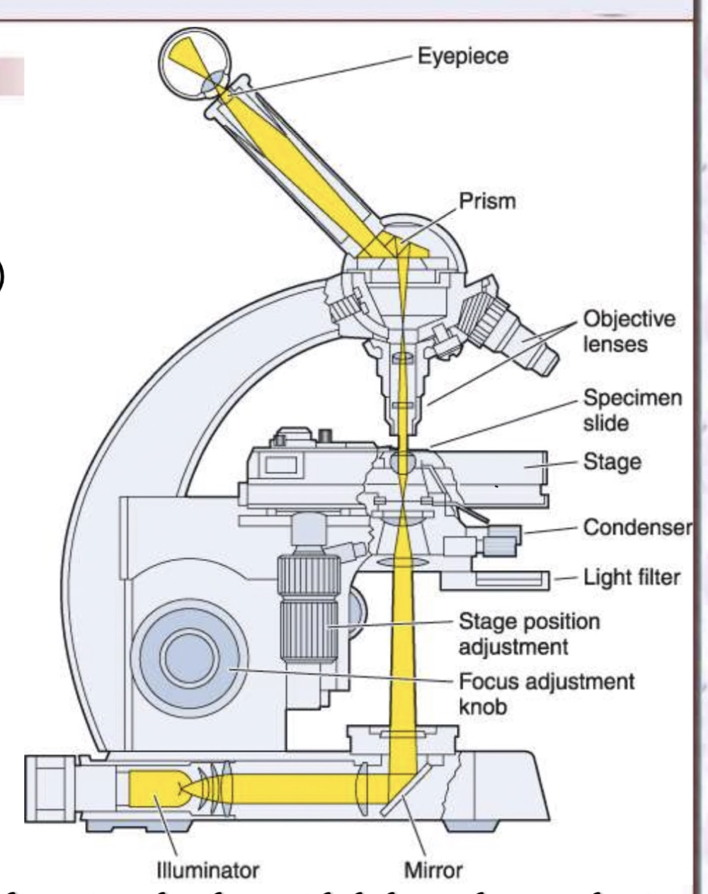
The light microscope is built from three parts: a mechanical part, an optical part and an illuminating part. Each of the three is described in the notes that follow.

Labelled diagram of a light microscope showing the eyepiece, prism, objective lenses, specimen slide, stage, condenser, light filter, stage position adjustment, focus adjustment knob, illuminator and mirror

The mechanical part

note

The mechanical part holds everything else in position:

  • the stand, made up of the arm and the base;
  • the stage, on which the slide or specimen is placed;
  • the tube.

The optical part

note

Three lens systems do the work of forming the image:

  • the ocular lens, or eyepiece, which magnifies the image and projects it onto the viewer's retina;
  • the objective lens, which collects light rays to create the image and project it towards the observer;
  • the condenser, which focuses the light on the object.

The illuminating part

note

The illuminating part is the light source, which illuminates the specimen.

Resolving power

note

The resolving power of the light microscope is 0.2 μm. Objects smaller than this cannot be distinguished, which is why ribosomes, membranes and structures of that order are beyond it.

This single number is the reason electron microscopy exists, and the reason a great deal of what the rest of this course describes was unknown before the twentieth century.

4. Fluorescence microscopy

note

The fluorescence microscope is used to display naturally occurring fluorescent molecules, such as neurotransmitters and vitamin A.

  • Fluorescent substances appear bright on a dark background.
  • The instrument has a source of ultraviolet light and filters that select rays of different wavelengths.
  • Tissue sections are usually irradiated with ultraviolet light, which lets the objects be seen more clearly.
  • Its application is mostly in the detection of antibodies and antigens, in immunohistochemistry.

5. Phase-contrast microscopy

note

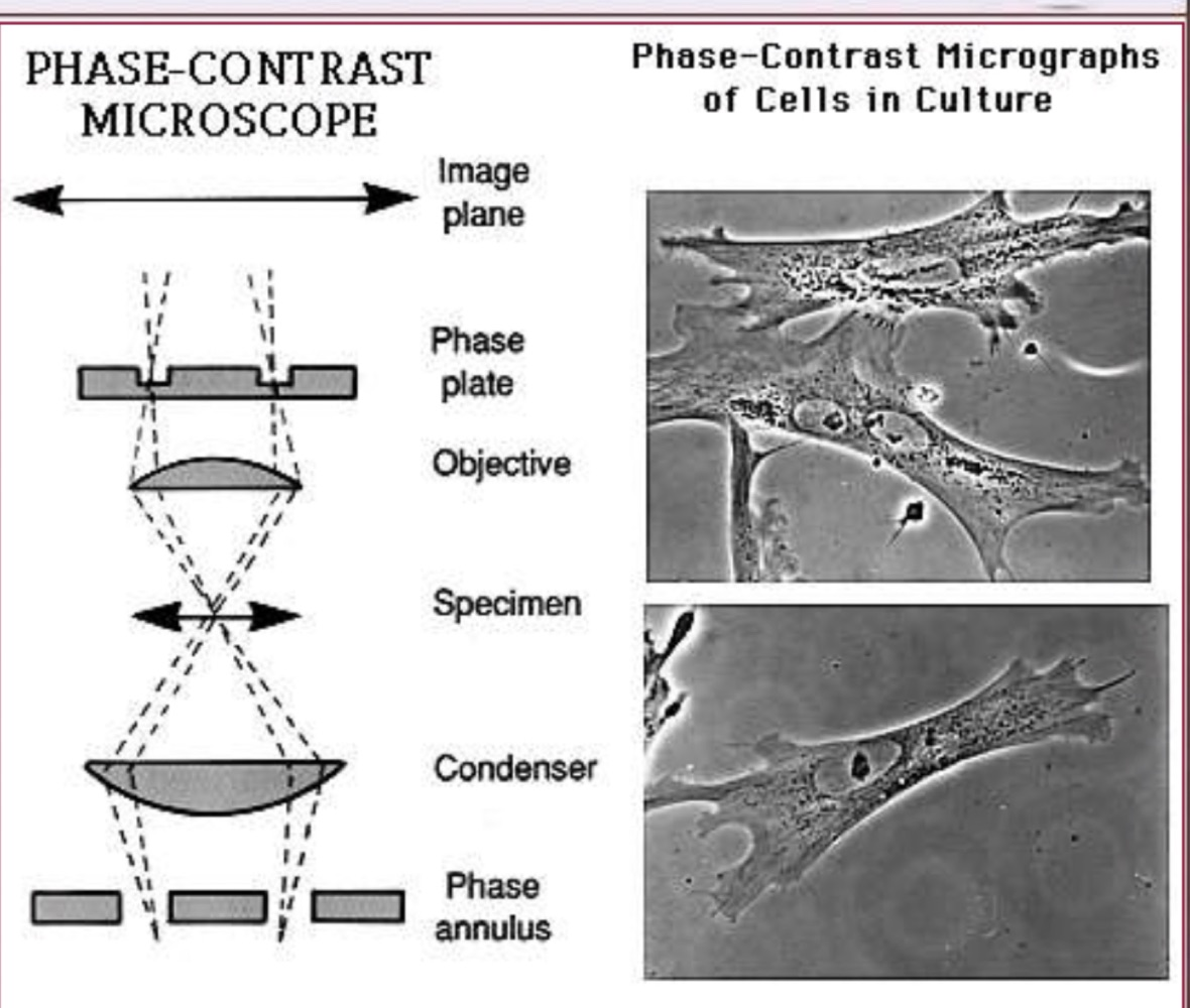
Phase-contrast microscopy does not require staining to view the slide. It uses a lens system that produces visible images from transparent objects, which is what allows us to view living cells and the cell cycle.

A variation of this method is differential interference contrast microscopy, which produces a three-dimensional version of living cells.

Diagram of a phase-contrast microscope beside phase-contrast micrographs of cells in culture

6. Confocal microscopy

note

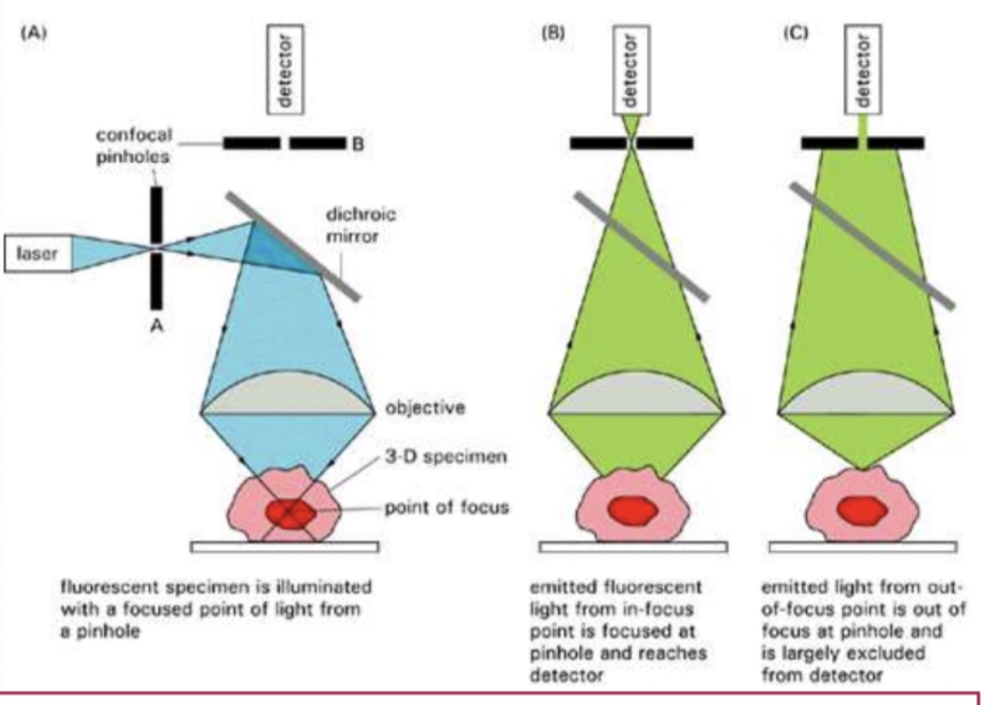
Confocal microscopy is a technique that allows the visualisation of a biological specimen in three dimensions. A mirror system moves a laser beam across the specimen.

High resolution and sharp focus are achieved by using two things together:

  1. sharply focused light, for example a laser;
  2. a plate with a pinhole.

The consequences are that resolution improves, that the specimen can be localised with much greater precision, and that it is possible to create multiple images.

Three diagrams comparing confocal optics, showing the specimen illuminated from a focused point of light through a pinhole and out-of-focus light blocked from the detector

7. Polarizing microscopy

note

The polarizing microscope is used to observe specimens that are visible primarily because of their optically anisotropic character. It allows tissue structures containing molecules such as cellulose, collagen, microtubules and microfilaments to be recognised.

It carries two filters: the polarizer and the analyzer.

8. Electron microscopy

note

In electron microscopy, objects are scanned and the images returned are based on the interaction of the tissue with beams of electrons.

There are two kinds:

  • the transmission electron microscope, TEM;
  • the scanning electron microscope, SEM.

The transmission electron microscope

note

In the TEM:

  • a cathode emits electrons;
  • the electrons are attracted towards an anode;
  • the electron beam passes through several electromagnetic lenses;
  • the condenser lens shapes the diameter of the electron beam;
  • the objective lens focuses and magnifies the beam;
  • the projector lenses magnify it again;
  • the final image is viewed on a fluorescent screen or on a photographic plate.

The contrast in the image comes from what the specimen does to the electrons. Portions of the specimen through which electrons have passed have a bright appearance. Portions which absorbed or scattered the electrons have a dark appearance.

To improve contrast, heavy metal ions are added.

The scanning electron microscope

note

The SEM works like the TEM, but the electrons do not pass through the object.

Instead the surface of the object is dried and spray-coated with a heavy metal, often gold. Electrons then reflect off the metal and are caught by the detector, and the resulting image is shown on a monitor.

These are three-dimensional black and white images.

All 95 Cytology topics · exam study support, not clinical guidance. SuperMed is not affiliated with the Medical University of Sofia.

Start free. No card needed.

Every account starts free, with free topics in Cytology and Medical Biology. Super opens the rest. Super is €15 a month.

Create a free account