



Cytology · Year 1 · Medical University of Sofia

17

Specialized structures of the cell membrane

Free notes for topic 17 of the Cytology syllabus, open without an account. Written by a senior student against the syllabus question and checked line by line by a second student before publishing. How content is made

Updated

The short version

note

There are four types of structural specialisation of the cell membrane: microvilli, stereocilia, cilia and flagella, and basal cell surface folds.

The dividing line to hold on to is what is inside them. Microvilli and stereocilia contain actin filaments and are not motile. Cilia and flagella contain microtubules and are motile. Everything else follows from that.

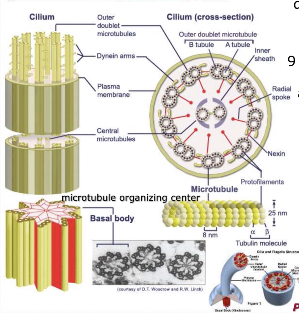
The microtubule arrangements are the examinable detail: a kinocilium's axoneme is 9 × 2 + 2, its basal body is 9 × 3 + 0, and a sensocilium is 9 × 2 + 0.
1. Microvilli

note

Microvilli are 1 μm high and 0.08 μm wide.

  • They are finger-like extensions from the surface of the cell.
  • They increase surface area for diffusion while minimising the increase of volume. Together they form the structure of the brush border.
  • Each microvillus contains actin filaments.
  • Their functions are absorption, cell movement, phagocytosis and motility.
  • They are found in the intestines and kidney.

2. Stereocilia

note

Stereocilia have no motility.

  • They contain actin filaments, like microvilli.
  • They increase surface area for absorption.
  • They are much longer than kinocilia but less motile, and resemble microvilli.
  • They are found in the ductus deferens and epididymis, both parts of the male reproductive system, and in the hair cells of the inner ear.

The name is misleading and is worth naming as such: a stereocilium is not a cilium. It contains actin, not microtubules, and it does not beat.

3. Cilia and flagella

note

Cilia and flagella are membrane extensions. Both contain microtubules, and both have motility.

Cilia

note

Cilia are short, numerous membrane extensions, and are highly motile. They are larger than microvilli.

  • The basal bodies, or kinetosomes, are provided by centrioles.
  • They are much shorter than flagella, but numerous.
  • ATP is the source of energy for ciliary motion, and dynein is the motor of movement.
  • They are found in the respiratory epithelium.

Kinocilia and the 9 × 2 + 2 arrangement

note

Kinocilia are the typical cilium: motile cilia, abundant in epithelial cells, whether cuboidal or columnar.

Their length is 5 to 10 μm and their diameter is 0.2 μm.

They have three parts:

The axoneme consists of 9 peripheral doublets of microtubules, the A and B tubules, and 2 central singlets of microtubules surrounded by an inner sheath. This gives the arrangement

9 × 2 + 2

The transitory part, between the axoneme and the basal body.

The basal body, which is the microtubule organising centre. It consists of 9 peripheral triplets of microtubules, the A, B and C microtubules, with no microtubules in the centre. This gives the arrangement

9 × 3 + 0

Diagram of a cilium in longitudinal and cross section showing the axoneme with nine peripheral doublets and two central microtubules, the microtubule organising centre and the basal body

Sensocilia

note

Sensocilia are non-motile, and are found in specialised cells of sense organs: the eye, nose and ear.

Their structure is an axoneme, the central core, with 9 peripheral doublets of microtubules and no singlets in the centre. This gives the arrangement

9 × 2 + 0

The single difference from a kinocilium is the missing central pair, and it is exactly the difference that makes it non-motile.

Flagella

note

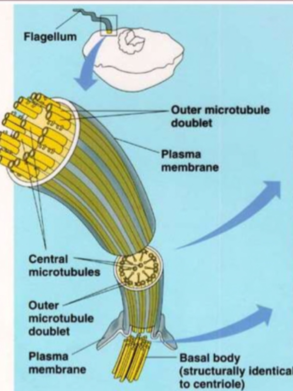
The flagellum is a long, singular membrane extension.

  • Its length is 100 to 200 μm.
  • There is a limit of one flagellum per cell.
  • It is supported and made up of microtubules.
  • In humans it occurs only in spermatozoa.
Diagram of a flagellum showing the outer microtubule doublets, the central microtubules, the plasma membrane and the basal body, which is structurally identical to a centriole

4. Basal cell surface folds

note

Basal cell surface folds increase the surface for absorption.

They facilitate the exchange of ions and metabolites between the cell and the underlying blood vessel.

All 95 Cytology topics · exam study support, not clinical guidance. SuperMed is not affiliated with the Medical University of Sofia.

Start free. No card needed.

Every account starts free, with free topics in Cytology and Medical Biology. Super opens the rest. Super is €15 a month.

Create a free account