Medical Biology · Year 1 · Medical University of Sofia

08

Proteins after translation: sorting, folding, modifications, degradation

Free notes for topic 08 of the Medical Biology syllabus, open without an account. Written by a senior student against the syllabus question and checked line by line by a second student before publishing. How content is made

Updated

What this topic covers

note

Chaperones and their rescue action; prions; the post-translational modifications of proteins, including partial proteolysis, glycosylation, phosphorylation and dephosphorylation, and polyproteins; protein degradation by ubiquitination and the proteasome; the distribution of proteins between cellular compartments, with transport into the nucleus, into mitochondria, and to the cell surface and lysosomes; the pathway for exporting proteins out of the cell and the three pathways after the Golgi complex; and the vesicle network for transport within the cell, including endosomes, the localisation of exocytosis, retrograde trafficking from endosomes to the trans-Golgi and the action of ricin, and exosomes.
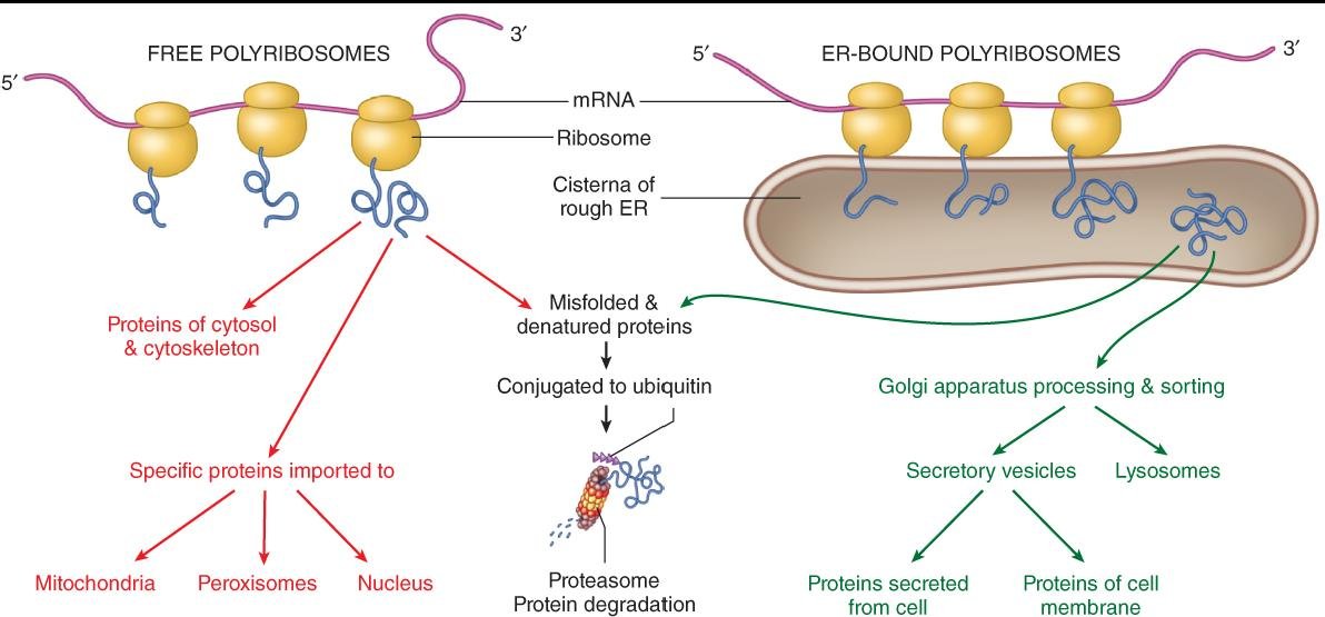
Protein fates after translation: free ribosomes give cytosolic proteins or import to mitochondria, peroxisomes and nucleus, misfolded proteins are ubiquitinated for proteasome degradation, and ER-bound ribosomes feed Golgi sorting to secretory vesicles, lysosomes and membrane proteins
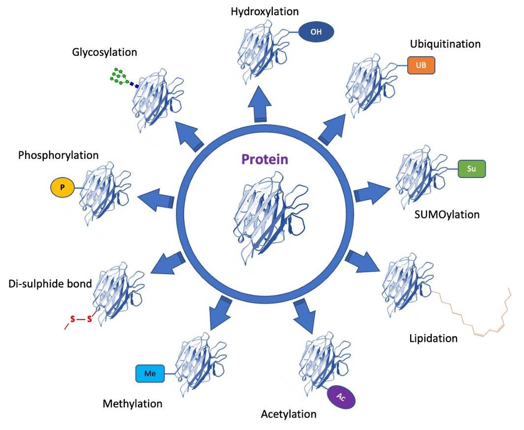
Wheel of post-translational modifications on a protein: hydroxylation, ubiquitination, SUMOylation, lipidation, acetylation, methylation, disulphide bond formation, phosphorylation and glycosylation

1. The native state is easily lost

note

The native structure is the spatial structure that is able to perform the biological function of the protein.

Two things can go wrong, and both are common.

  • Some newly synthesised proteins fail to acquire the native state in the first place.
  • Even correctly folded proteins can lose it. They can be denatured by a number of physical and chemical agents, including the normal body temperature of 37 °C.

Once denatured, a protein is unlikely to renature on its own. That is the whole reason the next section exists.

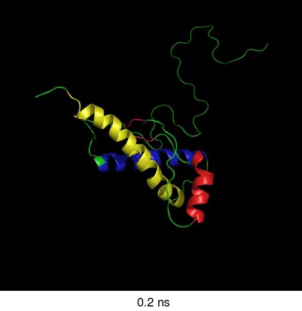
The hallmark of a denatured protein is the presence of hydrophobic side chains on the molecular surface. Those exposed hydrophobic patches make the protein less soluble and prone to aggregate by hydrophobic interactions - and they are also the signal that the cell reads.

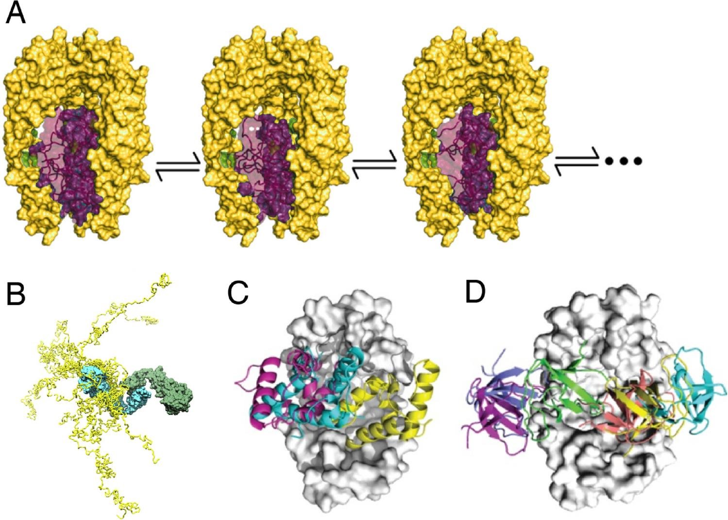
Molecular dynamics snapshot at 0.2 ns of a protein partly unfolded from its native state, with ordered helical segments and a long, extended unstructured tail, unlabelled

2. Chaperones

note

In the cell, special proteins help other proteins attain or regain their native state. These helpful proteins are called chaperones.

The word is of French origin, and its primary meaning is "a middle-aged woman accompanying a young unmarried woman in public" - the sense being that the chaperone does not do the work, it prevents the wrong associations.

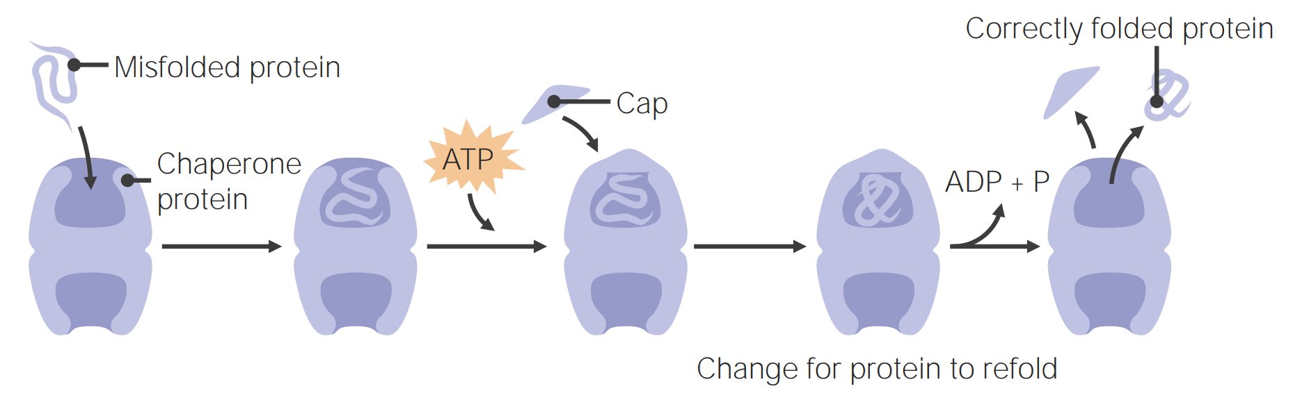
Chaperone cycle: a chaperone protein binds a misfolded protein, ATP binds and a cap closes over it, then ADP and phosphate are released and the correctly folded protein comes out

The rescue action of chaperones

note

Chaperones recognise the exposed hydrophobic parts of denatured proteins. From there they work in two ways:

  • some chaperones bind to those parts and help the protein fold properly;
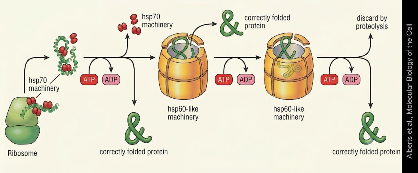
  • others form a cylindrical complex in which the protein is accommodated and refolds. It is easier to try different conformations inside this cylinder than in water solution, because the protein is protected from aggregating with its neighbours while it experiments.

Most chaperones are designated by hsp, for heat shock protein, plus a number showing the molecular mass. In fact they are related to any type of stress, not just heat, and the name is a historical accident.

Hsp70 and hsp60-like chaperone machinery: hsp70 binds the nascent chain from the ribosome, then ATP-driven cycles either fold it directly or commit it to a barrel-shaped hsp60-like chamber, ending in a correctly folded protein or discard by proteolysis

Misfolded proteins provoke the synthesis of more chaperones

note

The system is self-regulating, and the loop runs like this.

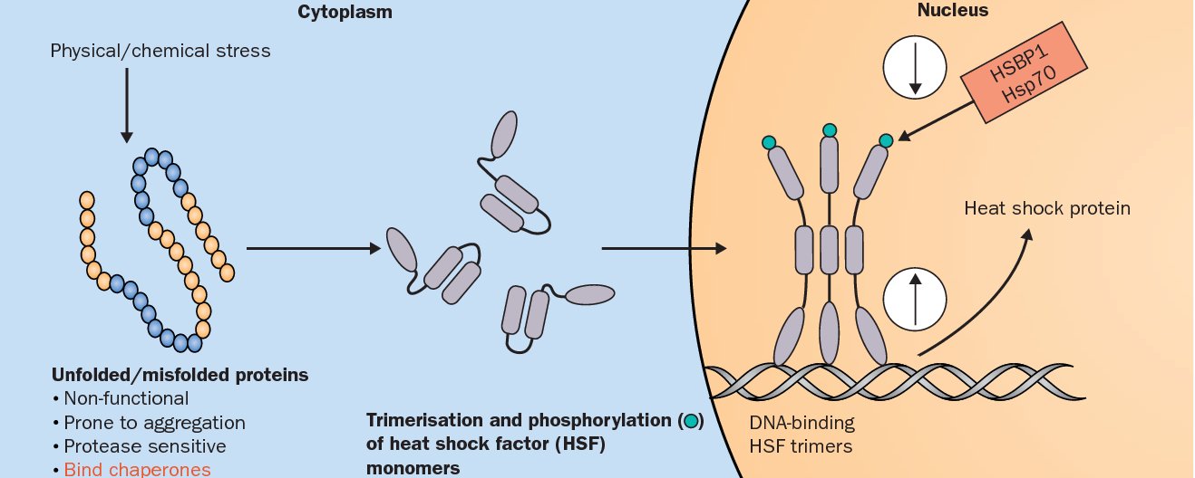
  1. Physical or chemical stress induces the production of unfolded or misfolded proteins.
  2. The presence of misfolded proteins provokes phosphorylation and aggregation of heat shock factor monomers in the cytoplasm.
  3. They form complexes - in the standard example, trimers - which are translocated into the nucleus.
  4. There they bind to the heat shock protein gene promoter, leading to induction of Hsp gene transcription.

So the damage is itself the signal that orders the repair crew.

Heat shock response: physical or chemical stress produces unfolded, aggregation-prone misfolded proteins that trigger trimerisation and phosphorylation of heat shock factor monomers, which enter the nucleus, bind DNA and induce heat shock protein transcription

Chaperone-client complexes

note

A client is a protein that needs repair. Structural studies show several arrangements:

  • the client protein sitting in the cavity of the chaperone;
  • a conformational ensemble of a disordered client bound to a chaperone;
  • a chaperone with a bound client in which the chaperone changes the conformation of the client.
Structural models of chaperone-client complexes, unlabelled: a client sitting inside the chaperone cavity, a disordered client ensemble bound to a chaperone, and complexes in which the chaperone reshapes the client's conformation

Chaperones in non-stressful situations: nucleoplasmin

note

The cell needs chaperones for many functions that have nothing to do with stress. Nucleoplasmin is the example, and its job is assembly rather than folding.

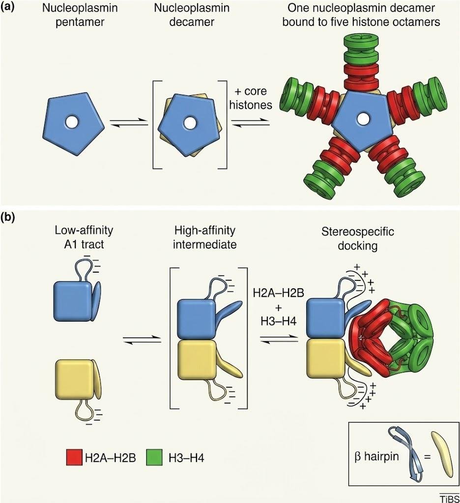
  • Nucleoplasmin is a pentamer, and two pentamers make a decamer.
  • It connects five H2A-H2B complexes, and the whole structure looks like a star.
  • The formation of decamers triggers a conformational change in nucleoplasmin: the β hairpins become extended outwards.
  • The H2A-H2B dimers bind to those β hairpins.
  • Then each of the five parts of the star connects an H3-H4 complex.

The resulting H2A-H2B-H3-H4 complexes are ready to be included in nucleosomes.

Nucleoplasmin assembly: a nucleoplasmin pentamer forms a decamer that binds five H2A-H2B and H3-H4 histone complexes through a low-affinity to high-affinity to stereospecific docking sequence, with the beta hairpin extending outward to bind H2A-H2B

3. Prions do the opposite: they turn normal protein into abnormal

note

Mammalian neurons have a membrane protein called the prion protein. Its normal function is still debated.

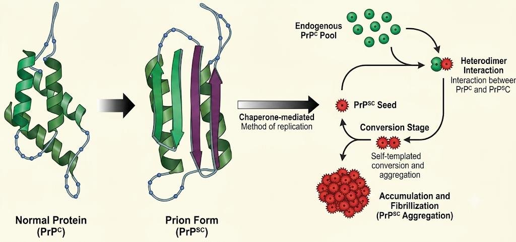
  • Its native conformation, PrPC, is rich in alpha helices.
  • A mutation, an error in synthesis, or some other factor can cause it to misfold into another conformation, PrPSc, which is rich in beta sheets.

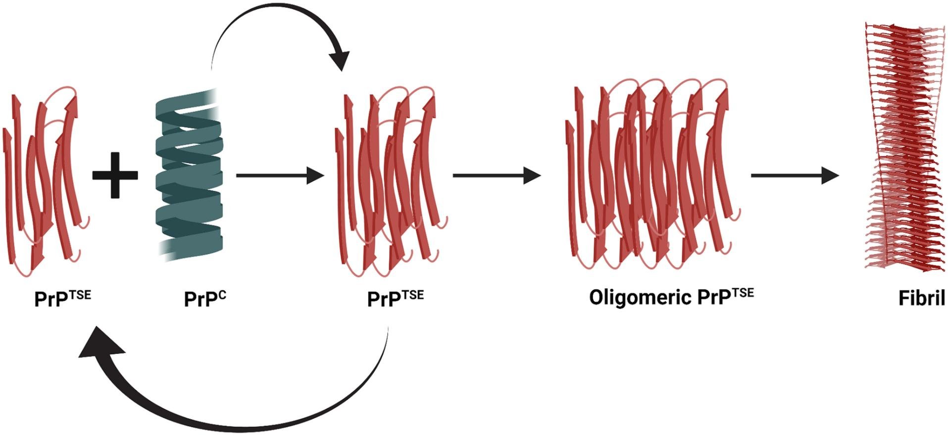
Worse, the PrPSc molecule binds to PrPC molecules and influences them to misfold as well. An aggregate of dangerous PrPSc molecules is formed, and it is called a prion, from proteinaceous infectious particle.

The consequence is exponential. If just single defective proteins are produced, they turn all the normal proteins into prions. The prion number grows and kills the cell. Then the prions invade neighbouring cells.

PrPC to PrPSc conversion: the alpha-helical normal prion protein converts to the beta-sheet-rich prion form, which forms a heterodimer with endogenous PrPC, seeds self-templated conversion and accumulates into a fibril aggregate

Spongiform encephalopathies

note

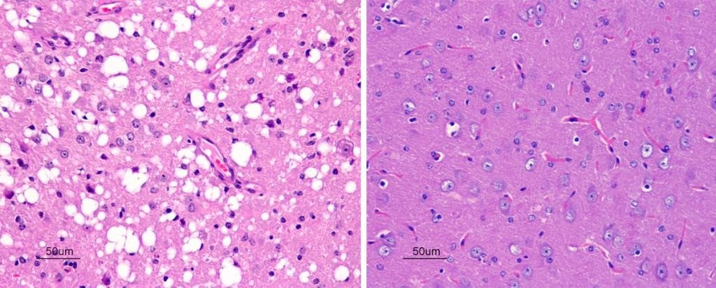
The prion penetrates another neuron and kills that one as well, and the process continues. Empty spaces appear in the brain at the place of the dead neurons, which is where the name comes from.

Two brain histology sections side by side: one shows spongiform vacuolation, many empty round spaces in the neuropil, the other shows normal-looking brain tissue for comparison

The resulting neurodegenerative condition is called a prion disease or spongiform encephalopathy. It is progressive and fatal.

Animals and humans can develop spongiform encephalopathy not only when a prion appears inside them, but also when one is injected or ingested from outside. Prions are unique infectious agents without DNA or RNA, and cooking and digestion destroy only part of them.

Examples of prion diseases:

  • in humans: Creutzfeldt-Jakob disease, fatal familial insomnia, and kuru;
  • in sheep: scrapie;
  • in cattle: bovine spongiform encephalopathy, "mad cow disease".

Both PrPC and PrPSc interact with membrane components through their polypeptide, their glycans, and their glycolipid anchors.

Amyloids and the wider family of neurodegenerative diseases

note

It is not only prion infections. Many other neurodegenerative diseases are characterised by the aggregation of misfolded proteins in the brain, and Alzheimer's disease and Parkinson's disease are grouped with the prion diseases for this reason.

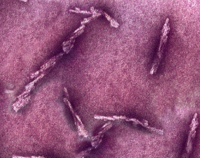
These diseases are caused by misfolded proteins which form fibres called amyloids. The fibres make abnormal contacts between neurons, disturb their function, and eventually kill them.

The newly formed abnormal protein further acts as a seed to form oligomers and fibrils - the same self-templating logic as the prion.

Electron micrograph of amyloid fibrils as elongated, twisted, rod-shaped filaments
Amyloid self-templating cycle: normal PrPC combines with a PrPTSE seed to give more PrPTSE, which assembles into oligomeric PrPTSE and then into a fibril

How protein aggregates spread between neurons

note

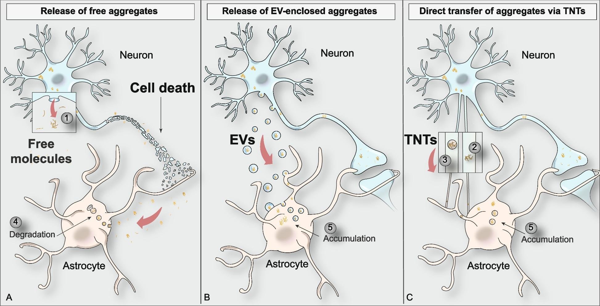
Protein aggregates can be transmitted from a sick neuron to healthy neurons, whether neighbouring or distant, by several mechanisms:

  • as free soluble complexes;
  • packed in exosomal membrane vesicles;
  • along nanotubes between the cells.
Three routes of aggregate spread from a dying neuron to an astrocyte: release of free aggregate molecules, release of aggregates enclosed in extracellular vesicles, and direct transfer through tunnelling nanotubes, ending in accumulation or degradation

Amyloid aggregates in Alzheimer's disease

note

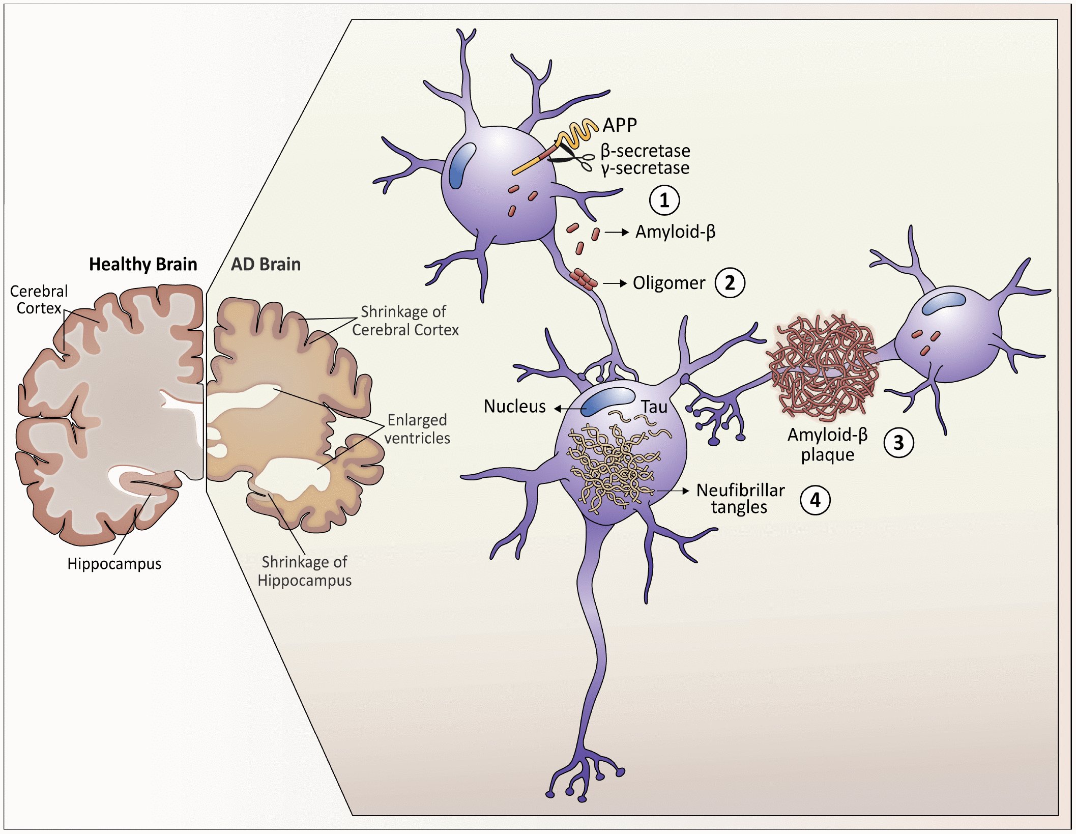
As Alzheimer's disease progresses, the brain tissue shrinks and the volume of the cerebrospinal fluid increases markedly.

At the molecular level, four things happen:

  1. Amyloid peptides are produced by the cleavage of the amyloid precursor protein (APP) on the neuron membrane.
  2. In the space between the neurons, the amyloids form oligomers that disrupt the function of the synapses.
  3. The amyloid fibrils form plaques, which interfere with the function of the neurons.
  4. In the cytoplasm, phosphorylation of the protein Tau causes the formation of tangles, displacing intracellular vesicular transport.
Alzheimer's pathway: healthy versus AD brain shows cortical shrinkage and enlarged ventricles; a neuron cleaves APP by beta- and gamma-secretase into amyloid-beta, which forms oligomers and a plaque, while intracellular Tau forms neurofibrillary tangles

4. Post-translational modifications

note

Unlike conformational changes, modifications are chemical changes: they include the breakage and formation of covalent bonds.

The ones named in this lecture, with what each is for:

  • Disulfide bond - it holds two polypeptide chains, or two parts of the same protein, together.
  • Glycosylation - increases solubility, prevents aggregation, and gives resistance to proteolytic degradation.
  • Phosphorylation - its main function is to regulate protein activity.
  • Ubiquitination - ubiquitin is a small peptide that tags proteins for degradation.
  • SUMOylation - the small peptide SUMO alters protein activity, stability, and interactions with other molecules.
  • Lipid modification - one of its functions is to facilitate interactions with membranes.

Others on the same figure contribute to protein stability, for instance mediating the creation of stable fibres; regulate protein-protein interactions, enzymatic activity, subcellular localisation and stability; or dramatically change the function of a protein by altering its hydrophobicity, solubility and surface properties.

Wheel of post-translational modifications on a protein: hydroxylation, ubiquitination, SUMOylation, lipidation, acetylation, methylation, disulphide bond formation, phosphorylation and glycosylation

Limited proteolysis

note

Special proteases cut selected proteins at specific sites, removing parts of the polypeptide chain. There are three reasons for doing so.

Activation of a precursor. Some proteins are produced as inactive precursors and are activated by proteolysis when needed. The examples are digestive enzymes, clotting factors, complement proteins, and the caspases that trigger apoptosis. The logic is the same in every case: a protein that would be dangerous if it were always active is made in a form that must be cut before it can work.

Removal of a signal peptide. Mitochondrial and secreted proteins contain signal peptides necessary for their transport; when their job is done, the signal peptides must be cut out.

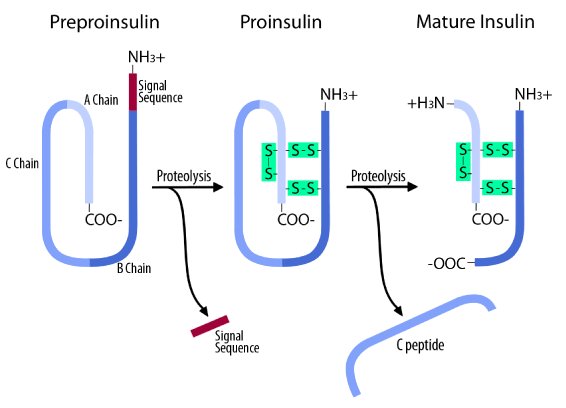
Removal of a folding aid. Some parts of the protein chain are needed only for correct folding and are then excised. The example is the C peptide of insulin.

Preproinsulin to mature insulin: proteolysis removes the signal sequence to give disulphide-bonded proinsulin, then a second proteolysis excises the C peptide, leaving the disulphide-linked A and B chains of mature insulin

Glycosylation

note

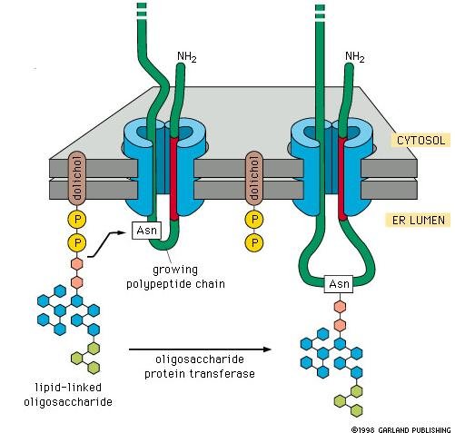
Glycosylation is the addition of a mono- or oligosaccharide. It is typical of export proteins: those transferred to the cell surface, those secreted, and the lysosomal proteins.

Glycosylation begins in the endoplasmic reticulum and continues in the Golgi complex.

The main function of the sugar part is to protect the polypeptide chain from proteases. It can also:

  • make the glycoprotein more hydrophilic;
  • give it a negative charge, as the sialic acid of glycophorin does;
  • allow recognition by a lectin receptor, as the mannose-6-phosphate of the lysosomal enzymes does.
Glycosylation at the ER membrane: a lipid-linked oligosaccharide on dolichol phosphate is transferred by oligosaccharide protein transferase onto an Asn residue of the growing polypeptide chain as it enters the ER lumen

Phosphorylation and dephosphorylation

note

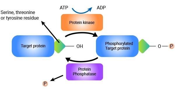
A phosphate residue taken from ATP can be added to the -OH group of serine, threonine or tyrosine. The enzymes catalysing this reaction are the protein kinases.

Unlike the previous modifications, phosphorylation is reversible. Other enzymes, the protein phosphatases, remove the phosphate group.

In this way many proteins exist in two forms, phosphorylated and dephosphorylated, of which only one is active - and which one it is depends on the protein. So the enzymes that add and remove phosphate are regulators of the activity of other proteins.

Protein phosphorylation is crucial for cell cycle progression.

Phosphorylation cycle: protein kinase uses ATP to add phosphate to the OH of a serine, threonine or tyrosine residue on the target protein, protein phosphatase removes it, interconverting target protein and phosphorylated target protein

Polyproteins

note

A polyprotein is any protein that is cleaved to produce a number of smaller polypeptides - for example hormones or viral proteins. In other words, polyproteins are chains of covalently conjoined smaller proteins.

After translation, polyproteins are cleaved into their constituent proteins by highly specific proteases.

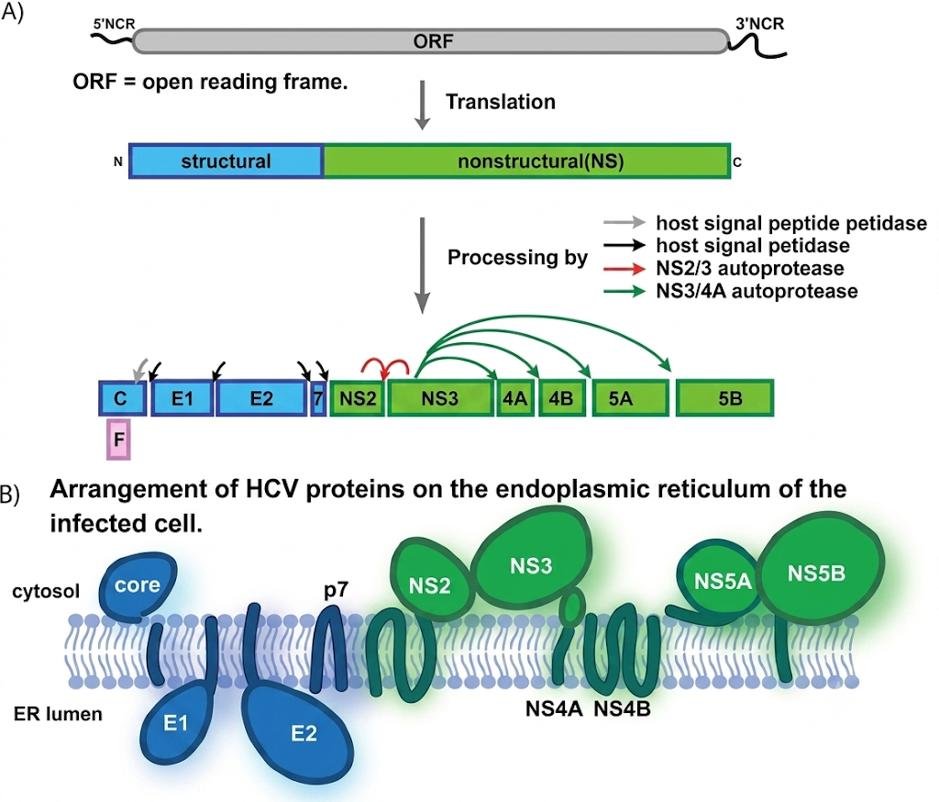
Example: hepatitis C virus (HCV).

  • The HCV genome is a single-stranded RNA encoding a single large open reading frame (ORF) of roughly 3,000 amino acids, flanked by 5' and 3' non-coding regions.
  • Translation of that open reading frame generates a large polyprotein consisting of several structural proteins in the amino-terminal third, plus a protein that regulates the replication of viral RNA.
  • The polyprotein undergoes a complex co- and post-translational series of cleavage events, catalysed by both host and viral proteases, to produce the 10 individual HCV proteins.
Hepatitis C virus polyprotein: the single ORF with 5' and 3' non-coding regions is translated into a structural-nonstructural polyprotein, cleaved by host and viral proteases into core, E1, E2, p7, NS2 to NS5B, arranged across the ER membrane

5. Protein degradation: ubiquitination

note

What is discussed here is not the mass-scale proteolysis of the digestive system and the lysosomes, but the selective degradation in the cytosol of proteins that are short-lived, have synthesis or folding errors, or are chemically damaged.

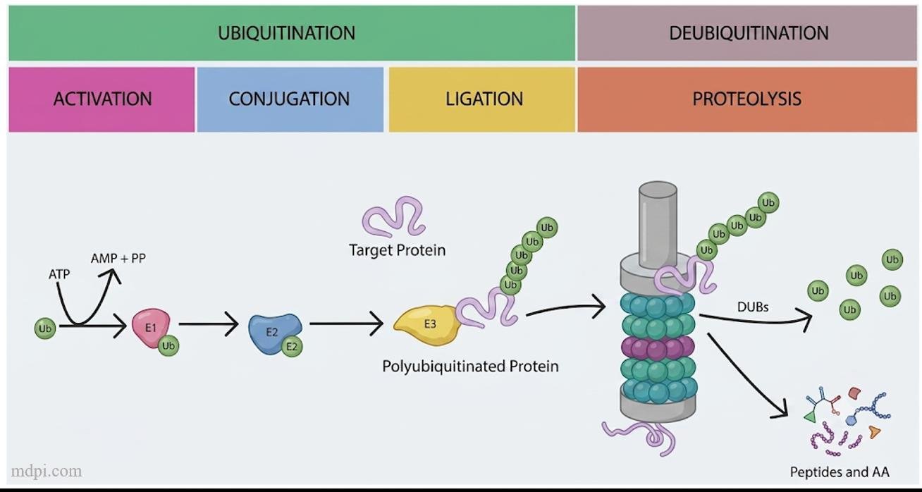
Special enzymes recognise the proteins that must be degraded and label them. The label is a small protein called ubiquitin, named for its wide distribution in eukaryotes.

The enzyme attaches ubiquitin covalently to a lysine residue of the protein substrate. After the first ubiquitin, several more are attached, forming a chain which is the "death sentence" - the signal for degradation.

Ubiquitination and deubiquitination cycle: E1 activates ubiquitin with ATP, E2 conjugates it, E3 ligates a polyubiquitin chain onto the target protein, the proteasome degrades it to peptides and amino acids, and DUBs release free ubiquitin

The proteasome

note

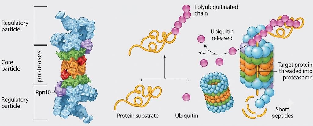
The poly-ubiquitinated protein is handled by a large 26S protein complex located in the cytosol, called the proteasome. It cuts the protein into small peptides.

The proteasome has the shape of a hollow cylinder where the protein enters, quite like some of the chaperones that assist in protein folding. The polyubiquitinated protein enters and meets lysis at the end of the tunnel.

Two details are worth keeping.

  • Ubiquitin is not degraded. The proteasome recognises it and releases it intact to be reused.
  • The released peptides may be used to show the immune system what proteins are being synthesised in the cell, though most often they are degraded by cytosolic peptidases to single amino acids.
26S proteasome with core and regulatory particles containing proteases; a polyubiquitinated protein has its chain removed, ubiquitin released, and the target protein threaded into the proteasome and cut into short peptides

6. Protein sorting to cellular organelles

note

The eukaryotic cell has many compartments, each with its own set of proteins. So newly made proteins must be sent where they belong.

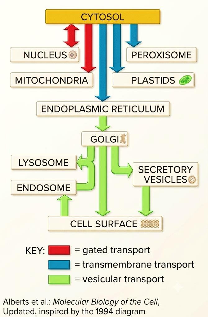
In the standard diagram of destinations and sorting pathways, a circular arrow means retention in the same compartment, and default pathways are shown by thick arrows.

All other pathways require specific sorting signals. A signal is a short peptide sequence included in the primary structure of the protein - the address is written into the protein itself.

Protein fates after translation: free ribosomes give cytosolic proteins or import to mitochondria, peroxisomes and nucleus, misfolded proteins are ubiquitinated for proteasome degradation, and ER-bound ribosomes feed Golgi sorting to secretory vesicles, lysosomes and membrane proteins

The three ways of protein transport

note

  • Gated transport - used only for nuclear proteins. The protein molecule passes through the nuclear pore complex.
  • Transmembrane transport - the protein, in a stretched conformation, is pushed through the lipid bilayer of a membrane.
  • Vesicular transport - the protein, already inside a membrane-bound organelle, is included in a vesicle which buds off, travels through the cytosol, then docks to another membrane-bound organelle and fuses with it.
Destinations from the cytosol with a colour key: red for gated transport to the nucleus, blue for transmembrane transport to mitochondria, peroxisomes, plastids and the ER, green for vesicular transport via the Golgi to lysosomes, endosomes, secretory vesicles and cell surface

7. Sorting of nuclear proteins

note

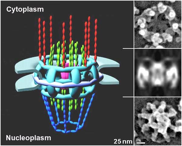
Nuclear pores are supported by a protein cylinder called the nuclear pore complex. Its large size and elaborate structure allow free diffusion of small molecules and gated transport of large ones.

If a protein carries a nuclear localisation signal, it is recognised by a receptor, and the pore opens to let it through.

3D structure of the nuclear pore complex spanning cytoplasm and nucleoplasm, with electron microscopy insets of the pore viewed from the front and side, unlabelled beyond cytoplasm and nucleoplasm

The nuclear localisation signal

note

The peptide sequence serving as the signal for nuclear localisation is short and can be located in any part of the polypeptide chain.

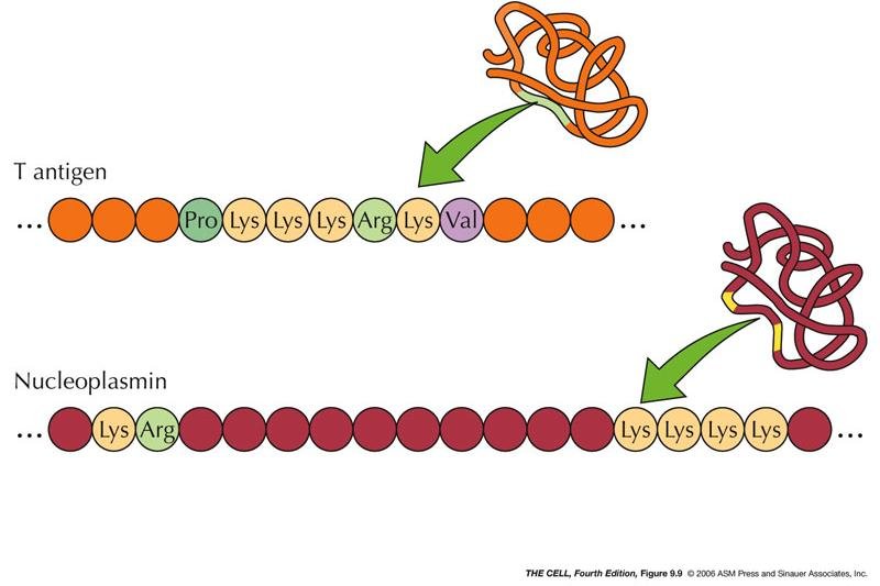
  • Usually, nuclear localisation signals are short stretches of basic amino acids such as lysine and arginine.
  • Sometimes those basic amino acids are not in one long stretch but in two halves separated by other amino acids. The example is the nuclear localisation signal of nucleoplasmin, the nuclear chaperone met above, where a lysine-arginine pair is separated from four lysines by a ten amino acid stretch.
Nuclear localisation signals: the SV40 T antigen signal is one stretch, Pro-Lys-Lys-Lys-Arg-Lys-Val, while the nucleoplasmin signal is bipartite, a Lys-Arg pair separated from a Lys-Lys-Lys-Lys stretch

The mechanism of gated transport

note

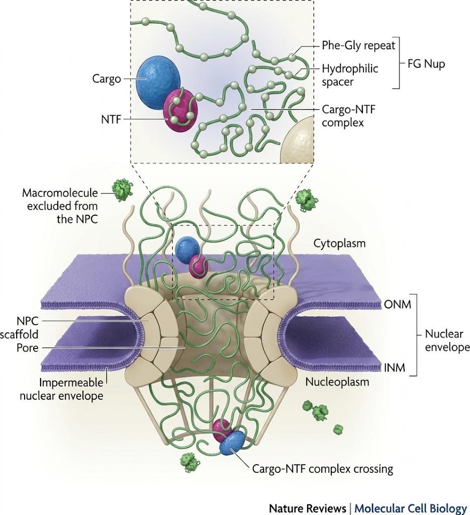
The outer and inner nuclear membranes join to form a ring-shaped pore where the nuclear pore complex (NPC) resides.

At the NPC, the nucleus and cytoplasm are connected by a channel filled with flexible, filamentous proteins, the Phe-Gly nucleoporins (FG Nups).

A nuclear transport factor (NTF) bound to its cargo enters the channel from either the cytoplasmic or the nucleoplasmic side, and then hops between binding sites on the FG Nups until it reaches the opposite side of the NPC.

Nuclear pore complex cross-section: cargo bound to a nuclear transport factor crosses through the FG Nup meshwork of Phe-Gly repeats and hydrophilic spacers between the outer and inner nuclear membranes, while other macromolecules are excluded

Why the nuclear signal is not removed

note

Unlike the other cases, the signal sequence is not removed after the protein is imported into the nucleus.

The reason is structural. In higher eukaryotes the nucleus is disassembled and reassembled at each cell division, and every time, the nuclear proteins have to be collected again from the cytoplasm. A protein that had lost its address after the first import could never be re-imported.

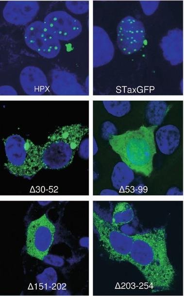
Example: Tax, an oncoprotein produced by the human T-cell leukaemia virus, which leads to the immortalisation of T cells. Tax functions in both the nucleus and the cytoplasm, and it contains nuclear localisation signals that enable shuttling between the two compartments. Mutations in the NLS lead to an inability to get into the nucleus - an illustration of how mutations in a viral genome can change the effectiveness of the virus.

Fluorescence images of Tax and NLS-deletion mutants, HPX, STaxGFP, delta30-52, delta53-99, delta151-202, delta203-254, showing different nuclear versus cytoplasmic localisation in green against blue-stained nuclei

8. Sorting of mitochondrial proteins

note

Although mitochondria have their own translation apparatus, most of their proteins are synthesised in the cytosol.

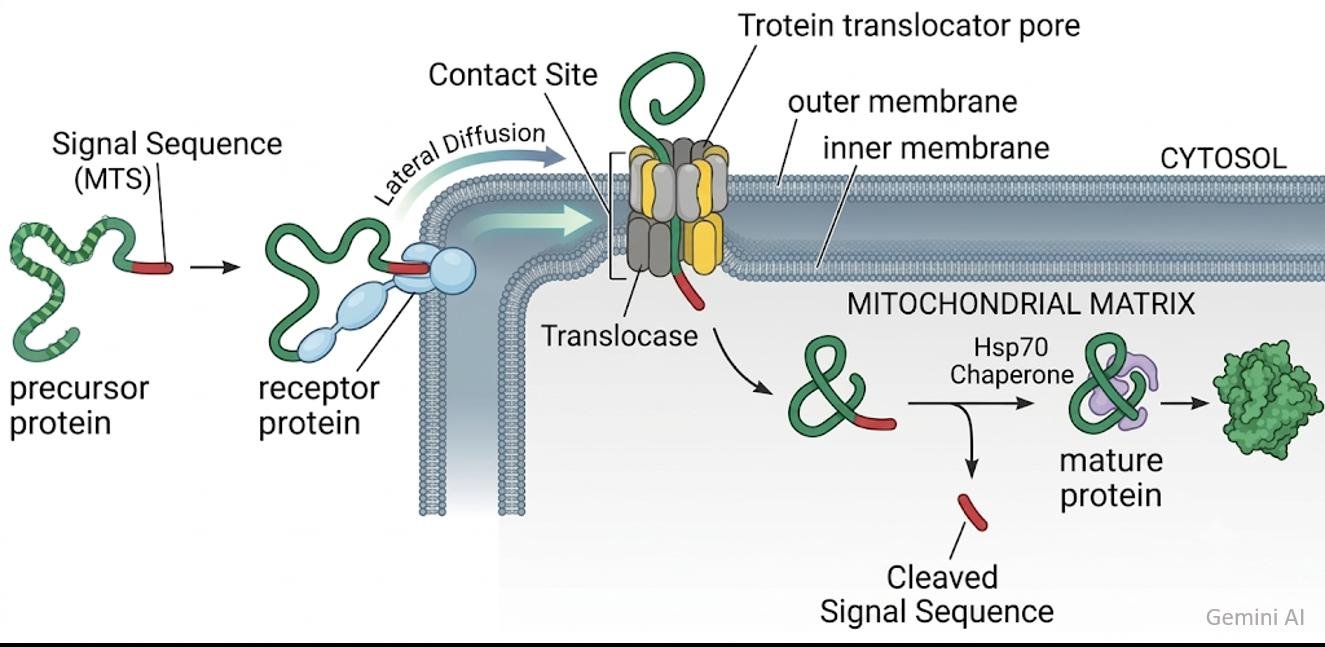
  1. These proteins have at their N terminus a relatively long signal peptide.
  2. It is recognised by a receptor in the outer mitochondrial membrane.
  3. The receptor directs it to other proteins called translocators, which form a channel through both mitochondrial membranes.
  4. The protein is pushed through the channel in a stretched conformation.
  5. Inside, a specific protease called signal peptidase cuts off the signal peptide, the address label which is no longer needed.
Mitochondrial protein import: the N-terminal signal sequence is bound by a receptor protein, the precursor is pushed through the outer and inner membrane translocator pore at a contact site, and Hsp70 chaperone folds it to a mature protein after the signal sequence is cleaved

9. Sorting of surface, secreted and lysosomal proteins: the export pathway

note

Most membrane organelles of eukaryotic cells - the endoplasmic reticulum, the Golgi complex, secretory vesicles, lysosomes and endosomes - are interconnected with the cell membrane and with the environment. Proteins travel between them, and this process is defined as the export pathway.

Proteins of the export pathway are synthesised on ribosomes attached to the rough, or granular, endoplasmic reticulum. In fact they are what makes that endoplasmic reticulum rough.

Polypeptide chains destined for export or for lysosomes are never found in the cytosol. They are translocated into the lumen of the ER while still being synthesised.

From there, proteins are sent by vesicular transport first to the Golgi complex, and then either to the cell surface or to the lysosomes.

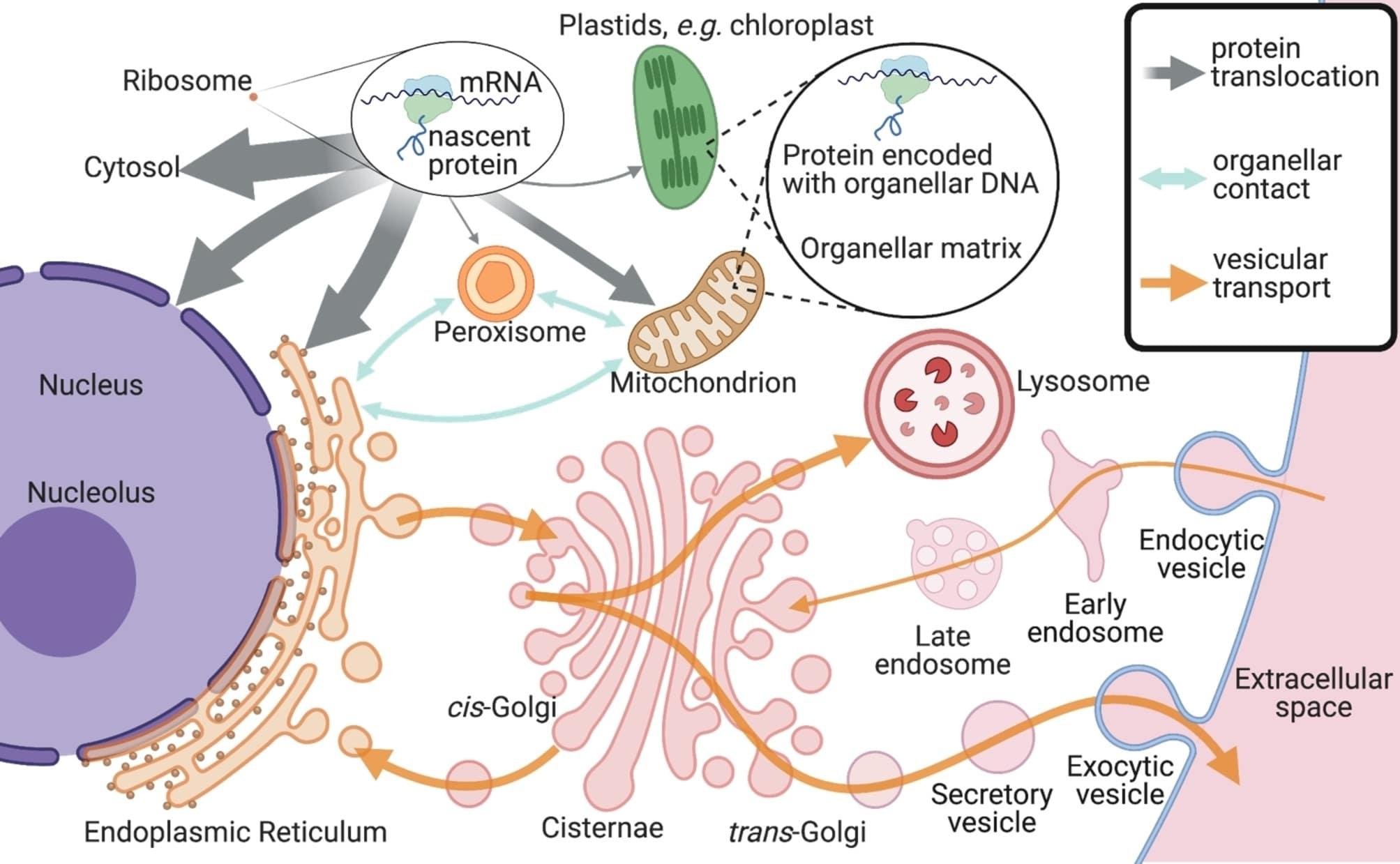
Cell-wide protein trafficking map: ribosomes make nascent proteins for the cytosol or organelles with their own DNA, while rough ER, cis- and trans-Golgi, lysosome and early and late endosomes exchange material by vesicular transport with the cell surface
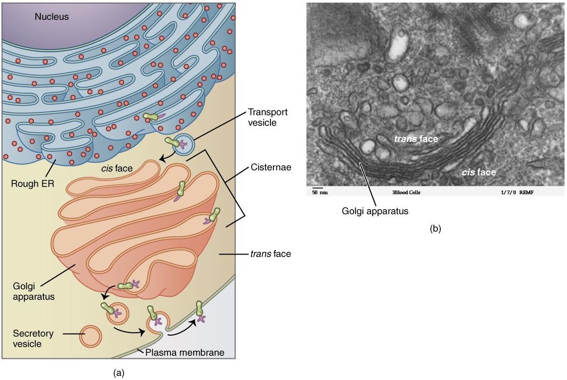
Rough ER studded with ribosomes feeding transport vesicles into the Golgi apparatus at its cis face, through the cisternae to secretory vesicles at the trans face, with an electron micrograph of the Golgi's cis and trans faces

Translocation to the endoplasmic reticulum

note

Export proteins, like mitochondrial ones, have a signal peptide at their N terminus.

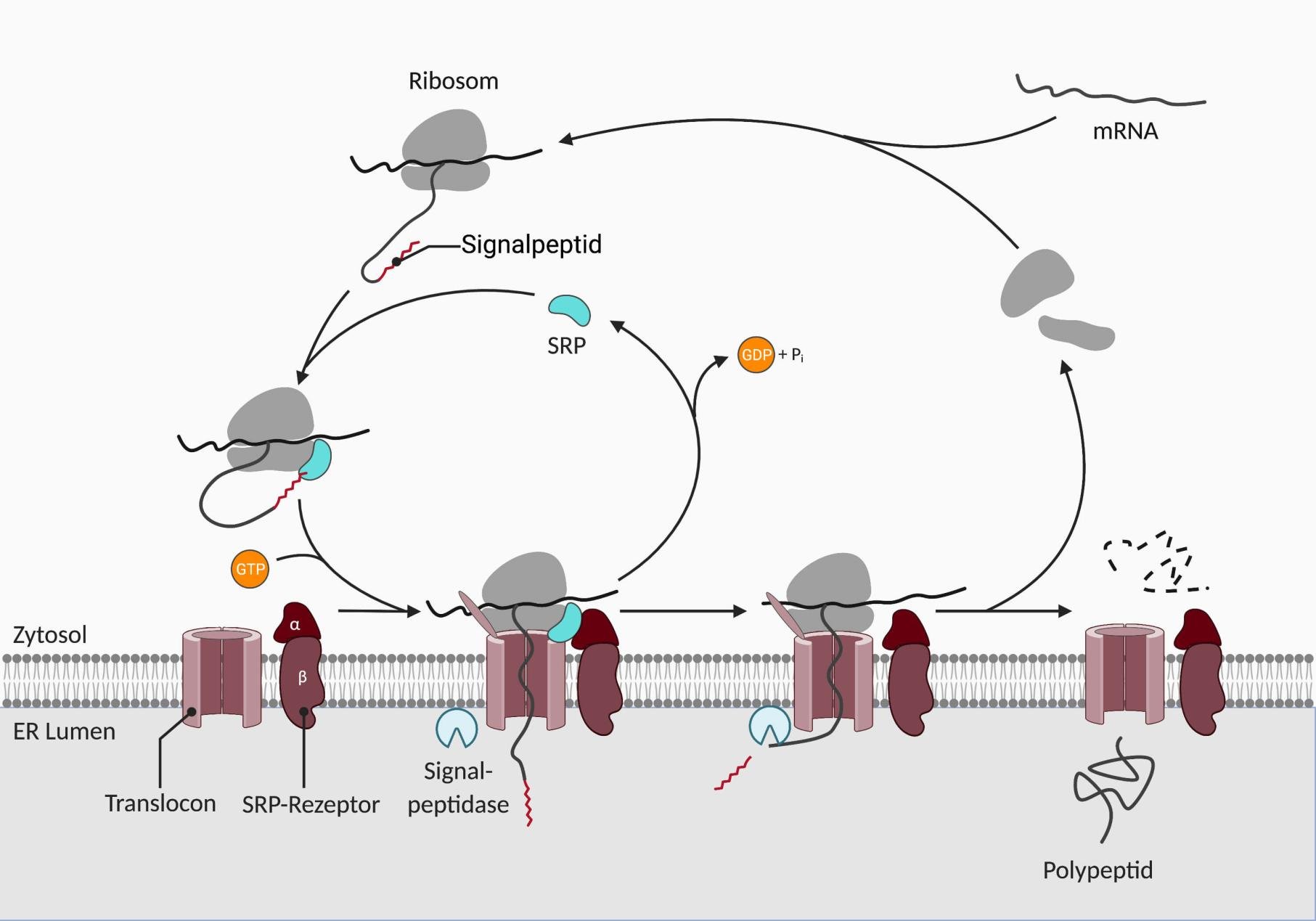
  1. The signal is recognised by a ribonucleoprotein called the signal recognition particle (SRP).
  2. SRP binds to the signal and then to a receptor in the ER membrane, docking the ribosome to the ER.
  3. The elongating polypeptide is pushed through the ER membrane by translocators.
  4. When this is finished, the signal is excised by a signal peptidase.
SRP cycle at the ER membrane: the ribosome's signal peptide is bound by SRP, which docks via GTP to the SRP receptor at the translocon so the growing polypeptide threads into the ER lumen, and the signal peptidase then cleaves the signal peptide

10. The three ways out of the Golgi

note

From the ER, proteins are sent to the Golgi and travel through its cisternae. The last cisterna dispatches proteins to three destinations.

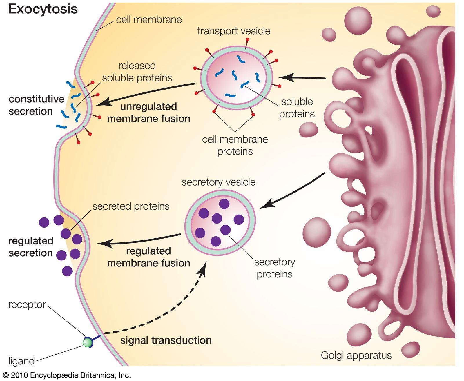
  1. Constitutive secretion. This is the default: small vesicles to the cell membrane, with no additional signal required.
  2. Regulated secretion. Proteins bud off in secretory vesicles that will be exocytosed only after a special signal. These vesicles are coated by the protein clathrin, which is the hallmark of regulated vesicular transport. The example is the protein hormones.
  3. To the lysosomes. These vesicles are labelled by mannose 6-phosphate.
Exocytosis at the Golgi: a transport vesicle releases soluble and cell membrane proteins by unregulated membrane fusion, constitutive secretion, while a secretory vesicle releases its proteins only after receptor-triggered signal transduction, regulated secretion

Vesicle transfer between the Golgi and the ER

note

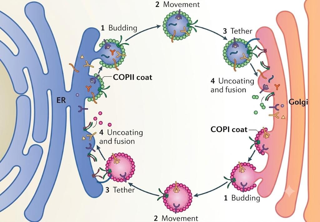
Traffic in both directions requires membrane receptors, and the coats differ by direction:

  • from ER to Golgi - vesicles labelled by coat protein II (COPII);
  • the opposite direction - vesicles labelled by coat protein I (COPI).
COPII and COPI vesicle cycles between ER and Golgi: COPII-coated vesicles bud, move, tether and uncoat from ER to Golgi, while COPI-coated vesicles bud, move, tether and uncoat in the reverse direction, from Golgi back to ER

The localisation of exocytosis

note

The topology of the sites of exocytosis may correlate with the functional polarisation of the cellular surfaces, as in epithelia or neurons.

The docking, membrane fusion and release of the contents of secretory granules can therefore also be polarised, and performed in a special active zone of the cell membrane.

Polarised exocytosis is achieved through the coordinated actions of the membrane trafficking machinery and the cytoskeleton.

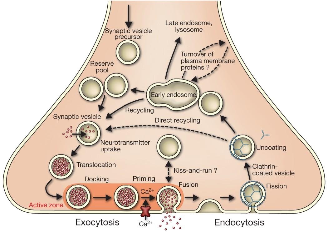
Synaptic vesicle cycle at the active zone: vesicles from the reserve pool dock, prime and fuse with Ca2+ influx to release neurotransmitter, exocytosis, while clathrin-coated vesicles form, uncoat and recycle directly or via the early endosome, endocytosis

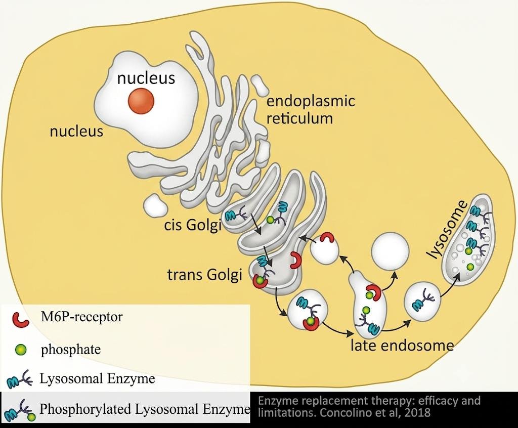
11. The mannose 6-phosphate pathway to the lysosomes

note

  1. Lysosomal hydrolases are synthesised in the endoplasmic reticulum.
  2. They are packed into vesicles that move to the cis Golgi network.
  3. The hydrolases are modified by the addition of mannose-6-phosphate (M6P) groups.
  4. At the trans Golgi network, the M6P signal allows the segregation of lysosomal hydrolases from all other types of protein, by means of M6P receptors.
  5. The resulting clathrin-coated vesicles bud off from the trans-Golgi network and fuse with late endosomes.
  6. At the low pH in the late endosome, the hydrolases dissociate from the M6P receptors, and the empty receptors are recycled to the Golgi apparatus for further rounds of transport.
  7. The enzymes are packed into vesicles for further transport to established lysosomes. The late endosome itself can eventually grow into a mature lysosome.
Mannose 6-phosphate pathway: lysosomal enzymes made in the ER are phosphorylated in the cis and trans Golgi, bind M6P receptors into a late endosome where low pH releases the enzyme and recycles the receptor to the Golgi, and the enzyme reaches the lysosome

When sorting to lysosomes fails: I-cell disease

note

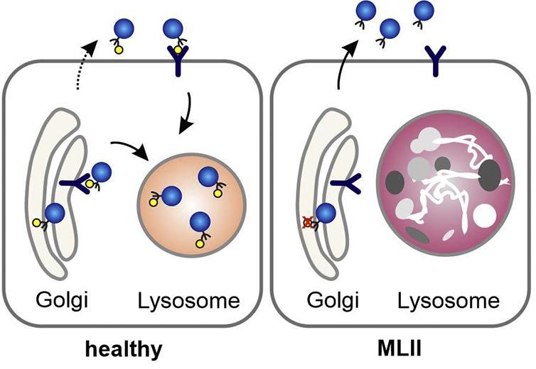
If mannose-6-phosphate cannot be synthesised because of a gene mutation, lysosomal enzymes cannot be sent where they belong. They are instead exocytosed by constitutive secretion - the default pathway - and everything that ought to be digested by the lysosomes fills the cell instead, forming abnormal, harmful inclusions.

Healthy versus MLII cell: in the healthy cell phosphorylated lysosomal enzymes are captured by M6P receptors from the Golgi into the lysosome, in the MLII cell the enzyme cannot be phosphorylated, is not captured, and is secreted instead

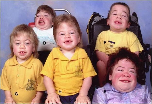
At the level of the organism this molecular defect is manifested as a severe autosomal recessive disorder called mucolipidosis II, or I-cell disease, where the I stands for "inclusions".

Like many other inborn errors of metabolism, I-cell disease is progressive and ultimately lethal. Children have:

  • short-trunk dwarfism and other skeletal abnormalities;
  • retarded psycho-motor development;
  • coarse facial features;
  • restricted joint movement.

Most of them die before the age of 7, of heart failure or respiratory tract infections.

Photograph of several children with I-cell disease, mucolipidosis II, showing coarse facial features, short stature and restricted mobility, one seated in a wheelchair

12. The vesicular transport network: endosomes

note

Endosomes are membrane vesicles. They play crucial roles in various physiological processes, such as nutrient uptake and the sorting and delivery of macromolecules.

After endocytosis, endosomes provide the environment in which material is sorted before it reaches the degradative lysosome.

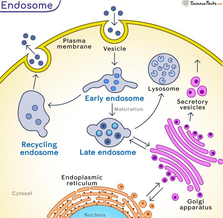
Endosomes have three major compartments - early, late, and recycling - distinguished by their morphology and localisation, the material they process, and the marker proteins on their surface.

Endosome compartments diagram: a plasma-membrane vesicle becomes an early endosome, which recycles via a recycling endosome back to the membrane or matures into a late endosome heading to the lysosome, with links to the ER and Golgi

Early, late and recycling endosomes

note

Early endosomes lie close to the plasma membrane. Endocytic vesicles fuse with them, and an early endosome processes the received material in order to form a late endosome.

Late endosomes lie deep in the cytoplasm. Molecules in the early endosomes are sorted into smaller vesicles that bud from the endosomal membrane, which turns them into multivesicular bodies, another name for late endosomes. Late endosomes eventually fuse with lysosomes for the degradation of their content.

Recycling endosomes. The set of membrane proteins a cell carries - receptors, ion channels, ion pumps and adhesion molecules - depends on the function of the cell. When the cell must change its function, many of these proteins are internalised by endocytosis, and the vesicles are then called recycling endosomes. Once internalised, the membrane proteins undergo sorting that determines their fate: trafficking to degradation pathways, or processing for return to the membrane.

All these types of endosome communicate with each other by vesicles.

Endosome maturation: a plasma-membrane vesicle forms an early endosome near the membrane, which matures into a late endosome deeper in the cytoplasm, while membrane proteins recycle back via a recycling endosome

13. Retrograde trafficking, and ricin as its example

note

Ricin is a lectin, a carbohydrate-binding protein, produced in the seeds of the castor oil plant, Ricinus communis. It is highly toxic: a dose of purified ricin powder the size of a few grains of table salt can kill an adult human.

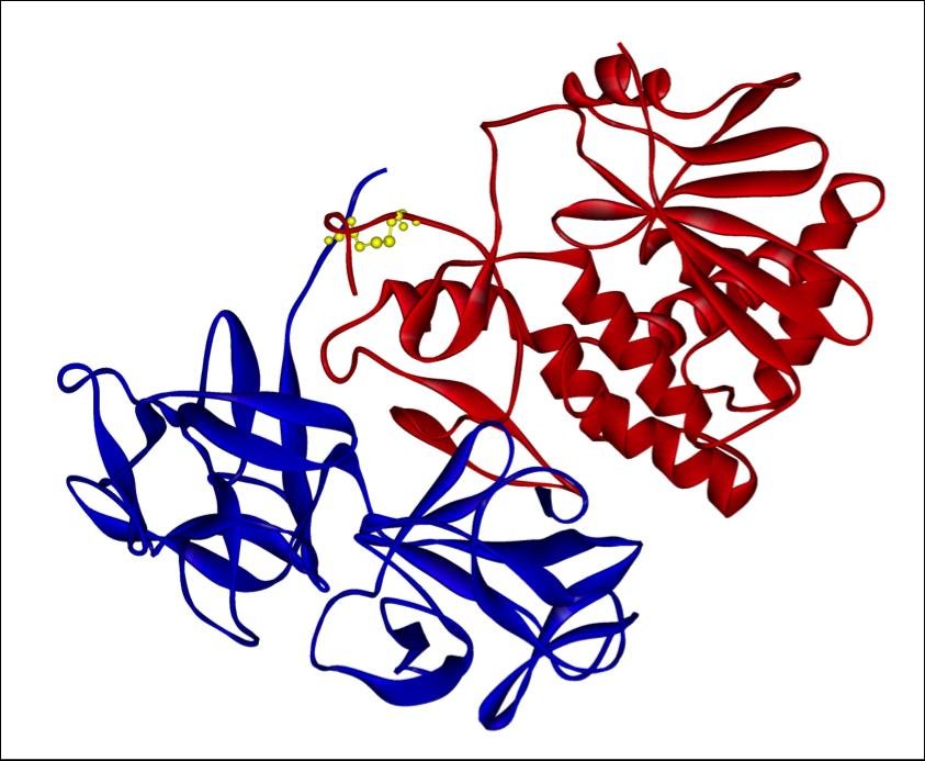
The ricin molecule consists of two polypeptide chains, joined by a disulfide bond:

Ribbon structure of the ricin heterodimer, unlabelled: the smaller A chain in blue is linked to the larger lectin B chain in red
  • Chain A (ricin toxin A, RTA) is responsible for ribosome inactivation. It breaks the glycosidic bond within the large rRNA of the 60S subunit of eukaryotic ribosomes and completely inactivates them.
  • Chain B (ricin toxin B, RTB) is responsible for entry into the cytoplasm. It binds specific carbohydrates on the membrane of eukaryotic cells.

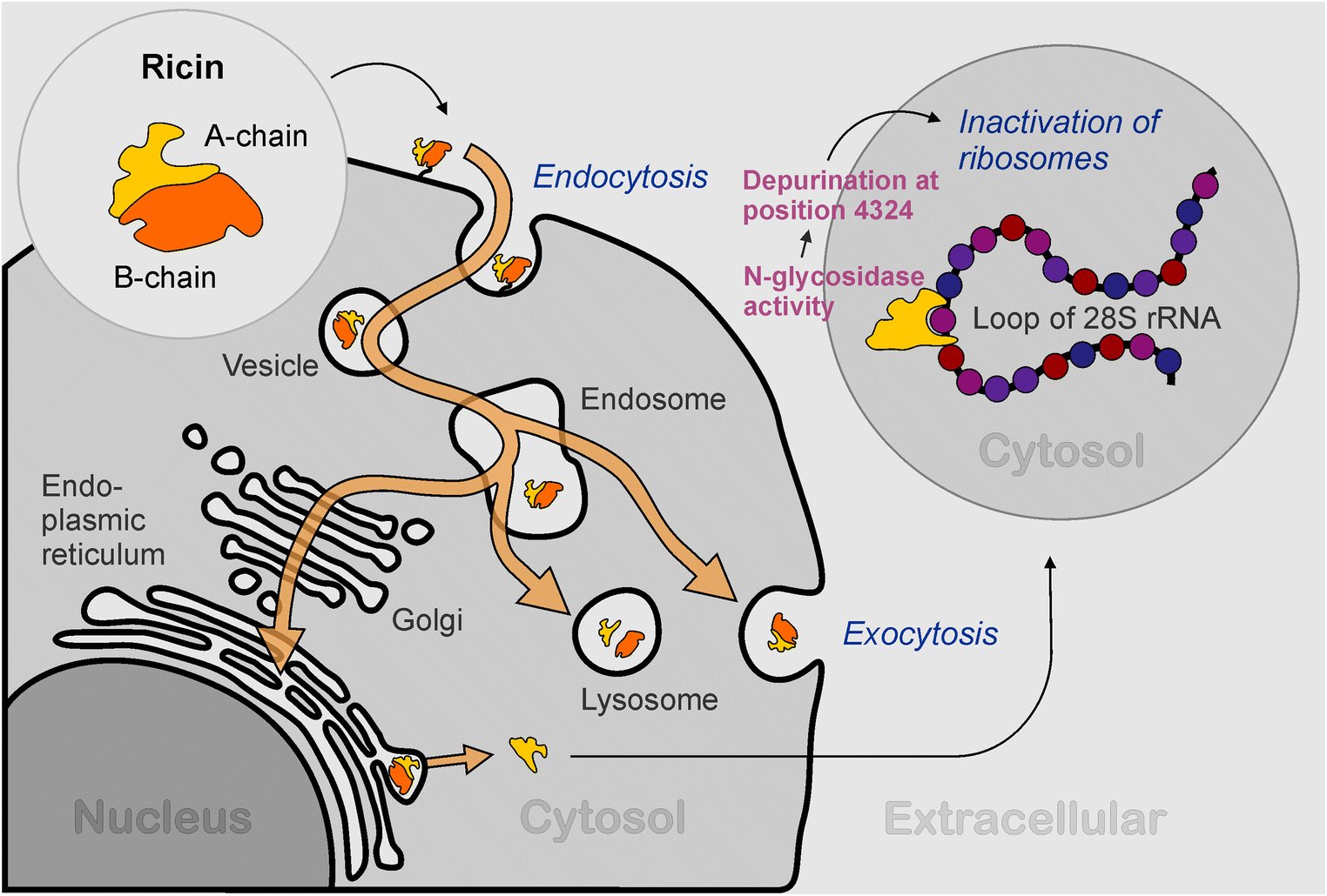
The route it takes. Ricin binds to cell surface receptors by its B chain and enters the cell by endocytosis. From there it can take three possible ways:

  • it can be destroyed in lysosomes;
  • it can leave the cell by exocytosis;
  • it can be transferred to the Golgi complex and then, by retrograde trafficking, to the endoplasmic reticulum.

Only the third route is productive for the toxin. In the lumen of the ER, the ricin A and B chains are separated, and in the cytosol the A chain inactivates ribosomes by depurination of the 28S rRNA.

Ricin's route through the cell: after endocytosis the A and B chain toxin reaches an endosome, then the lysosome or exocytosis, or via the Golgi to the ER, where the A chain reaches the cytosol and depurinates the 28S rRNA loop at position 4324, inactivating ribosomes

Retrograde trafficking from the endosome to the trans-Golgi network

note

Usually vesicles are transferred from ER to Golgi and then to the cell membrane. Sometimes the transfer runs in the opposite direction, and that is retrograde trafficking.

The cell's own use for it is to return to the Golgi and the ER their own proteins, which were "washed away" during normal trafficking.

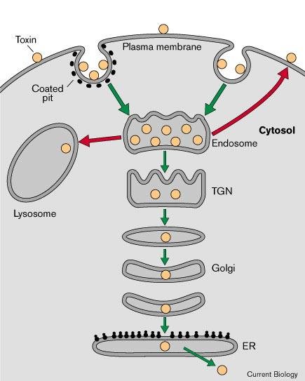
Some toxins press the cell to do retrograde trafficking, and this is how they reach their target molecules. Most of the toxin in the endosomes is either recycled back to the surface or transported to lysosomes for degradation, but a small proportion is transported to the trans-Golgi network (TGN), and that is the productive route for subsequent intoxication. From the TGN the toxin travels by retrograde vesicular transport through the Golgi cisternae to the ER, and can then cross the ER membrane into the cytosol.

This pathway may be very inefficient, but the extreme potency of these toxins ensures that a lethal amount reaches the cytosol.

Generic retrograde toxin trafficking: toxin enters by a coated pit into the endosome, most goes to the lysosome or recycles to the plasma membrane, but some travels via the trans-Golgi network and Golgi to the endoplasmic reticulum

14. Exosomes

note

Most living cells release an array of extracellular vesicles (EVs).

The nomenclature depends on the cell of origin, the function and the size of the vesicle, and it has generated confusion about the definition of exosomes. The names in use include ectosomes, microvesicles, microparticles, prostasomes, tolerosomes (which induce immunological tolerance to some antigens), apoptotic bodies (released by apoptotic cells), and nanovesicles.
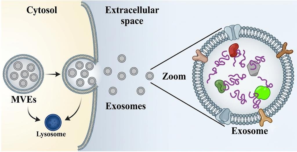
Functions. Cells deliver mRNA, miRNA, proteins and other biomolecules between organelles by means of membrane vesicles, which contain receptors to ensure traffic specificity. Sometimes these vesicles are secreted into body fluids, including blood, saliva, breast milk and sperm.

Animal cells use exosomes for intercellular communication, and each tissue produces specific exosomes. For example, exosomes released from cancer cells contribute to the development of metastases.
For the same reason exosomes are used in the diagnosis of cancer: they carry biomarkers specific to the cell they came from.

Multivesicular endosome releasing its internal vesicles as exosomes into the extracellular space, or fusing with the lysosome for degradation, with a zoomed exosome showing membrane-anchored and luminal cargo proteins

Exosome biogenesis

note

Microvesicles are formed by direct blebbing from the plasma membrane, and the result is a multivesicular endosome (MVE).

The MVE can then take one of two fates:

  • fuse with the plasma membrane, releasing the exosomes into the extracellular matrix;
  • fuse with the lysosome for degradation.

The exosomal membrane contains proteins specific to the cell of origin, which is what makes exosomes readable as a diagnostic signal.

Exosome biogenesis: a multivesicular endosome either fuses with the lysosome for degradation or fuses with the plasma membrane to release its internal vesicles as exosomes, with a zoomed exosome carrying membrane and luminal proteins

The most important things to know

note

  • A denatured protein displays hydrophobic side chains on its surface. That is what makes it aggregate, and it is also what chaperones recognise.
  • Chaperones (hsp + mass) either bind the exposed patches or provide a cylinder in which refolding is protected. Misfolded protein induces heat shock factor trimers, which switch on more hsp genes.
  • Prions are the inverse: PrPC (α-helical) converts to PrPSc (β-sheet), which then converts its neighbours. Infectious with no DNA or RNA.
  • Modifications are covalent changes. Only phosphorylation is reversible, through kinases and phosphatases, and it is the main regulator of activity.
  • Limited proteolysis has three uses: activating a dangerous precursor, removing a used signal peptide, and removing a folding aid (insulin's C peptide).
  • Glycosylation protects from proteases and begins in the ER, continuing in the Golgi.
  • Degradation: a polyubiquitin chain on a lysine is the death sentence; the 26S proteasome shreds the protein and recycles the ubiquitin.
  • Three transport mechanisms: gated (nucleus only), transmembrane (stretched, e.g. mitochondria), vesicular (the export pathway).
  • Only the nuclear signal is kept, because the nucleus is rebuilt at every division.
  • Out of the Golgi: constitutive (default, no signal), regulated (clathrin, needs a signal), and lysosomal (mannose 6-phosphate).
  • Losing M6P causes I-cell disease: the hydrolases are secreted by default and the undigested material forms inclusions.
  • Retrograde trafficking normally recovers the cell's own proteins; ricin hijacks it to reach the cytosol, where RTA depurinates the 28S rRNA.
Protein fates after translation: free ribosomes give cytosolic proteins or import to mitochondria, peroxisomes and nucleus, misfolded proteins are ubiquitinated for proteasome degradation, and ER-bound ribosomes feed Golgi sorting to secretory vesicles, lysosomes and membrane proteins
Wheel of post-translational modifications on a protein: hydroxylation, ubiquitination, SUMOylation, lipidation, acetylation, methylation, disulphide bond formation, phosphorylation and glycosylation

All 105 Medical Biology topics · exam study support, not clinical guidance. SuperMed is not affiliated with the Medical University of Sofia.

Start free. No card needed.

Every account starts free, with free topics in Cytology and Medical Biology. Super opens the rest. Super is €15 a month.

Create a free account