


Medical Biology · Year 1 · Medical University of Sofia

78

Protozoa. Phylum Sarcomastigophora. Subphylum Mastigophora (Flagellata). Genus Trypanosoma

Free notes for topic 78 of the Medical Biology syllabus, open without an account. Written by a senior student against the syllabus question and checked line by line by a second student before publishing. How content is made

Updated

What this topic covers

note

What protozoa are and which groups of them the course deals with; the taxonomic ranks used to name them; how protozoans reproduce, by closed mitosis and by sexual processes, and what definitive, intermediate and reservoir hosts are; the phylum Sarcomastigophora and its two kinds of members, flagellates and amoeboid protozoans; the kinetoplastids, their kinetoplast and undulating membrane, and their four morphological forms; and the genus Trypanosoma, with Trypanosoma brucei (African sleeping sickness, its vector, life cycle, distribution and antigenic variation) and Trypanosoma cruzi (Chagas disease, its amastigotes, intracellular parasitism, life cycle and routes of transmission).
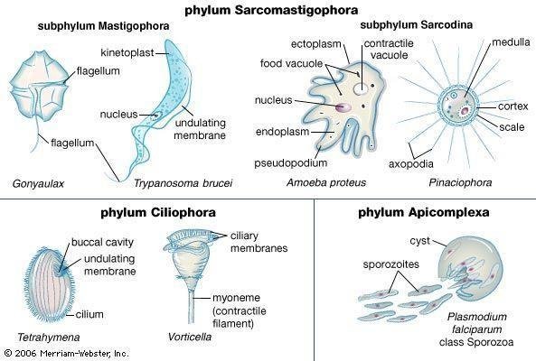
Four protozoan groups compared, one genus shown for each: Sarcomastigophora-Mastigophora (Trypanosoma brucei) with kinetoplast, flagellum, undulating membrane; Sarcodina (Amoeba proteus) with pseudopodium, food and contractile vacuole; Ciliophora (Tetrahymena, Vorticella); Apicomplexa (Plasmodium falciparum) with sporozoites

1. What is Protozoa

note

Protozoa is an informal umbrella name of unicellular motile eukaryotes. It is a convenience, not a natural group.

  • Being motile, capable of engulfing particles (including other cells) and devoid of a cell wall, they resemble animal cells, but few of them are indeed closely related to animals.
  • Some protozoans are free-living, others are parasites.

Phylum Sarcomastigophora includes flagellated and amoeboid protozoans. Today, it is obsolete and has been subdivided into several distinct phyla, but is still used in medical parasitology for convenience. That is why the name still appears in the syllabus and on the examination paper.

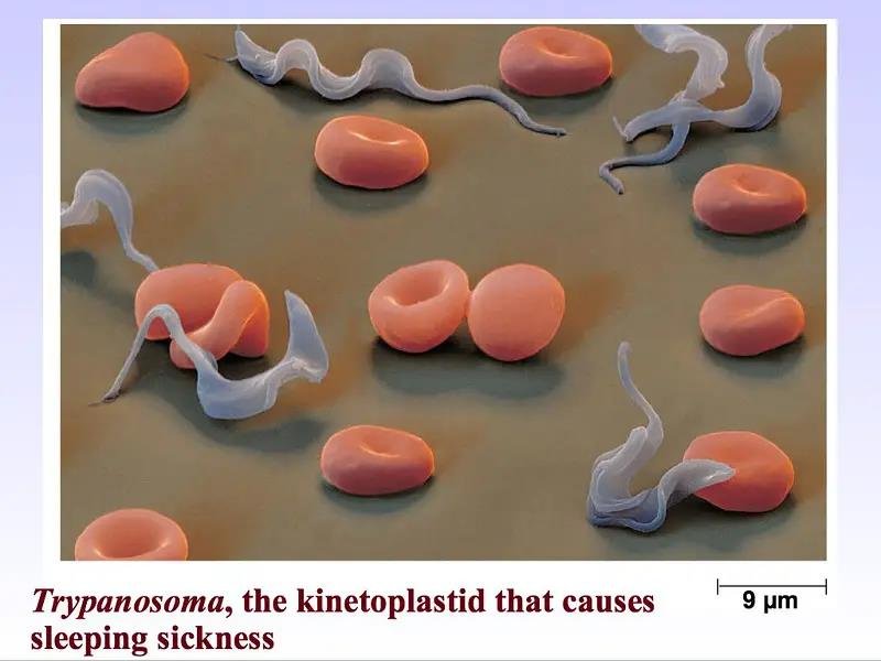
The cover image of the lecture shows Trypanosoma, the kinetoplastid that causes sleeping sickness, among red blood cells, with a scale bar of 9 μm - which is a useful reminder of how small these cells are.

Trypanosoma trypomastigotes coiled among red blood cells, unlabelled, with a 9 μm scale bar shown at bottom right

The protozoan groups discussed in this course

note

The opening figure sets out the four groups the course will deal with, with a representative of each:

GroupRepresentatives shownStructures labelled
Phylum Sarcomastigophora, subphylum Mastigophora (the flagellates)*Gonyaulax, *Trypanosoma brucei**kinetoplast, flagellum, nucleus, undulating membrane
Phylum Sarcomastigophora, subphylum Sarcodina (the amoeboid protozoans)*Amoeba proteus, *Pinaciophora**ectoplasm, endoplasm, food vacuole, contractile vacuole, nucleus, pseudopodium; and in Pinaciophora, axopodia, cortex, medulla and scale
Phylum Ciliophora (the ciliates)*Tetrahymena, *Vorticella**buccal cavity, undulating membrane, cilium; ciliary membranes and myoneme (contractile filament)
Phylum Apicomplexa*Plasmodium falciparum, *class Sporozoa**cyst, sporozoites

The first two of these, the flagellates and the amoeboid parasites, are the subject of this lecture, which is Protozoa, Part 1: flagellates and amoeboid parasites.

Four protozoan groups with a representative genus each, labelled: Mastigophora (Trypanosoma brucei) - kinetoplast, flagellum, nucleus, undulating membrane; Sarcodina (Amoeba proteus, Pinaciophora); Ciliophora (Tetrahymena, Vorticella); Apicomplexa (Plasmodium falciparum)

2. But what is actually a phylum? Systematics of organisms

note

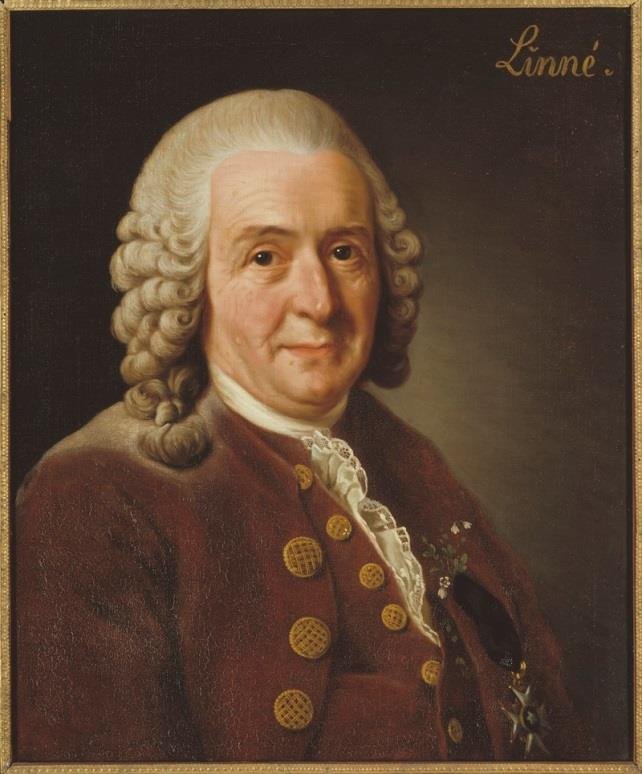
Systematics, introduced by Carl Linnaeus (1707-1778), classifies organisms into a hierarchical system of taxonomic ranks. Ideally, it is a phylogenetic tree, a "pedigree" of the living world reflecting the evolutionary origin and relationships of the groups of organisms.

Portrait of Carl Linnaeus, the eighteenth-century founder of systematics, in period dress, unlabelled

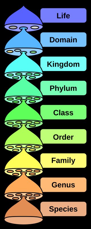
Each group (taxon) has a scientific Latin name. By convention, genus and species names are written in italic, while names of higher taxons are not. In addition to the taxonomic ranks shown, there are intermediate ones (e.g. subphylum or superfamily) as well as unofficial ones. Study the Latin names!

The human is shown as the example, running from the top of the hierarchy downwards:

Taxonomic rank pyramid, top to bottom: Life, Domain, Kingdom, Phylum, Class, Order, Family, Genus, Species, each rank as its own coloured tier
RankTaxon
Life
DomainEukaryota
KingdomMetazoa
PhylumChordata
ClassMammalia
OrderPrimates
FamilyHominidae
Genus*Homo*
Species*Homo sapiens*

3. Reproduction of Protozoa: mitosis

note

In many sources, the division of protozoans is called binary fission. However, this term does not describe the actual process. Like all eukaryotic cells, protozoans divide by mitosis.

However, unlike our mitosis, they reproduce by closed mitosis, i.e. the nuclear envelope remains intact throughout division and all movements of chromosomes happen within the nucleus.

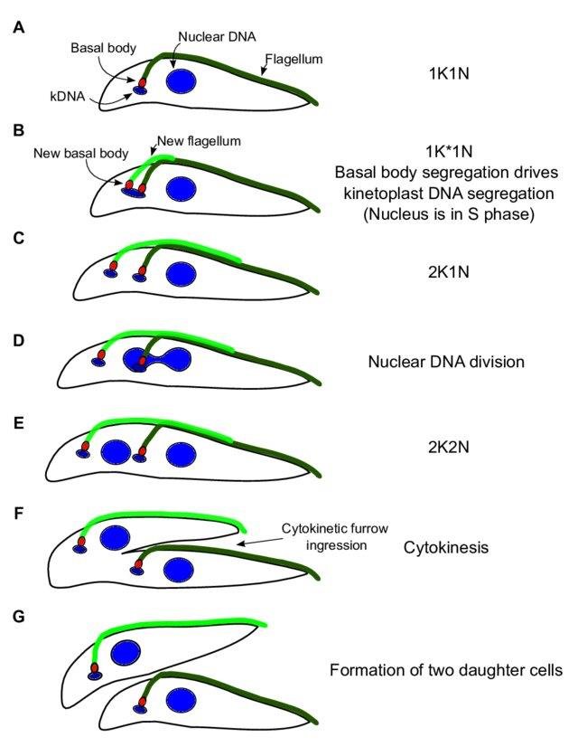
The cytoplasmic structures, which are present in the cell in a single copy or in a precisely defined small number - flagella, basal body, etc., are also duplicated in advance and are distributed precisely.

The figure shows the division of Trypanosoma, where K is the kinetoplast and N the nucleus, in seven steps:

  1. 1K1N - the resting cell, with kinetoplast DNA (kDNA), basal body, nuclear DNA and flagellum labelled.
  2. 1K*1N - a new basal body and a new flagellum appear; basal body segregation drives kinetoplast DNA segregation, while the nucleus is in S phase.
  3. 2K1N - two kinetoplasts, one nucleus.
  4. Nuclear DNA division.
  5. 2K2N - two kinetoplasts and two nuclei.
  6. Cytokinesis, by cytokinetic furrow ingression.
  7. Formation of two daughter cells.

The order matters: the kinetoplast divides before the nucleus, and each daughter cell is guaranteed one of each organelle rather than a random share.

Trypanosoma closed mitosis in seven labelled steps: 1K1N, 1K*1N with new basal body and flagellum, 2K1N, nuclear DNA division, 2K2N, cytokinesis by furrow ingression, two daughter cells

Reproduction of Protozoa: sexual processes

note

Unicellular organisms also have sexual processes, although in many species meiosis and fertilisation occur rarely and are poorly studied. Male and female gametes are more often called micro- and macrogametes, respectively.

In Protozoa, the haploid-diploid cycle has different variations. There are:

  • Protozoans in which, like in us, meiosis is part of gametogenesis and the organism is diploid during most of its life cycle.
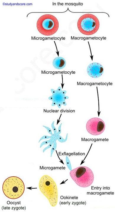
  • Protozoans in which the zygote undergoes meiosis and the organism is haploid during most of its life cycle (an example is Plasmodium shown in the diagram).

A special case are the ciliates Ciliophora, in which haploid nuclei are formed and fuse instead of haploid cells. Their sexual process will be discussed later.

The diagram shows the sexual process in Plasmodium, which takes place in the mosquito: microgametocyte and macrogametocyte are taken up in the blood; the microgametocyte undergoes nuclear division and exflagellation to give microgametes, the macrogametocyte becomes a macrogamete, then comes entry into the macrogamete, producing the ookinete (early zygote) and then the oocyst (late zygote).

For parasites, when there are two or more hosts and it is known where the parasite sexual process takes place, this host is called the definitive or final, and the other hosts are called intermediate.

Sexual cycle of Plasmodium in the mosquito, labelled: microgametocyte and macrogametocyte, nuclear division, exflagellation, microgamete, macrogamete, entry into macrogamete, ookinete, oocyst

4. Phylum Sarcomastigophora

note

It includes free-living inhabitants of terrestrial and aquatic environments as well as parasites in animals and humans. It has two kinds of members, distinguished by how they move.

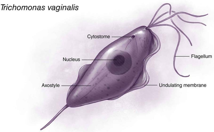
Flagellates. These protozoans move by one or several flagella. Some species have a flagellum that, instead of coming out freely, is connected to the cell body by an undulating membrane - a thin layer of cytoplasm, surrounded by a cell membrane, which the flagellum moves. It is used as an extended fin for faster swimming. The flagellate drawn as the example is Trichomonas vaginalis, with its cytostome, nucleus, axostyle, flagellum and undulating membrane labelled.

Trichomonas vaginalis diagram labelled: cytostome, flagellum, nucleus, axostyle, undulating membrane

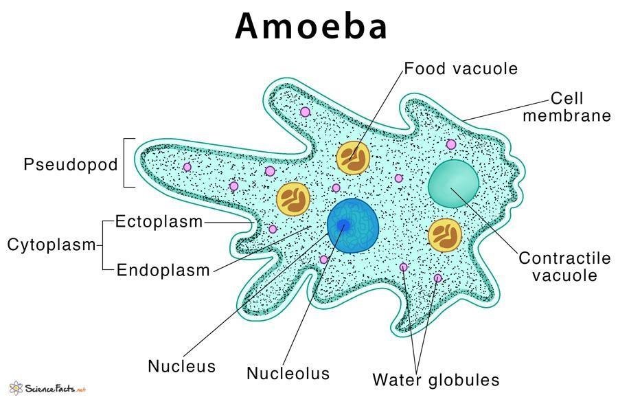
Amoebozoans. In these protozoans, the cytoplasm forms temporary protrusions - pseudopodia, for locomotion and food intake. Movement by pseudopodia is called amoeboid. The amoeba drawn as the example has its pseudopod, cytoplasm divided into ectoplasm and endoplasm, food vacuole, contractile vacuole, water globules, cell membrane, nucleus and nucleolus labelled.

Amoeba diagram labelled: pseudopod, cell membrane, cytoplasm divided into ectoplasm and endoplasm, food vacuole, contractile vacuole, water globules, nucleus, nucleolus

5. Parasites with a single flagellum and a kinetoplast (Kinetoplastida)

note

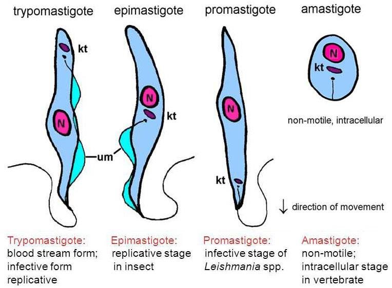
These flagellates are distinguished by a kinetoplast (kt on the diagram) - a cluster of multiple copies of mitochondrial DNA at a specific location in the single large mitochondrion. So the kinetoplast is not an organelle of its own: it is a concentration of mitochondrial DNA inside the one big mitochondrion these cells have.

There is one flagellum which sometimes supports an undulating membrane (um on the diagram).

Trypomastigote, epimastigote, promastigote and amastigote of a kinetoplastid drawn side by side, labelled kt (kinetoplast), N (nucleus) and um (undulating membrane)

The four morphological forms

note

According to the location of the flagellum and the undulating membrane (if any), we distinguish the following morphological forms:

FormShapeBasal bodyFlagellum and undulating membraneWhere it occurs
Trypomastigoteelongated, spindle-shapedat the posterior endthe flagellum supports a long undulating membraneblood stream form; infective form; replicative
Epimastigoteelongatedin the middlethe flagellum supports a short undulating membranereplicative stage in the insect
Promastigoteelongatedat the anterior endthe flagellum is free, there is no undulating membraneinfective stage of Leishmania spp.
Amastigotesmall, oval-there is no flagellum or undulating membranenon-motile; intracellular stage in the vertebrate

The diagram also gives the direction of movement: the flagellated forms swim with the flagellum leading.

The structure of the trypomastigote of Trypanosoma (drawing by Markova and Kavaldzhieva) is labelled with the basal body, the axial apparatus (axoneme), the nucleus, the cell membrane, the flagellum, the undulating membrane, and a cross section through the cell showing the flagellum lying alongside the body.

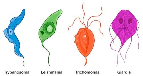
Such simple drawings help memorise the morphology of flagellated parasites, and the lecture gives a row of four of them side by side - Trypanosoma, Leishmania, Trichomonas and Giardia - as the picture to keep in mind for the whole of this material.

Trypomastigote, epimastigote, promastigote and amastigote compared, labelled kt, N and um, each captioned with where it occurs: blood stream, insect, Leishmania infective stage, intracellular
Trypanosoma, Leishmania, Trichomonas and Giardia drawn as a row of coloured silhouettes, named but not internally labelled

6. Genus Trypanosoma

note

  • Different species of trypanosomes infect a variety of vertebrates, including humans.
  • With rare exceptions, trypanosomes are transmitted by blood-sucking insects.
  • Sexual reproduction takes place in the insect but is poorly studied.
  • All Trypanosoma species look very similar.

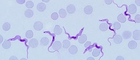
In a blood smear of Trypanosoma brucei, the trypomastigotes are seen among the red blood cells with the undulating membrane, the nucleus, the flagellum and the kinetoplast visible. The kinetoplast is seen as a dot with the same colour as the nucleus. Our mitochondrial DNA is not so easily observed - a reminder of how much mitochondrial DNA is packed into that one spot.

Trypanosoma brucei trypomastigotes among red blood cells in a Giemsa-stained blood smear, unlabelled

Structure of the trypomastigote form

note

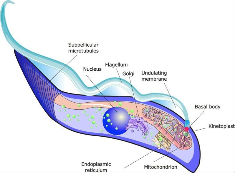
The kinetoplast is located near the basal body of the flagellum, at the posterior end; the flagellum then runs forward along the cell to the anterior end. The detailed drawing labels, from posterior to anterior, the kinetoplast, the basal body, the mitochondrion, the endoplasmic reticulum, the Golgi, the nucleus, the subpellicular microtubules, the flagellum and the undulating membrane. The subpellicular microtubules are what hold the spindle shape of the cell.

A video of living trypanosomes in diluted blood shows Trypanosoma equipersum, a relation of T. brucei which evolved into a sexually transmitted horse parasite.

Trypomastigote of Trypanosoma labelled: subpellicular microtubules, nucleus, flagellum, Golgi, undulating membrane, basal body, kinetoplast, endoplasmic reticulum, mitochondrion

7. Trypanosoma brucei - the disease

note

Trypanosoma brucei causes African trypanosomiasis (sleeping sickness).

  • T. brucei variant gambiense - chronic sleeping sickness in humans, West and Central Africa.
  • T. brucei variant rhodesiense - acute sleeping sickness in humans, East and Southeast Africa.

T. brucei is an extracellular parasite. It is found in the blood, skin, lymph nodes, spleen, and in the advanced stage also in cerebrospinal fluid. It never enters the host cells at all, which is what separates it from the next species in this lecture.

Main symptoms: sleeping disturbances, alteration of mental state, abnormal reflexes, coordination, and sensory disorders. Untreated disease is 100% fatal. The "chronic" form takes longer to kill than the acute form.

Over 60 million people at risk.

The parasite infects also a number of domestic and wild mammals. Cattle herds have been implicated in the spread of the disease to new areas. Wild animals are thought to be responsible for sporadic transmission to park visitors. Infection of international travellers is rare, but it occasionally occurs.

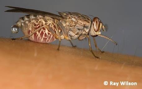
Tsetse fly (Glossina) feeding on human skin with its abdomen engorged with blood, unlabelled - the vector of Trypanosoma brucei

The vector

note

T. brucei is transmitted by blood-sucking dipteran insects of genus Glossina (tse-tse fly). Such hosts that infect other hosts are called vectors.

Meiosis, gametogenesis, and fertilisation of the parasite occur in the salivary glands of the fly. By the definition given earlier, that makes the tsetse fly the definitive host and the mammal the intermediate one.

Tsetse fly (Glossina), the vector of Trypanosoma brucei, shown feeding on human skin, unlabelled

Trypanosoma brucei - life cycle

note

In humans (or other mammals), T. brucei exists in a form called blood trypomastigote, adapted to multiply in the blood plasma and cerebrospinal fluid.

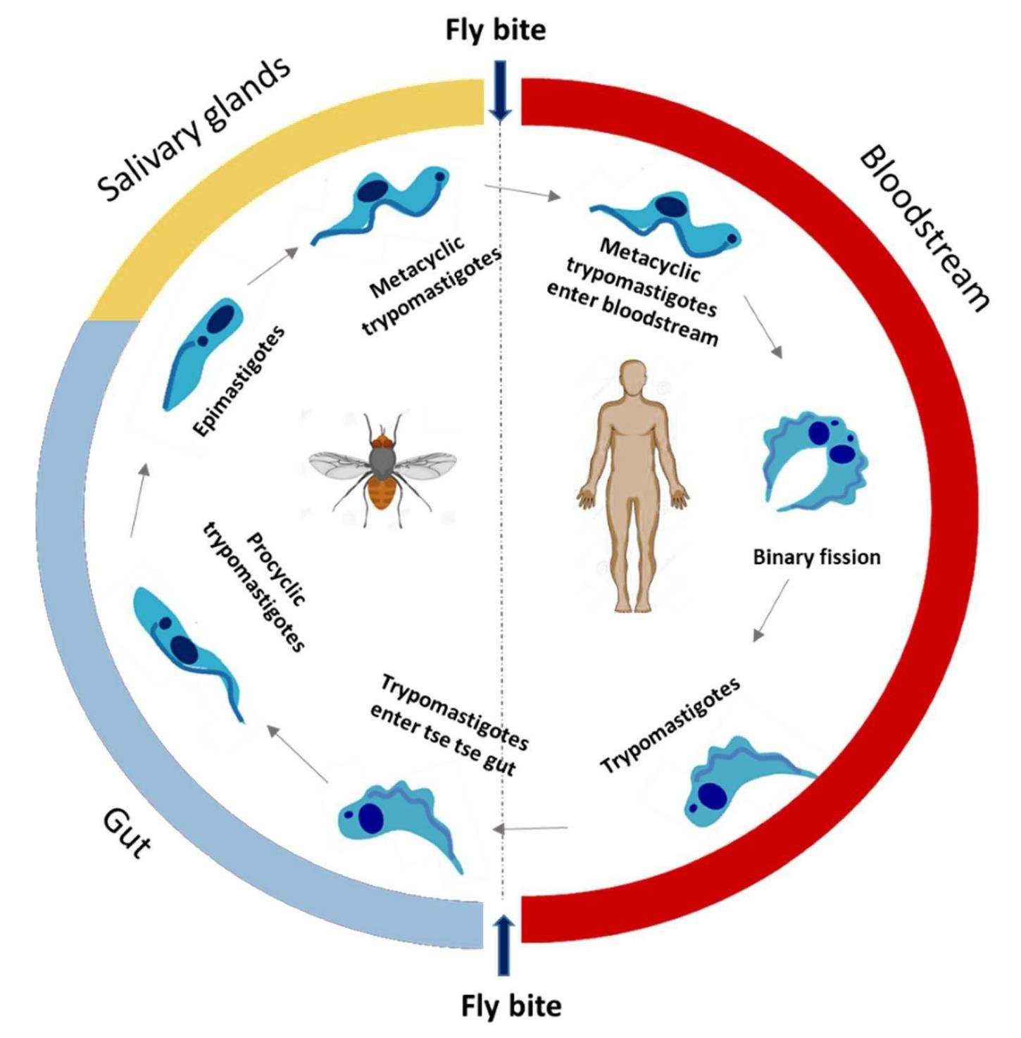
If ingested by a tsetse fly, they become procyclic trypomastigotes and divide in the fly's midgut. Then they leave the midgut, become epimastigotes, and travel to the salivary glands, where they continue to multiply. After a while, they stop dividing and become metacyclic trypomastigotes, which are the infective stage for mammals. If they enter the human body by bloodsucking, they turn into blood trypomastigotes.

Pro- and meta- (from Greek "before" and "after", respectively) refer to the reproduction in the vector. The procyclic form comes before that multiplication, the metacyclic form after it.

The cycle drawn as a circle reads: fly bite → metacyclic trypomastigotes enter the bloodstream → trypomastigotes multiply in the bloodstream by binary fission → trypomastigotes enter the tsetse gut when the fly bites again → procyclic trypomastigotes in the gut → epimastigotes → metacyclic trypomastigotes in the salivary glands → fly bite.

Trypanosoma brucei life cycle: fly bite, metacyclic trypomastigotes enter bloodstream, binary fission, trypomastigotes enter tsetse gut, procyclic trypomastigotes, epimastigotes, metacyclic trypomastigotes in salivary glands

Trypanosoma brucei - distribution

note

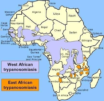
  • T. brucei gambiense causes West African ("chronic") sleeping sickness. Humans are the most common vertebrate host, although other mammals (primates and ungulates) are also infected. On the map, West African trypanosomiasis covers a broad band of West and Central Africa.
  • T. brucei rhodesiense causes East African ("acute") sleeping sickness. It is a zoonosis - it affects various domestic and wild mammals, with humans as an incidental (accidental) host. The most important reservoir is cattle. On the map, East African trypanosomiasis appears as scattered foci down the eastern side of the continent.

Hosts that occupy the same place as humans in the parasite life cycle are called reservoir hosts.

Notice that the same lecture writes these two both as variants and as subspecies-style names (T. brucei gambiense); both forms appear in the slides and either is acceptable.

Map of Africa: West African trypanosomiasis as a broad band across West and Central Africa, East African trypanosomiasis as scattered orange foci down the eastern side

Trypanosoma brucei - antigenic variation

note

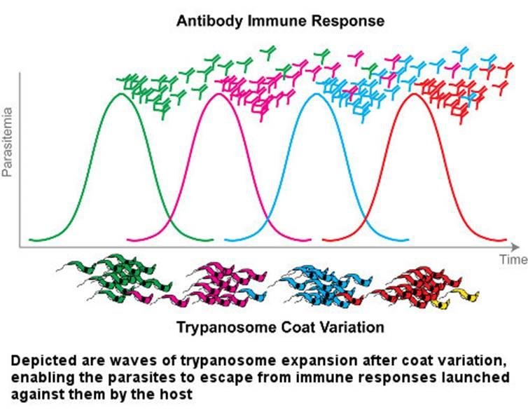
Trypanosomes periodically change the major glycoprotein covering their surface. This protects them from the host immune response, because as soon as antibodies are produced against the surface antigen, its change makes them useless.

The figure shows waves of trypanosome expansion after coat variation, enabling the parasites to escape from immune responses launched against them by the host: each peak of parasitaemia is brought down by antibody against the current coat, and each new coat starts a new peak. This is why the infection is not cleared and why a vaccine against the surface antigen is not a practical proposition.

Graph of antibody immune response to Trypanosoma coat variation: four sequential parasitaemia peaks, each cleared by a differently coloured antibody as the surface coat changes
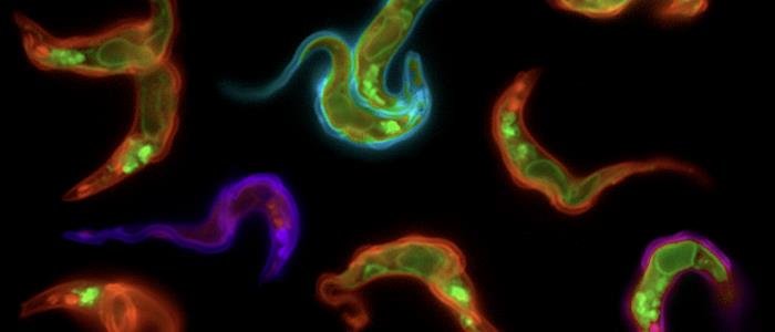
Fluorescence micrograph of trypanosomes with differently coloured surface coats, illustrating antigenic variation, unlabelled

8. Trypanosoma cruzi - the disease

note

Trypanosoma cruzi causes American trypanosomiasis (Chagas disease).

  • T. cruzi is found in vectors and wildlife reservoirs throughout the Americas from the southern half of the United States down to Argentina.
  • Chagas disease cases have been reported from South and Central American countries, particularly in rural, impoverished areas.
  • Vertebrate hosts: humans and a number of other mammals, e.g. armadillos, opossums, raccoons, rats and other rodents, and domestic dogs.
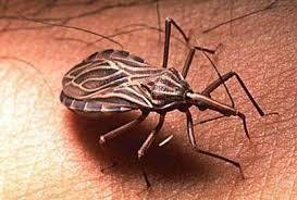
  • Vector: Triatoma ("kissing bug"), insect of the bedbug order.
Triatoma, the kissing bug vector of Trypanosoma cruzi, feeding on human skin, unlabelled

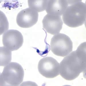
In a blood smear the parasite is seen as a trypomastigote among the red blood cells, exactly as T. brucei is.

Trypanosoma cruzi trypomastigote among red blood cells in a Giemsa-stained blood smear, unlabelled

Trypanosoma cruzi - amastigotes

note

Unlike T. brucei, which is entirely extracellular, T. cruzi exists in two forms in the vertebrate host: trypomastigote (extracellular) and amastigote (intracellular).

The amastigote is formed when the parasite enters the host cells (becoming an intracellular parasite) and loses its flagellum and part of the cytoplasm. Intracellular parasites are characterised by a small size and lack of a flagellum; even if other forms of the same parasite move with a flagellum, it is useless in the highly viscous intracellular environment. That is a general rule of parasitism, not a peculiarity of this species.

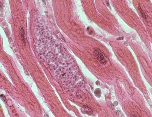
In the micrograph, numerous granules - amastigote forms of the trypanosome - are visible in the cytoplasm of a cardiomyocyte.

Histology of cardiac muscle showing a pseudocyst packed with Trypanosoma cruzi amastigotes inside a cardiomyocyte, haematoxylin and eosin stain

Trypanosoma cruzi - intracellular parasitism

note

Trypomastigotes enter a mammalian cell by attaching to the cell membrane and making the cell take them up by endocytosis.

They remain in the endosome, which is called the parasitophorous (i.e. parasite-carrying) vacuole.

There they transform into amastigotes and multiply until they fill the host cell. They then transform back into trypomastigotes, lyse the cell, and escape into the extracellular environment, where they can infect new cells or be taken up by the vector.

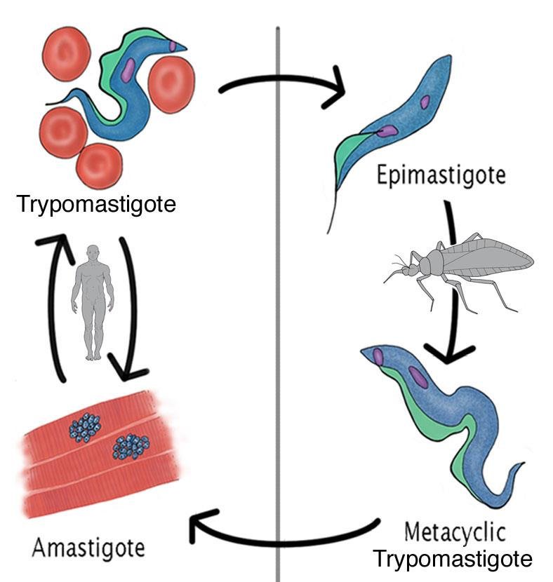
The accompanying diagram runs trypomastigote → amastigote in the human, and epimastigote → metacyclic trypomastigote in the bug, which is the same alternation of forms as in T. brucei with the amastigote added.

Trypanosoma cruzi form cycle, labelled: trypomastigote among red blood cells, amastigote clusters in muscle, epimastigote and metacyclic trypomastigote stages in the bug

Chagas disease - acute and chronic phase

note

Chagas disease has an acute and chronic phase.

  • The acute phase is often asymptomatic, but may present with nonspecific symptoms of varying severity and type.
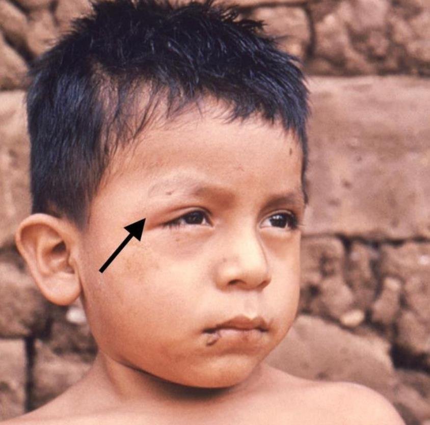
  • In the acute phase, swelling may be observed around the site where bedbug faeces were rubbed into skin or mucosa. Such local lesions are called shagomas.
  • The chronic phase occurs years or decades later. Cardiac and gastrointestinal problems occur.

Symptoms are due to the amastigote forms of the parasite. They parasitise mainly in muscle tissue and accumulate in the cardiac muscle and smooth muscles of the oesophagus, small intestine, and large intestine. That is the direct explanation of the two chronic syndromes: the heart disease and the dilated hollow organs of the gut.

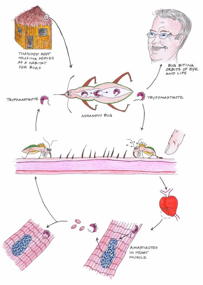
The bite is usually on the face, close to the mouth or the eyes (hence the common name "kissing bug"). A hand-drawn summary of the same story shows that thatched roof housing serves as a habitat for the bugs, that the bug bites the orbits of the eye and the lips, that trypomastigotes pass in both directions between the assassin bug and the human skin, and that the amastigotes end up in the heart muscle.

Young boy with periorbital swelling, arrow marking a Chagas disease shagoma from bug faeces rubbed into the skin near the eye
Hand-drawn Chagas cycle: thatched roof housing as bug habitat, assassin bug biting the orbit of the eye and lips, trypomastigotes passing between bug and skin, amastigotes in heart muscle

Trypanosoma cruzi - life cycle

note

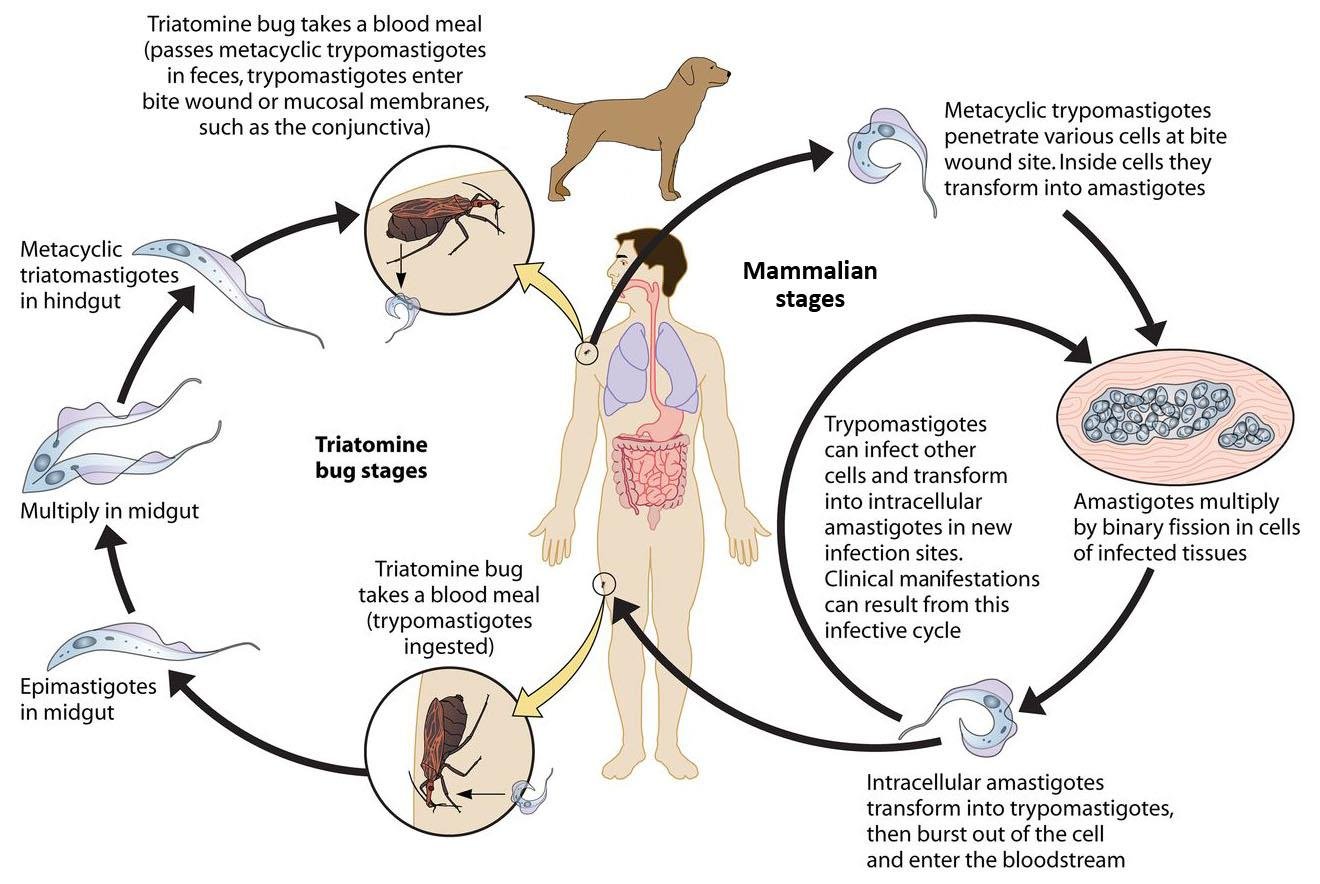
Note that the parasite in the vector infects the mammal not in the usual way - from the salivary glands during the bite itself, but by being excreted with the faeces that the mammal rubs into its skin or mucosa when scratching. This is the single most examinable difference between the two trypanosome cycles.

The cycle in the drawing reads:

  • The triatomine bug takes a blood meal and passes metacyclic trypomastigotes in its faeces; the trypomastigotes enter the bite wound or the mucosal membranes, such as the conjunctiva.
  • Metacyclic trypomastigotes penetrate various cells at the bite wound site. Inside the cells they transform into amastigotes.
  • Amastigotes multiply by binary fission in cells of infected tissues.
  • Intracellular amastigotes transform into trypomastigotes, then burst out of the cell and enter the bloodstream.
  • Trypomastigotes can infect other cells and transform into intracellular amastigotes in new infection sites. Clinical manifestations can result from this infective cycle.
  • A triatomine bug takes a blood meal and the trypomastigotes are ingested.
  • In the bug: epimastigotes in the midgut, which multiply in the midgut, then metacyclic trypomastigotes in the hindgut.

Dogs are drawn in the cycle beside the human, because they are one of the domestic reservoirs.

Trypanosoma cruzi life cycle, mammalian and triatomine bug stages labelled: metacyclic trypomastigotes penetrating skin, amastigotes multiplying in tissue, trypomastigotes bursting into bloodstream, epimastigotes and metacyclic forms in the bug gut, dog shown as a reservoir

Trypanosoma cruzi - other transmission routes

note

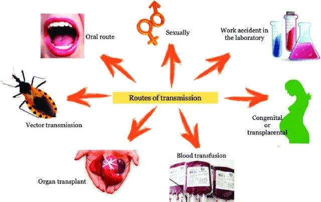
Though vector transmission is most important, T. cruzi can be transmitted by other routes as well. The figure gives them as: vector transmission, the oral route, sexually, work accident in the laboratory, congenital or transplacental, blood transfusion, and organ transplant.

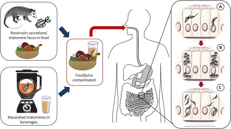
The oral route is through ingestion of food or drinks contaminated with triatomine bugs or their faeces. Most dangerous are homemade fresh fruit juices. Infection contracted this way tends to be severe. The accompanying figure shows reservoir secretions or triatomine faeces on food, and macerated triatomines in beverages, reaching the gastric and intestinal mucosa where the parasites invade the epithelium.

Routes of transmission of Trypanosoma cruzi as icons around a central label: vector transmission, oral route, sexually, laboratory accident, congenital or transplacental, blood transfusion, organ transplant
Oral transmission of Trypanosoma cruzi in three steps: reservoir secretions or macerated triatomines contaminating food or juice, then parasites invading the gastric and intestinal epithelium

Who described the disease

note

The disease was described in 1909 by Carlos Chagas, a Brazilian physician and scientist. The parasite is named after the institute where he worked (Oswaldo Cruz, in Rio de Janeiro). So the disease carries the name of the discoverer and the parasite the name of his institute.

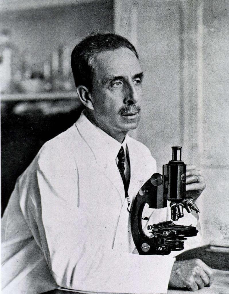
Historical photograph of Carlos Chagas at a microscope in his laboratory, unlabelled

The two human trypanosomes side by side

note

*Trypanosoma brucei**Trypanosoma cruzi*
DiseaseAfrican trypanosomiasis (sleeping sickness)American trypanosomiasis (Chagas disease)
DistributionWest and Central Africa (gambiense); East and Southeast Africa (rhodesiense)the Americas, from the southern half of the USA to Argentina
VectorGlossina, the tse-tse flyTriatoma, the "kissing bug", of the bedbug order
How the vector infectsby the bite, from the salivary glandsby faeces rubbed into skin or mucosa
Forms in the vertebrateblood trypomastigote only; entirely extracellulartrypomastigote (extracellular) and amastigote (intracellular)
Forms in the vectorprocyclic trypomastigote, epimastigote, metacyclic trypomastigoteepimastigote in the midgut, metacyclic trypomastigote in the hindgut
Where the parasite sitsblood, skin, lymph nodes, spleen, later cerebrospinal fluidmuscle tissue, especially cardiac muscle and smooth muscle of the oesophagus and intestines
Immune evasionantigenic variation of the surface glycoproteinhiding inside the cell, in the parasitophorous vacuole
Trypanosoma brucei life cycle showing infection by fly bite from the salivary glands, shown here against T. cruzi's different route
Trypanosoma cruzi form cycle showing both an extracellular trypomastigote and an intracellular amastigote stage, unlike T. brucei's blood-only form

The most important things to know

note

  • Protozoa is an informal name for unicellular motile eukaryotes without a cell wall; some are free-living, some are parasites. Phylum Sarcomastigophora is obsolete but kept in medical parasitology, and contains the flagellates and the amoeboid protozoans.
  • Protozoans divide by closed mitosis, with the nuclear envelope intact, and duplicate flagella and basal bodies in advance; in Trypanosoma the kinetoplast divides before the nucleus (1K1N → 2K1N → 2K2N).
  • Micro- and macrogametes are the male and female gametes; the host in which the sexual process occurs is the definitive (final) host, the others are intermediate; a host occupying the same place as humans in the cycle is a reservoir host; a host that infects other hosts is a vector.
  • Kinetoplastids have one flagellum and a kinetoplast, a mass of mitochondrial DNA inside the single large mitochondrion. Their four forms are trypomastigote (basal body posterior, long undulating membrane), epimastigote (basal body in the middle, short undulating membrane), promastigote (basal body anterior, free flagellum) and amastigote (small, oval, no flagellum, intracellular).
  • *T. brucei* causes sleeping sickness, is entirely extracellular, is transmitted by the tse-tse fly (Glossina) from its salivary glands, and is 100% fatal untreated; gambiense is the chronic West and Central African form, rhodesiense the acute East African zoonosis with cattle as reservoir.
  • Antigenic variation of the surface glycoprotein lets T. brucei escape antibodies in repeated waves of parasitaemia.
  • *T. cruzi* causes Chagas disease, has trypomastigote and amastigote forms in the vertebrate, multiplies inside the parasitophorous vacuole, and is transmitted by Triatoma faeces rubbed into the skin or mucosa, as well as orally, sexually, congenitally, by transfusion, by transplant and by laboratory accident.
  • Acute Chagas disease may show a shagoma at the site of entry; chronic disease appears years or decades later as cardiac and gastrointestinal problems, caused by amastigotes in cardiac and smooth muscle.
  • The disease was described by Carlos Chagas in 1909; the parasite is named after the Oswaldo Cruz institute.
Trypanosoma, Leishmania, Trichomonas and Giardia drawn as a row of coloured silhouettes, the summary picture for the flagellated parasites in this topic

All 105 Medical Biology topics · exam study support, not clinical guidance. SuperMed is not affiliated with the Medical University of Sofia.

Start free. No card needed.

Every account starts free, with Medical Ethics, Urology and Computer Science open in full and free topics in Cytology, Latin Language, Medical Chemistry, Medical Physics, Medical Biology, Anatomy, Biophysics, Medical Psychology, MCS (Disaster Medicine), Physiology, Biochemistry, Social Medicine, Medical Genetics, Pharmacology, Pathophysiology, Microbiology, Internal Medicine, Nephrology, Cardiology, Pulmonology, Gastroenterology, Internal Medicine (Clinical), Obstetrics and Gynecology, Physiotherapy and Rehabilitation, Dental Materials, Preclinics of Prosthetic Dental Medicine, Medical Psychology (Dental Medicine), SMME (Social Medicine & Medical Ethics), Preclinics of Pediatric Dental Medicine, Hygiene and Epidemiology, General Surgery, Preclinics of Conservative Dental Medicine, Preclinics of Oral and Maxillofacial Medicine, Dental Public Health, Dental Prophylaxis, Diagnostic Imaging, Dental Allergology, Dental Implantology, Physiotherapy, Cytology & Histology, Medical Chemistry, Medical Physics, Anatomy I, Biophysics and Medical Biology. Super opens the rest. Super is €14.99 a month.

Create a free account