


Microbiology · Year 3 · Medical University of Sofia

01

Scope and objectives of Medical Microbiology - Historical stages

Free notes for topic 01 of the Microbiology syllabus, open without an account. Written by a senior student against the syllabus question and checked line by line by a second student before publishing. How content is made

Updated

Summary

note

  • Medical microbiology studies the microorganisms that cause infectious disease in humans, and the beneficial microbes that form part of the human microbiota (normal flora).
  • Microbiology is the study of organisms too small to be seen with the naked eye: bacteria, archaea, viruses, fungi, prions, protozoa and algae.
  • Most bacteria are about 1 μm across; the smallest, Chlamydia and Rickettsia, are 0.1 to 0.2 μm. Viruses measure 18 to 300 nm and are seen only with an electron microscope.
  • Microorganisms are either part of the normal flora, opportunists (harmless normally, but they cause disease in immunocompromised people or in sterile organs) or obligate pathogens (they cause disease in a healthy host).
  • The key historical steps are Hooke's microscope (1665), Leeuwenhoek's first sight of microbes, Spallanzani's boiling experiments, Jenner's cowpox vaccination (1796 or 1798), Cohn's shape classification, Pasteur's work against spontaneous generation, Koch's link between a microbe and a disease, and Ivanovski's filterable agents (1892).
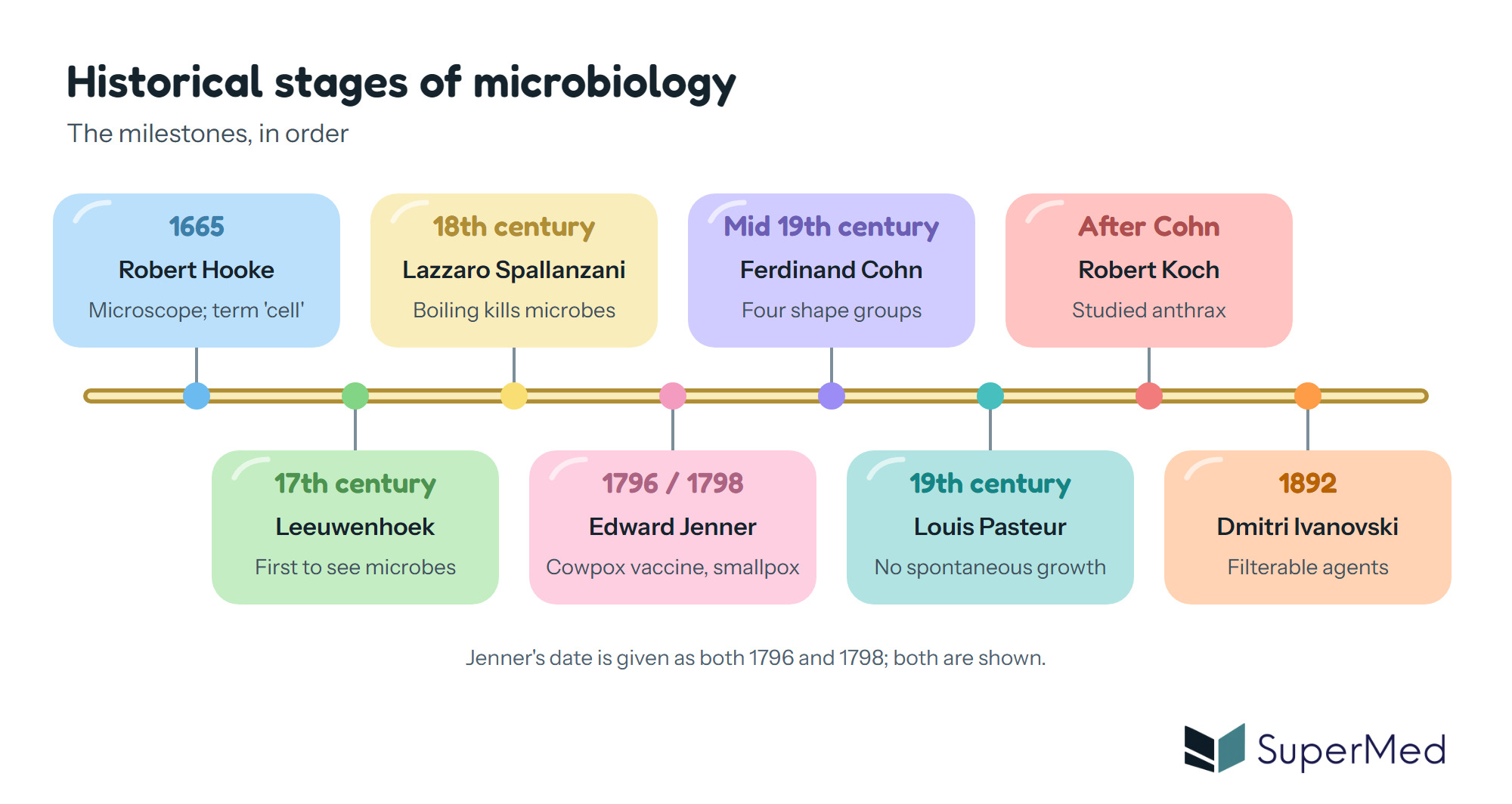
Historical stages of microbiology: Hooke 1665, Leeuwenhoek, Spallanzani, Jenner, Cohn, Pasteur, Koch and Ivanovski 1892, in order

1. Scope of medical microbiology

note

Microbiology is the branch of science that deals with microorganisms, which are organisms too small to be seen with the naked eye. The field includes bacteria, archaea, viruses, fungi, prions, protozoa and algae. Microbes can be unicellular, form cell clusters or be multicellular. They have important roles in almost every ecosystem where there is water, including decomposition and nitrogen fixation. Medical microbiology is largely concerned with the microbes that are pathogenic to humans.

The objectives of the subject are:

  • to study the morphology, structure and physiology of microorganisms;
  • to understand how microorganisms interact with the environment, and their effects on health, industry and agriculture;
  • to investigate how infection develops, the host immune response, and the diagnosis, prevention and treatment of infectious diseases.

Branches of microbiology

note

  • General microbiology focuses on the morphology, structure and general characteristics of microorganisms.
  • Infection and immunity studies the immune responses to infections and the basic principles of immunology.
  • Special microbiology focuses on specific pathogens (bacteria, viruses, fungi and parasites), including their diagnosis, treatment and prevention.
  • Applied fields are medical microbiology (human health), veterinary microbiology (animal health), industrial microbiology (pharmaceuticals and food production) and agricultural microbiology (health of plants and farm animals).

Microbiota and microbiome

note

The microbiota is the community of microorganisms, mainly bacteria, that live on the skin and mucous membranes, especially in the gastrointestinal tract. The microbiome is the total genetic material of all the microorganisms that inhabit the human body.

Human skin and mucosa are colonised by a large number of microorganisms, which make up the normal flora. The lecture gives more than 500 bacterial species, with occasional fungi and protozoa, and methane-producing archaea in the large intestine. The lecture also states that there are about 10¹⁴ bacteria in the body, ten times as many as the body's own cells, most of them in the oral cavity and the large intestine.

The microbiota has three main functions:

  • it aids digestion;
  • it protects against pathogens;
  • it modulates the immune system.

Dysbiosis is an imbalance of the microbiota. It is often caused by antibiotics, cytostatics (cytotoxic drugs) or immunosuppressants, and it can lead to opportunistic infections.

Normal flora is also called indigenous, natural or autochthonous flora. It is residential when the microbes live permanently on the body. It is transitory when microbes enter the body periodically and colonise the mucosa for a time. Transitory microbes can multiply in the host or come from the environment as saprophytes.

Types of microorganisms related to human health

note

Exposure of a person to a microorganism can have one of three outcomes. The organism can transiently colonise the person, permanently colonise them, or produce disease. Colonisation does not disturb normal body functions, whereas disease is a pathological process that damages the human host.

  • Normal flora (microbiota) coexists with the human body without causing disease under normal conditions.
  • Pathogenic microorganisms are not part of the normal flora. They enter a healthy host and cause an infectious process, from mild to lethal. Examples given in the lectures are Mycobacterium tuberculosis (tuberculosis), Neisseria gonorrhoeae (gonorrhoea), Francisella tularensis (tularaemia), Plasmodium spp. (malaria) and rabies virus. The lectures also name Salmonella Typhi (typhoid fever), Vibrio cholerae (cholera) and poliovirus (poliomyelitis) as obligate pathogens.
  • Opportunistic microorganisms are normally harmless. They cause disease in immunocompromised people, or when they reach normally sterile organs and tissues. They come from the normal flora or are saprophytes from the environment. Typical opportunists include members of the Enterobacteriaceae (Klebsiella, Proteus, Morganella, Pseudomonas aeruginosa, Burkholderia cepacia), anaerobes such as Bacteroides, Porphyromonas, Prevotella and Fusobacterium, Staphylococcus spp., Streptococcus spp., and the fungi Candida albicans, Aspergillus niger, Cryptococcus neoformans and Pneumocystis (called P. carinii in the lecture).
  • Some opportunists have a higher pathogenic potency: Staphylococcus aureus, Streptococcus pyogenes and Streptococcus pneumoniae, and the fungi Histoplasma capsulatum and Coccidioides immitis.

Exam tip: Know the three groups (normal flora, opportunists and pathogens) and the difference between colonisation and disease.

2. Size of the organisms that cause human disease

note

Human pathogens differ greatly in size. A light microscope with an immersion objective has a resolution of 0.2 μm and a magnification of about ×1240, and bacteria can be seen with it. The smallest bacteria, Chlamydia and Rickettsia, are 0.1 to 0.2 μm in diameter. Larger bacteria are 5 to 10 μm, and some are visible to the naked eye. The average bacterium is about 1 μm across.

For comparison, eukaryotic cells range from 7 μm (a red blood cell) to several feet long (the length of certain nerve cells). Viruses are filterable agents of 18 to 300 nm, from parvoviruses at the small end to poxviruses at the large end. They can be seen only with an electron microscope.

The lecture also includes prions, which are infectious proteins.

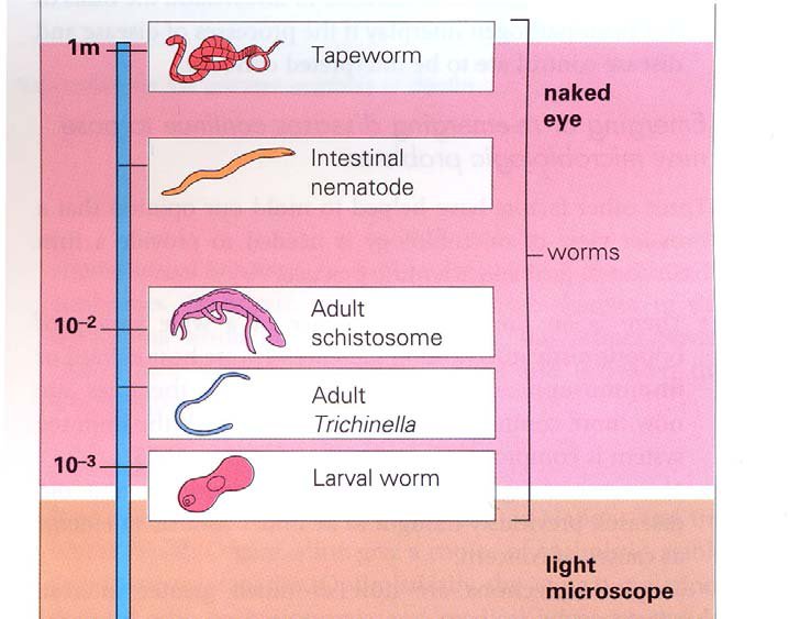
Scale of worms from a 1 m tapeworm down to a 10⁻³ m larval worm, with the naked eye and light microscope ranges marked

The worms in the first scale are large enough to be seen with the naked eye. The larval worm, at about 10⁻³ m, is at the limit of the light microscope.

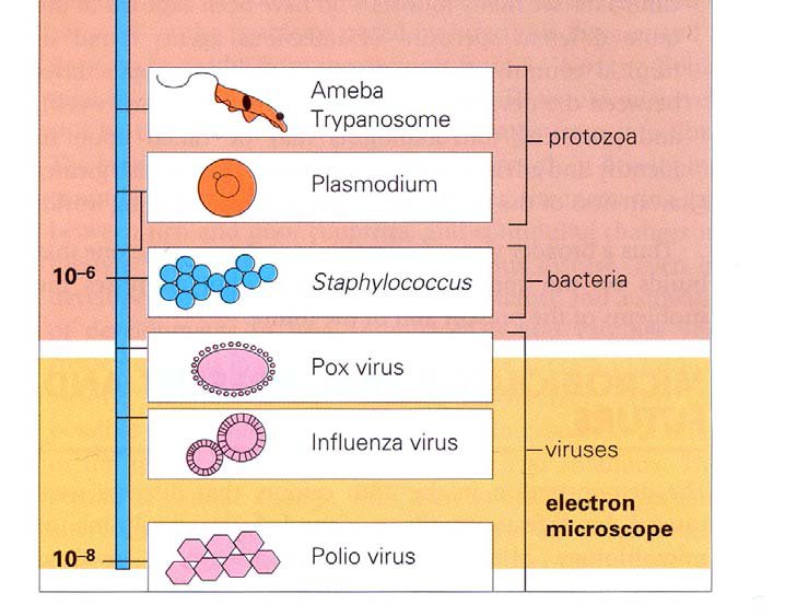
Scale of microbes from amoeba, trypanosome and Plasmodium (protozoa) to Staphylococcus (bacteria, about 10⁻⁶ m) and pox, influenza and polio viruses (about 10⁻⁸ m, electron microscope)

On the microbial scale, protozoa are the largest group, bacteria are about 10⁻⁶ m and viruses are seen only in the electron microscope. Polioviruses are about 10⁻⁸ m.

3. Historical stages of microbiology

note

Microbiology began with the microscope. The historical stages below are named in the lectures.

Historical stages of microbiology: Hooke 1665, Leeuwenhoek, Spallanzani, Jenner, Cohn, Pasteur, Koch and Ivanovski 1892, in order

Early observers and the first ideas

note

  • Robert Hooke (17th century) made a microscope by changing the lens of a telescope. In 1665 he published his observations and coined the term cell for biological organisms.
  • Antonie van Leeuwenhoek was the first person to observe microorganisms with a microscope. He called them animalcules and saw that they moved. In one experiment (1684) he placed a toothpick in his mouth, prepared a slide from it, and observed what he saw. He did not understand what he had seen.
  • Lazzaro Spallanzani (18th century) showed that boiling fluids sterilised them and killed the microorganisms in them, and that the microorganisms had a relationship with air. This was the start of the science of sterilisation.
  • Louis Pasteur proved that microorganisms could not arise spontaneously. This removed the theory of spontaneous generation and supported germ theory.
  • Ferdinand Cohn (mid-19th century) is described as the founder of bacteriology. He studied Bacillus, its life cycle and its endospore, which allows survival in harsh environments. He classified bacteria by shape into four groups: spherical (cocci), rods, threads and spirals. This shape classification still survives.
  • Robert Koch showed that microorganisms cause disease, by studying anthrax (Bacillus anthracis). From this point, medical microbiology was born.

Koch's postulates

note

Koch's postulates are the criteria used to show that a particular microorganism causes an infectious disease. The lecture states that the criteria apply to the obligate pathogens. In their classic form they require that the organism is found in every case of the disease, that it can be grown in pure culture, that it causes the disease when introduced into a healthy susceptible host, and that it can be re-isolated from that host.

Milestones in vaccination

note

  • 1796 or 1798: Edward Jenner. Jenner noticed that the mild cattle disease cowpox protected against the deadly human disease smallpox. He inoculated an eight-year-old boy with exudate from a cowpox pustule, and the boy was fully protected against smallpox. The lectures give the discovery as 1796 on one slide and the history date as 1798 on another.
  • 1885: Louis Pasteur made the first attenuated viral vaccine, against rabies.
  • Early 1900s: toxoid vaccines against diphtheria and tetanus.
  • 1936: influenza vaccine.
  • 1950s: tissue-culture attenuated poliovirus vaccine. The work was recognised with a Nobel Prize for Enders, Robbins and Weller.
  • 1960s: live attenuated vaccines against measles, mumps and rubella (MMR).

Virology begins

note

Dmitri Ivanovski (1864 to 1920) described viruses in 1892 as filterable agents, because they passed through filters that retained bacteria. He discovered the agent of tobacco mosaic virus. Viruses lack cellular organisation, cannot make their own proteins, and cannot replicate independently of the host cell. They are obligate intracellular parasites.

4. Course organisation and examination

note

The course runs over two semesters. In the first semester there is one practical and one lecture each week. In the second semester there is one practical and one lecture each week, or two lectures for medical students. Medical students take two obligatory colloquiums and three obligatory tests. The final examination has three parts: a test, one practical question and three theoretical questions.

5. Key facts and comparison tables

note

Table 1. Historical figures named in the lectures

NameDate givenContribution
Robert Hooke1665Microscope; coined the term cell
Antonie van Leeuwenhoek17th century (no date given)First to observe microorganisms
Lazzaro Spallanzani18th centuryBoiling sterilises fluids and kills microbes
Edward Jenner1796 (lecture slide) or 1798 (lecture slide)Cowpox vaccination against smallpox
Ferdinand CohnMid-19th centuryFounder of bacteriology; four shape groups
Louis Pasteur19th century (rabies vaccine 1885)Disproved spontaneous generation; first rabies vaccine
Robert KochAfter Cohn (no date given)Linked a microbe to a disease (anthrax)
Dmitri Ivanovski1892Filterable agents; tobacco mosaic virus

Table 2. Sizes of organisms relevant to human disease

OrganismApproximate sizeSeen with
Chlamydia, Rickettsia0.1 to 0.2 μmLight microscope at high resolution
Typical bacteriumAbout 1 μmLight microscope
Larger bacteria5 to 10 μm and moreLight microscope; some with the naked eye
Red blood cell (eukaryotic cell)About 7 μmLight microscope
Viruses (parvoviruses to poxviruses)18 to 300 nmElectron microscope only

Table 3. Three groups of microorganisms in human health

GroupPart of normal flora?Causes disease whenExamples from the lectures
Normal floraYesUsually neverSkin and mucosal bacteria in the gut and mouth
OpportunistsOften yes, or environmental saprophytesThe host is immunocompromised, or the organism reaches a sterile siteStaphylococcus spp., Candida albicans, Pseudomonas aeruginosa
Obligate pathogensNoAlways, in a healthy hostMycobacterium tuberculosis, Salmonella Typhi, Vibrio cholerae, rabies virus

Exam tip: Remember Cohn's four shape groups (cocci, rods, threads, spirals), the 1892 date of Ivanovski and the rule that viruses are seen only with the electron microscope.

Clinical link: Antibiotics and immunosuppressants can cause dysbiosis, which allows opportunistic infections to develop from organisms that were previously harmless.

All 153 Microbiology topics · exam study support, not clinical guidance. SuperMed is not affiliated with the Medical University of Sofia.

Start free. No card needed.

Every account starts free, with Medical Ethics, Urology and Computer Science open in full and free topics in Cytology, Latin Language, Medical Chemistry, Medical Physics, Medical Biology, Anatomy, Biophysics, Medical Psychology, MCS (Disaster Medicine), Physiology, Biochemistry, Social Medicine, Medical Genetics, Pharmacology, Pathophysiology, Microbiology, Internal Medicine, Nephrology, Cardiology, Pulmonology, Gastroenterology, Internal Medicine (Clinical), Obstetrics and Gynecology, Physiotherapy and Rehabilitation, Dental Materials, Preclinics of Prosthetic Dental Medicine, Medical Psychology (Dental Medicine), SMME (Social Medicine & Medical Ethics), Preclinics of Pediatric Dental Medicine, Hygiene and Epidemiology, General Surgery, Preclinics of Conservative Dental Medicine, Preclinics of Oral and Maxillofacial Medicine, Dental Public Health, Dental Prophylaxis, Diagnostic Imaging, Dental Allergology, Dental Implantology, Physiotherapy, Cytology & Histology, Medical Chemistry, Medical Physics, Anatomy I, Biophysics and Medical Biology. Super opens the rest. Super is €14.99 a month.

Create a free account