



Cytology · Year 1 · Medical University of Sofia

19

Endoplasmic reticulum. Ribosomes. Annulate lamellae

Free notes for topic 19 of the Cytology syllabus, open without an account. Written by a senior student against the syllabus question and checked line by line by a second student before publishing. How content is made

Updated

The short version

note

The membrane-limited organelles are the endoplasmic reticulum, annulate lamellae, Golgi apparatus, secretory vesicles, lysosomes, peroxisomes, mitochondria and the nucleus.

The endoplasmic reticulum is an interconnected network extending from the surface of the nucleus, and it has two modifications. Rough ER carries ribosomes and does protein synthesis and protein folding. Smooth ER lacks ribosomes and does lipid, phospholipid and steroid hormone synthesis, carbohydrate metabolism, and the regulation of calcium in muscle.

Ribosomes are small, non-membranous organelles that link amino acids in the order specified by the codons of mRNA. They are either free or bound.

Annulate lamellae are stacks of flat membranous cisternae derived from the nuclear envelope, whose function is largely unknown.
1. The membrane-limited organelles

note

The membrane-limited organelles are:

  • Endoplasmic reticulum, smooth and rough;
  • Annulate lamellae;
  • Golgi apparatus;
  • Secretory vesicles, or granules;
  • Lysosomes;
  • Peroxisomes, also called microbodies;
  • Mitochondria;
  • Nucleus.

2. The endoplasmic reticulum

note

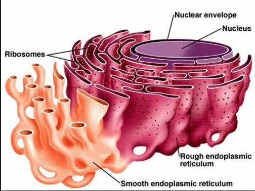
The endoplasmic reticulum is an interconnected network, a reticulum, which extends from the surface of the nucleus and encloses a series of intercommunicating channels and flattened sacs, the cisternae and vacuoles.

  • Its chemical composition is protein 60 to 65 per cent and lipids 35 to 40 per cent.
  • It is surrounded by a biomembrane.
  • It has two modifications: smooth and rough.
Diagram of the nucleus and its envelope continuous with the rough endoplasmic reticulum studded with ribosomes and the smooth endoplasmic reticulum

Rough ER

note

Rough ER is composed of cisternae and has a basophilic cytoplasmic surface with ribosomes, which are the site of protein synthesis. Because of the ribosomes it is called rough.

Its surface is basophilic for the reason given in the histochemistry topics: ribosomes are rich in RNA, RNA is negatively charged, and a negatively charged structure takes up basic dyes.

Protein synthesis. mRNA moves from the nucleus to a ribosome on the surface of the RER. The ribosome builds the amino acid chain, and the chain is pushed into the cisternal space of the RER until the protein is complete.

Protein folding. When the protein is complete it undergoes posttranslational modifications. Proteins are folded in the cisternae by chaperone proteins and enzymes. Only correctly folded proteins are transported to the Golgi apparatus or the cell membrane.

Rough ER is also involved in the manufacture of lysosomes, and of secreted and integral membrane proteins.

Smooth ER

note

Smooth ER has a structure of tubular cisternae and lacks ribosomes.

Its functions:

Synthesis of lipids, phospholipids and steroid hormones. The phospholipids are transferred from the SER to other membranes in three ways:

  1. by direct communication with the RER;
  2. by vesicles that move along the cytoskeleton and fuse with other membranous organelles;
  3. by phospholipid transfer proteins.

Metabolism of carbohydrates, that is glycogen.

Regulation of calcium ion concentration in muscles.

3. Ribosomes

note

Ribosomes are small, non-membranous organelles, measuring 20 × 30 nm.

They link amino acids together in the order specified by the codons of mRNA molecules, to form polypeptide chains.

The two subunits

note

The ribosome has two major components:

  • The smaller ribosomal subunit is a highly folded rRNA plus 30 unique proteins. It reads the RNA.
  • The larger ribosomal subunit has 3 rRNA molecules and 50 other basic proteins. It joins the amino acids to form a polypeptide chain.

A useful way to hold the division: the small subunit reads, the large subunit builds.

Free and bound ribosomes

note

Free ribosomes are not attached to organelles. They occur as monosomes, freely in the cytosol, or as free polysomes, which synthesise cytoskeletal proteins for use within the cytosol, or for import into the nucleus, mitochondria and peroxisomes.

Bound ribosomes are attached to an organelle, such as the RER or the outer nuclear membrane. They translate mRNAs coding for:

  • membrane proteins of the ER, Golgi apparatus and cell membrane;
  • enzymes to be stored in lysosomes;
  • secretory proteins undergoing exocytosis, for example collagen or antibody.

The rule underneath is about destination: a protein that stays in the cytosol is made free, and a protein that must end up inside a membrane compartment or outside the cell is made bound.

4. Annulate lamellae

note

Annulate lamellae are composed of stacks of flat, membranous cisternae.

  • They contain numerous pore complexes.
  • They are considered to be extensions of the RER cisternae, an intermediate stage between the ER and the nuclear envelope.
  • Their origin is that they are derived from the nuclear envelope.
  • They are abundant in human oocytes, spermatozoa and tumour cells.
  • Their function is largely unknown. Some biologists say they are attachment sites for stored RNA.

All 95 Cytology topics · exam study support, not clinical guidance. SuperMed is not affiliated with the Medical University of Sofia.

Start free. No card needed.

Every account starts free, with free topics in Cytology and Medical Biology. Super opens the rest. Super is €15 a month.

Create a free account