



Cytology · Year 1 · Medical University of Sofia

06

Immunohistochemistry

Free notes for topic 06 of the Cytology syllabus, open without an account. Written by a senior student against the syllabus question and checked line by line by a second student before publishing. How content is made

Updated

The short version

note

Immunohistochemistry is the process of using antibodies to identify the presence of antigens in a section. Where the other histochemical methods detect a whole class of molecule, this one detects a single named protein, because that is what an antibody binds.

Two things have to be chosen. The antibody: polyclonal, from different clones of plasma B cells and binding different epitopes, or monoclonal, from a single clone and binding one particular epitope. And the method: direct, where the label sits on the primary antibody, or indirect, where a labelled secondary antibody binds the primary one.

Indirect is the one used in practice, and the reason is worth remembering: more than one labelled secondary antibody can bind each primary antibody, so the signal is amplified.
1. What immunohistochemistry is

note

Immunohistochemistry is the process of using antibodies to identify the presence of antigens.

Antibodies belong to the immunoglobulin family of glycoproteins.

The closely related technique immunocytochemistry is used to determine the localisation of specific proteins or antigens.

2. Types of antibodies

note

There are two types of antibody, and they differ in how many clones of cell produced them.

Polyclonal antibodies are produced by different clones of plasma B cells. They interact with different epitopes on the same antigen.

Monoclonal antibodies are produced by the same clone of plasma B cells. They interact with a particular epitope on the same antigen. Their advantage is highly specific binding.

3. Labelling the antibody

note

The process runs as follows:

  • The tissue section, carrying the antigen, is placed in a solution containing the antibody.
  • The antibody binds to the antigen.
  • After a rinse, the antigen's location can be seen under a light or electron microscope by visualising the antibody.

An antibody on its own is invisible, so it has to carry a label. Antibodies are commonly coupled with:

  • a fluorescent compound, giving the immunofluorescence method;
  • an enzyme, peroxidase, giving the PAP method;
  • gold particles, for the transmission electron microscope.

4. Two methods of localising antigen

note

The two methods differ in whether the label is carried by the antibody that binds the antigen, or by a second antibody that binds the first.

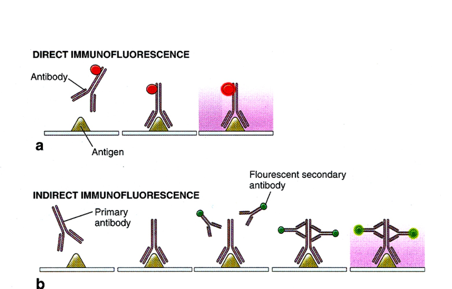
Diagram comparing direct immunofluorescence, where a labelled antibody binds the antigen directly, with indirect immunofluorescence, where an unlabelled primary antibody binds the antigen and several labelled secondary antibodies bind the primary

Direct immunohistochemistry

note

Direct immunohistochemistry uses a primary antibody with a label, for example a fluorescent compound. It binds specifically to the antigen.

  • Advantage: it needs only the primary antibody, so it is faster and less complex.
  • Disadvantage: low intensity.

Indirect immunohistochemistry

note

Indirect immunohistochemistry was devised to improve intensity.

The primary antibody binds to the antigen. Then a labelled secondary antibody, carrying for example a fluorescent compound or peroxidase, binds to the primary antibody, indirectly labelling the antigen of interest.

Because more than one labelled secondary antibody can bind each primary antibody, the sensitivity and signal intensity are much greater.

All 95 Cytology topics · exam study support, not clinical guidance. SuperMed is not affiliated with the Medical University of Sofia.

Start free. No card needed.

Every account starts free, with free topics in Cytology and Medical Biology. Super opens the rest. Super is €15 a month.

Create a free account