Cytology · Year 1 · Medical University of Sofia

07

Hybridization techniques

Free notes for topic 07 of the Cytology syllabus, open without an account. Written by a senior student against the syllabus question and checked line by line by a second student before publishing. How content is made

Updated

The short version

note

Hybridization techniques detect a specific sequence by letting a labelled probe find and bind its complementary partner. Three blotting methods are named after the three molecules of the central dogma.

Southern blotting detects a specific DNA sequence, and is named after Edwin M. Southern. Northern blotting detects RNA fragments, or isolated mRNA. Western blotting detects specific proteins.

In situ hybridization, ISH, does the same thing without extracting anything: the probe is applied to the tissue section, so the signal appears where the sequence actually is. It comes in radioactive and nonradioactive forms. The nonradioactive method was originally developed by Pardue and Gall in 1969, and independently by John and colleagues in the same year.

Its procedure has six steps: preparation of slides and fixation, choice of the probe and its labeling, denaturation of the in situ target DNA, in situ hybridization, immunocytochemical visualization, and microscopy.

Its medical applications divide into fundamental research, that is gene mapping, localization of gene expression, systematization of nuclear DNA and RNA, replication and cell sorting; and clinical research, that is cytogenetics, prenatal diagnostics, gene disorders, diagnostics of infectious and malignant diseases, and biological dosimetry.
1. Where hybridization sits among the methods

note

Hybridization is one of the techniques of cell and molecular biology, alongside cell fractionation, autoradiography, X-ray crystallography, histochemistry and cytochemistry, immunohistochemistry, and human brain connectivity.

It belongs with immunohistochemistry rather than with the staining methods, and the reason is worth stating at the outset. A stain reports a class of molecule: PAS finds carbohydrate, Sudan finds lipid. Immunohistochemistry finds one named protein by its antigen. Hybridization finds one named sequence of nucleic acid by its complementary base pairing. Each step down that list is a step in specificity.

2. The three blotting techniques

note

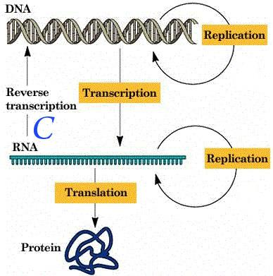
The three are named for the three molecules of the central dogma, and each detects the molecule at one point in it.

TechniqueDetectsNamed after
Southern blottingA specific DNA sequenceEdwin M. Southern
Northern blottingRNA fragments, or isolated mRNA
Western blottingSpecific proteins

Only Southern is a surname. Northern and Western were coined afterwards as jokes on it, which is why they are not named after anyone, and it is the reason the set is easy to remember once the joke is known: DNA, RNA, protein follow the compass from the one real name.

Diagram of the central dogma showing DNA with replication, transcription to RNA with reverse transcription running back, and translation to protein

3. In situ hybridization

note

In situ means in place. The blotting methods extract the molecule and run it on a gel; ISH applies the probe to the tissue section itself, so the result shows where in the tissue the sequence is, not merely that it is present.

There are two forms:

  • radioactive in situ hybridization;
  • nonradioactive in situ hybridization.

Radioactive ISH

note

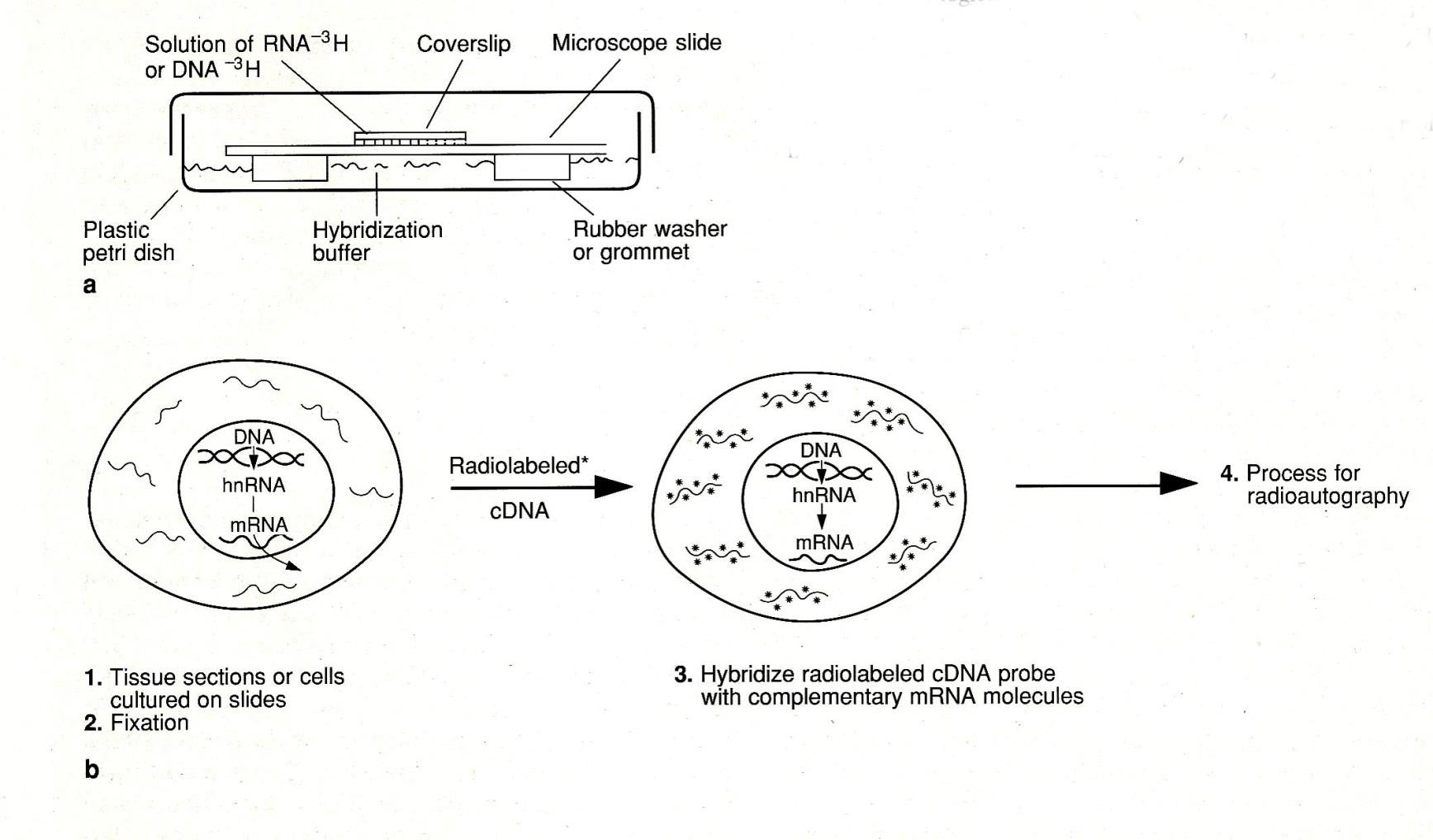
The tissue is set up on a microscope slide under a coverslip, in a plastic petri dish, with hybridization buffer held in by a rubber washer or grommet. The solution applied is RNA-³H or DNA-³H, that is probe labelled with tritium.

The sequence of the method:

  1. Tissue sections or cells cultured on slides.
  2. Fixation.
  3. Hybridize radiolabelled cDNA probe with complementary mRNA molecules.
  4. Process for radioautography.

The final step is the autoradiography met earlier in the same list of techniques. Hybridization puts the label where the sequence is; autoradiography is what makes the label visible.

Diagram of radioactive in situ hybridization showing the slide, coverslip, hybridization buffer and rubber washer in a petri dish, and below it a cell whose mRNA is hybridized with a radiolabelled cDNA probe before processing for radioautography

Nonradioactive ISH

note

It was originally developed by Pardue and Gall in 1969, and independently by John and colleagues in 1969.

Two groups arriving at the same method in the same year is a detail worth keeping, because it is the kind of thing an examiner asks: the answer is both, not one.

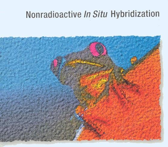
Photomicrograph labelled Nonradioactive In Situ Hybridization showing tissue with strongly coloured labelled cells against a contrasting background

The probe

note

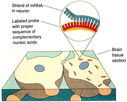
The labelled probe carries the proper sequence of complementary nucleic acids, and it binds the strand of mRNA in the cell, for example in a neuron within a brain tissue section.

That is the whole principle in one sentence. The probe is built to be the complement of the target, so it will bind that target and nothing else, and because it is labelled, wherever it binds becomes visible.

Diagram of a brain tissue section with an enlargement showing a labelled probe with the proper sequence of complementary nucleic acids binding a strand of mRNA in a neuron

4. The ISH procedure: six steps

note

The flow diagram for the ISH procedure runs:

  1. Preparation of slides and fixation of material.
  2. Choice of the probe and its labeling.
  3. Denaturation of the in situ target DNA, both probe and target.
  4. In situ hybridization.
  5. Immunocytochemical visualization.
  6. Microscopy.

Step 3 is the one that makes the rest possible. Double-stranded DNA has no free bases to pair with; denaturation separates the strands so that the probe has something to bind. Both the probe and the target must be denatured, which is why the step names them both.

Step 5 explains why this method sits beside immunohistochemistry. The probe is usually not visible in itself: it carries a marker that is then detected immunocytochemically, so the two techniques are used one after the other.

5. Medical applications

note

In fundamental research:

  • gene mapping;
  • localization of gene expression;
  • systematization of nuclear DNA and RNA;
  • replication;
  • cell sorting.

In clinical research:

  • cytogenetics;
  • prenatal diagnostics;
  • gene disorders;
  • diagnostics of infectious and malignant diseases;
  • biological dosimetry.

Reading the two lists

note

The division is between finding out where a sequence is and finding out whether a particular sequence is present in this patient.

Localization of gene expression is the research use that matters most, because it answers a question no other method on the list can: not which proteins a tissue contains, but which genes that tissue is currently reading. A protein may have been made elsewhere and transported; mRNA in a cell means that cell is transcribing that gene now.

Biological dosimetry is the odd item in the clinical list and worth a note: it estimates the radiation dose a person has received by counting chromosome damage in their cells, which is why it belongs to the cytogenetic methods rather than to diagnosis of a disease.

All 95 Cytology topics · exam study support, not clinical guidance. SuperMed is not affiliated with the Medical University of Sofia.

Start free. No card needed.

Every account starts free, with free topics in Cytology and Medical Biology. Super opens the rest. Super is €15 a month.

Create a free account