


Medical Biology · Year 1 · Medical University of Sofia

80

Genus Trichomonas. Giardia lamblia (Lamblia intestinalis)

Free notes for topic 80 of the Medical Biology syllabus, open without an account. Written by a senior student against the syllabus question and checked line by line by a second student before publishing. How content is made

Updated

What this topic covers

note

The metamonads, the flagellates with several flagella and no mitochondria, and what a commensal is; the genus Trichomonas, its general structure and the three species found in humans, Trichomonas vaginalis in the genital tract, Trichomonas tenax in the mouth and Trichomonas hominis in the large intestine; and Giardia lamblia (Lamblia intestinalis), its trophozoite and cyst, its adhesive disk and the damage it does to the intestinal mucosa, its life cycle and the faecal-oral transmission of giardiasis, together with what is known of the sexual process in Giardia and Trichomonas.
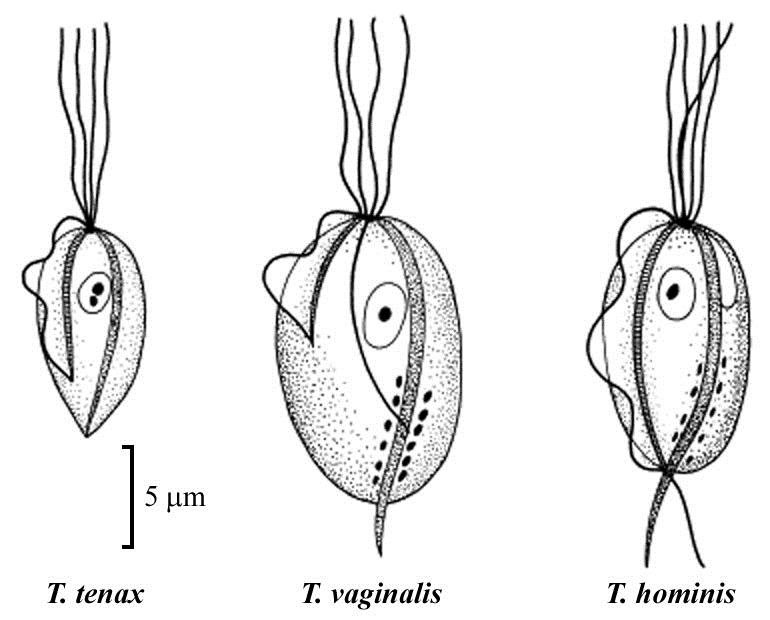
Trichomonas tenax, Trichomonas vaginalis and Trichomonas hominis drawn side by side at the same magnification with a 5 μm scale bar, each with anterior flagella, undulating membrane, nucleus and axostyle

1. Parasites with more than one flagellum and no mitochondria (Metamonada)

note

Metamonads have pear-shaped cells with more than one flagellum (usually at least 4).

Unlike kinetoplastids, which are remarkable with their mitochondrial DNA, metamonads have lost their mitochondria. The two groups are therefore opposites in this respect: one is defined by an enormous mass of mitochondrial DNA, the other by having no mitochondrion at all.

Most of them live as commensals or parasites on the mucosa of the intestine or another hollow organ, i.e. in anaerobic conditions. Having no mitochondria and living without oxygen go together.

Commensal is an organism which lives inside another organism and obtains food from it without harming it (if it harms the host, it is a parasite). The line between the two is therefore whether harm is done, not where the organism lives.

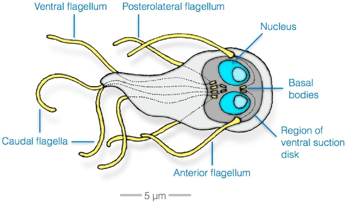
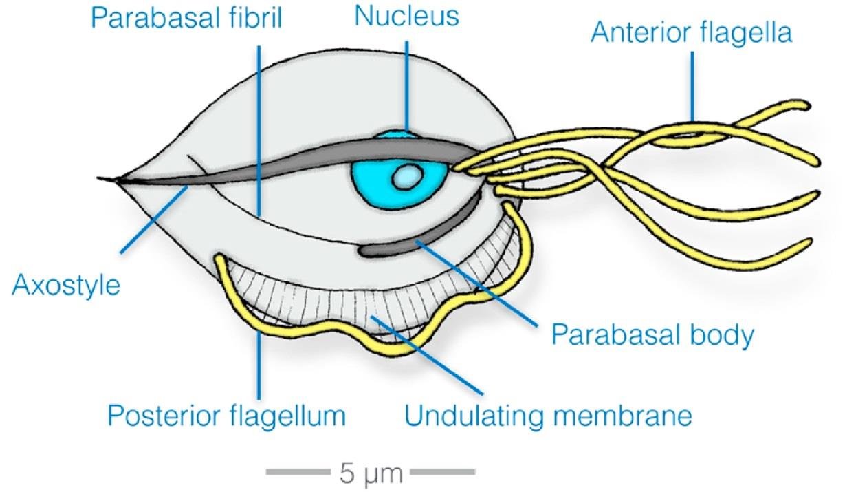
The drawings show the structure of two metamonads of medical importance, Trichomonas and Giardia, both of them with a 5 μm scale bar:

  • *Trichomonas vaginalis: *anterior flagella, posterior flagellum, undulating membrane, axostyle, parabasal fibril, parabasal body and nucleus**.
  • *Giardia lamblia: *anterior flagellum, ventral flagellum, posterolateral flagellum, caudal flagella, two nuclei, basal bodies and the region of the ventral suction disk**.
Trichomonas vaginalis structure with a 5 μm scale bar, labelled: anterior flagella, posterior flagellum, undulating membrane, axostyle, parabasal fibril, parabasal body, nucleus
Giardia lamblia structure with a 5 μm scale bar, labelled: anterior flagellum, ventral flagellum, posterolateral flagellum, caudal flagella, two nuclei, basal bodies, region of the ventral suction disk

2. Genus Trichomonas

note

  • Its species inhabit the human mouth, intestines and genitourinary tract.
  • The cell is pear-shaped.
  • From its anterior end, 5 flagella protrude. Of them, 4 are free and one supports an undulating membrane.

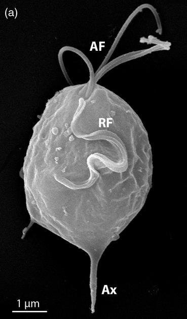
In the scanning electron micrograph, with a 1 μm scale bar: AF = anterior flagella; RF = recurrent flagellum (forming the undulating membrane); Ax = axostyle (forming the posterior tip of the cell). The axostyle is a firm stick that supports the shape of the cell.

Scanning electron micrograph of Trichomonas with a 1 μm scale bar, labelled AF (anterior flagella), RF (recurrent flagellum forming the undulating membrane) and Ax (axostyle)

Trichomonas vaginalis

note

  • It inhabits the human genital tract: the urethra of both sexes and the vagina of women.
  • Transmission is sexual or by genital contact.
  • Infected men are usually asymptomatic but in women the parasite causes colpitis (inflammation of the vagina).

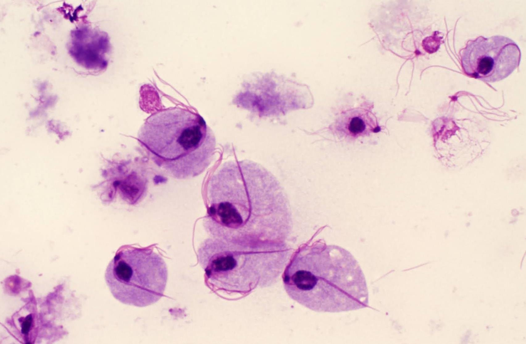
A diagnostic microscopic image shows the parasites in a smear; the spike-like axostyle and the flagella can be seen. Since the male infection is usually silent, an infected but symptomless partner is what keeps the infection circulating.

Stained smear of Trichomonas vaginalis, pear-shaped cells with a visible spike-like axostyle and trailing flagella, unlabelled

Other species of Trichomonas in humans

note

Trichomonas tenax

  • Commensal in the mouth.
  • Found around tartar in the teeth or in defects of carious teeth, particularly in patients with poor oral hygiene and advanced periodontal disease.
  • Transmission is through saliva, droplet spray, and kissing or use of contaminated dishes or water.

Trichomonas hominis

  • Commensal in the large intestine.
  • It is thought to be non-pathogenic although it has been associated with diarrhoeic stools.
  • Transmission by contaminated dishes, food and water.

The three species are drawn side by side at the same magnification, with a 5 μm scale bar: T. tenax is the narrowest, T. vaginalis the broadest, T. hominis intermediate, and all three show the same anterior flagella, undulating membrane and axostyle.

SpeciesWhere it livesPathogenic?Transmission
*Trichomonas vaginalis*urethra of both sexes, vagina of womenyes - colpitis in women; men usually asymptomaticsexual or by genital contact
*Trichomonas tenax*the mouth: around tartar, in carious defects, with poor oral hygiene and advanced periodontal diseaseno, a commensalsaliva, droplet spray, kissing, contaminated dishes or water
*Trichomonas hominis*the large intestinethought to be non-pathogenic, but associated with diarrhoeic stoolscontaminated dishes, food and water
Trichomonas tenax, Trichomonas vaginalis and Trichomonas hominis drawn side by side at the same magnification with a 5 μm scale bar: tenax narrowest, vaginalis broadest, hominis intermediate, each with anterior flagella, undulating membrane and axostyle

3. Giardia lamblia (Lamblia intestinalis)

note

The parasite has two names in common use, and the lecture gives both.

  • They are parasites in the small intestine of humans and many other mammals.
  • The intestinal form, called trophozoite, swims actively. It has two nuclei, 8 flagella, and an adhesive disk.
  • The parasite attaches to the intestinal epithelium by the adhesive disc, using it as a sucker.
  • In the lower parts of the intestine, trophozoites turn into resistant, oval-shaped cysts. The cyst is the infective stage.
  • The disease is called giardiasis (diarrhoea, excess gas, stomach or abdominal cramps).
  • Humans are infected by cysts in contaminated water or food - fruit or vegetables. This transmission route is called faecal-oral (written faecal-oral on the slides).

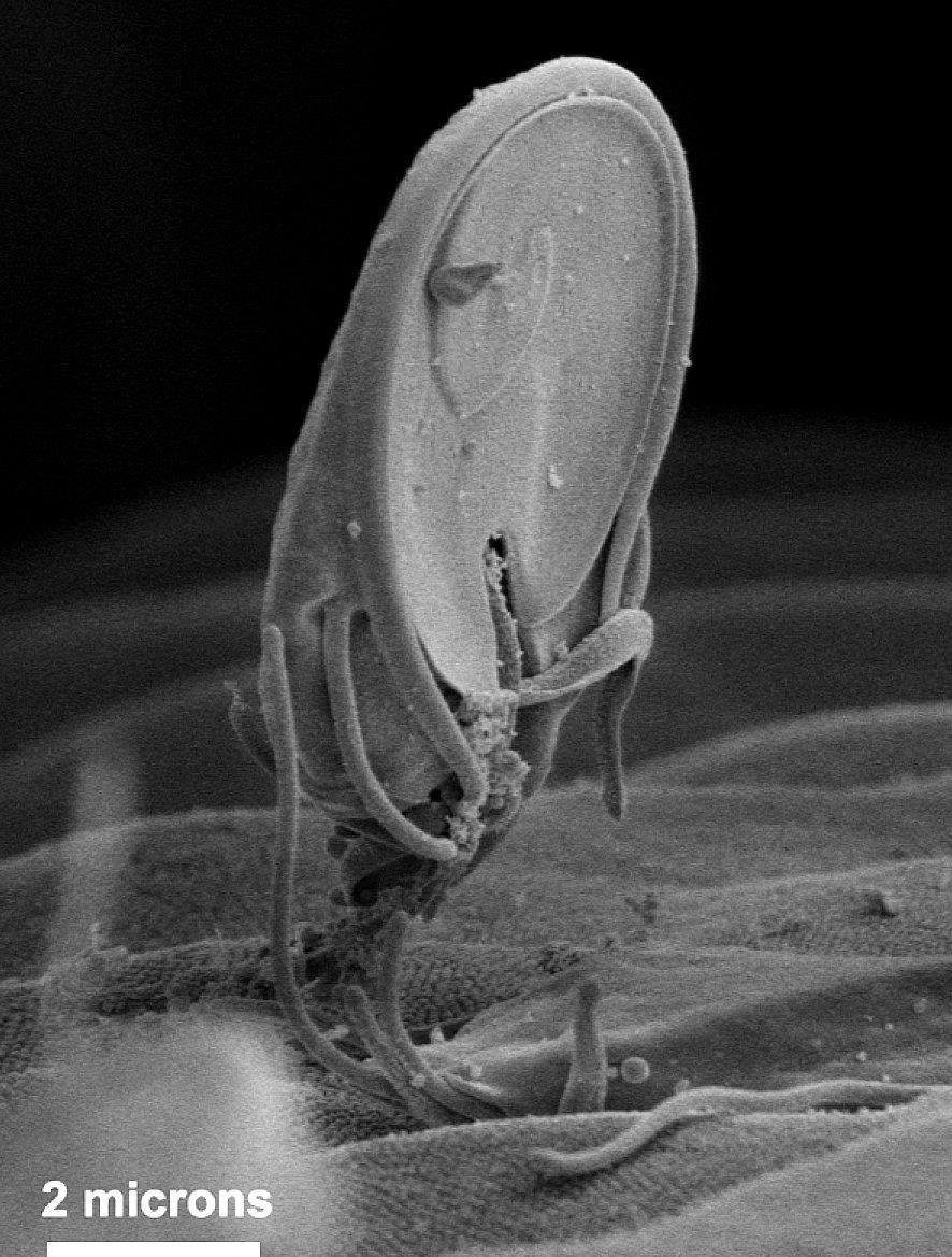
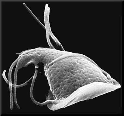
Scanning electron micrographs show a trophozoite of Giardia, one of them standing on the intestinal surface with a 2 micron scale bar, and one seen from the side showing the pear-shaped body and the flagella trailing behind it.

Scanning electron micrograph of a Giardia trophozoite standing on the intestinal surface by its adhesive disk, 2 micron scale bar
Scanning electron micrograph of a Giardia trophozoite seen from the side, showing the pear-shaped body and flagella trailing behind it, unlabelled

The structure of the trophozoite

note

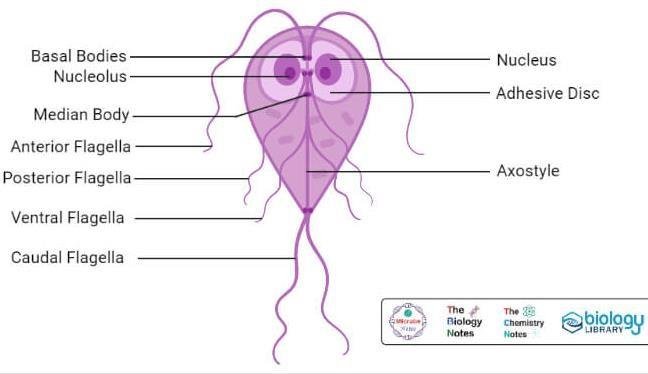
The labelled drawing of the trophozoite names, from the front of the cell backwards: the basal bodies, the nucleolus, the median body, the anterior flagella, the posterior flagella, the ventral flagella and the caudal flagella, together with the two nuclei, the adhesive disc and the axostyle. The eight flagella are therefore in four pairs, and the two nuclei sit symmetrically on either side of the midline, which is what gives the cell its "face-like" appearance in a stained smear.

Giardia trophozoite labelled front to back: basal bodies, nucleolus, median body, anterior flagella, posterior flagella, ventral flagella, caudal flagella, two nuclei, adhesive disc, axostyle

Giardia lamblia - illustration of the damage

note

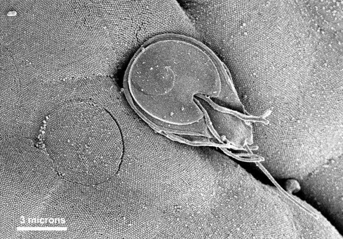
The adhesive disk of the Giardia trophozoite attaches to the intestinal mucosa like a vacuum suction cup.

The photo (scanning electron microscopy) shows how, after the detachment of the trophozoite, a circular imprint of the disk is visible on the dense microvilli. (CDC / Dr. Stan Erlandsen.) The scale bar is 3 microns. The imprint is the visible evidence of the damage: where the disc has been, the microvilli are flattened, and a mucosa covered with such marks absorbs less.

Scanning electron micrograph, 3 micron scale bar, showing a Giardia trophozoite attached by its adhesive disk beside a circular imprint left on the microvilli after an earlier trophozoite detached

Giardia lamblia - life cycle

note

Encystation is the transformation of a trophozoite into a cyst, excystation the reverse process.

Cysts are characteristic of intestinal protozoa transmitted by the faecal-oral route. They are immotile, metabolically quiescent, protected by a cyst wall, and oval or spherical to minimise contact with the environment. Every part of that description follows from the job the cyst has to do, which is to survive outside a host and be swallowed.

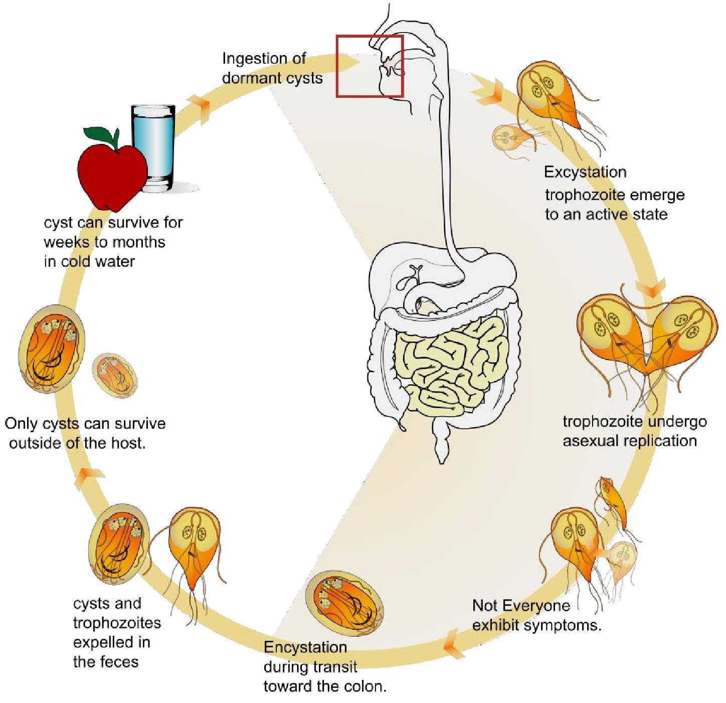
The cycle in the drawing runs:

  • Ingestion of dormant cysts, in contaminated water or on food such as an apple.
  • Excystation: the trophozoite emerges to an active state.
  • The trophozoites undergo asexual replication.
  • Not everyone exhibits symptoms.
  • Encystation during transit toward the colon.
  • Cysts and trophozoites are expelled in the faeces.
  • Only cysts can survive outside of the host. The cyst can survive for weeks to months in cold water.

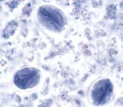
A microscopic image shows cysts in a faecal specimen, which is how the diagnosis is made.

Giardia life cycle: ingestion of dormant cysts in water or food, excystation to an active trophozoite, asexual replication, encystation during transit to the colon, cysts and trophozoites expelled in faeces, only cysts surviving outside the host
Giardia cysts in a stained faecal specimen, oval bodies with faint internal structure, unlabelled

4. Sexual process in Giardia and Trichomonas

note

  • Giardia has been shown to carry out genetic recombination between the two nuclei. It occurs during the process of encystation.
  • Some genes responsible for meiosis and crossing over have been identified in the Giardia genome. Homologs of the same genes have been found in Trichomonas.
  • Although the sexual process itself has not yet been studied, these findings suggest that it exists in metamonads. In fact, it seems to have been present in the common ancestor of all eukaryotes.

The evidence here is genetic rather than observed: nobody has watched these organisms mate, but they carry the machinery for it, and the fact that a group as ancient as the metamonads has it argues that sex is as old as the eukaryotic cell itself.

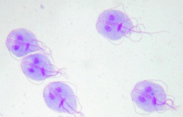
A stained preparation of Giardia trophozoites (Eva Nohynkova, Wikipedia) shows the two nuclei in each cell - which is the pair of nuclei between which that recombination happens.

Stained preparation of Giardia trophozoites, each cell showing its two nuclei side by side, unlabelled

The most important things to know

note

  • Metamonads are pear-shaped cells with more than one flagellum (usually at least 4) which have lost their mitochondria and live in anaerobic conditions on the mucosa of the intestine or another hollow organ.
  • A commensal lives inside another organism and takes food from it without harming it; if it harms the host it is a parasite.
  • *Trichomonas* has a pear-shaped cell with 5 anterior flagella, 4 free and 1 forming the undulating membrane, and an axostyle that stiffens the cell and forms its posterior tip.
  • *T. vaginalis* lives in the urethra of both sexes and the vagina, is sexually transmitted, causes colpitis in women and is usually asymptomatic in men. *T. tenax* is a mouth commensal of patients with poor oral hygiene and periodontal disease; *T. hominis* is a large-intestine commensal, probably non-pathogenic.
  • *Giardia lamblia* (*Lamblia intestinalis) lives in the *small intestine. The **trophozoite has two nuclei, 8 flagella and an adhesive disk with which it sticks to the epithelium like a suction cup, leaving a circular imprint on the microvilli.
  • The cyst is the infective stage: immotile, metabolically quiescent, protected by a cyst wall, oval, able to survive weeks to months in cold water, and the only stage that survives outside the host.
  • Giardiasis gives diarrhoea, excess gas and abdominal cramps, and is caught faecal-orally from contaminated water, fruit or vegetables; encystation happens on the way to the colon and excystation in the intestine of the next host.
  • Giardia recombines genetically between its two nuclei during encystation, and both Giardia and Trichomonas carry meiosis and crossing over genes, so a sexual process probably exists and probably dates back to the common ancestor of all eukaryotes.
Giardia trophozoite labelled with basal bodies, nucleolus, median body, four pairs of flagella, two nuclei, adhesive disc and axostyle, summarising the trophozoite structure

All 105 Medical Biology topics · exam study support, not clinical guidance. SuperMed is not affiliated with the Medical University of Sofia.

Start free. No card needed.

Every account starts free, with Medical Ethics, Urology and Computer Science open in full and free topics in Cytology, Latin Language, Medical Chemistry, Medical Physics, Medical Biology, Anatomy, Biophysics, Medical Psychology, MCS (Disaster Medicine), Physiology, Biochemistry, Social Medicine, Medical Genetics, Pharmacology, Pathophysiology, Microbiology, Internal Medicine, Nephrology, Cardiology, Pulmonology, Gastroenterology, Internal Medicine (Clinical), Obstetrics and Gynecology, Physiotherapy and Rehabilitation, Dental Materials, Preclinics of Prosthetic Dental Medicine, Medical Psychology (Dental Medicine), SMME (Social Medicine & Medical Ethics), Preclinics of Pediatric Dental Medicine, Hygiene and Epidemiology, General Surgery, Preclinics of Conservative Dental Medicine, Preclinics of Oral and Maxillofacial Medicine, Dental Public Health, Dental Prophylaxis, Diagnostic Imaging, Dental Allergology, Dental Implantology, Physiotherapy, Cytology & Histology, Medical Chemistry, Medical Physics, Anatomy I, Biophysics and Medical Biology. Super opens the rest. Super is €14.99 a month.

Create a free account