


Medical Biology · Year 1 · Medical University of Sofia

81

Subphylum Sarcodina. Genus Entamoeba

Free notes for topic 81 of the Medical Biology syllabus, open without an account. Written by a senior student against the syllabus question and checked line by line by a second student before publishing. How content is made

Updated

What this topic covers

note

The amoeboid parasites, which move and feed by pseudopodia, and the genus Entamoeba within them; Entamoeba histolytica, its trophozoite and cyst, its faecal-oral transmission, the amoebiasis it causes and how it invades the intestinal wall and reaches the liver, lungs and brain; the morphology of its two forms, including the ingested erythrocytes, the four nuclei of the mature cyst, the chromatoid body and the karyosome; its life cycle and what is suspected about its sexual process; and the other species of Entamoeba found in humans, Entamoeba dispar, Entamoeba coli and Entamoeba gingivalis.
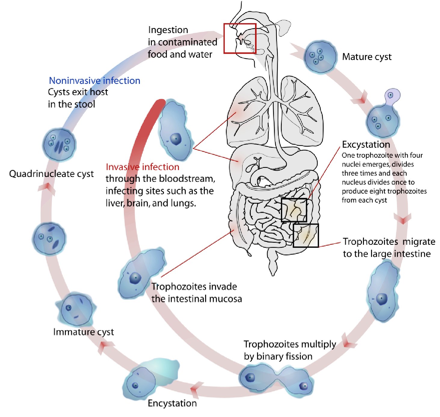
Entamoeba histolytica life cycle: ingestion of contaminated food and water, mature cyst, excystation into eight trophozoites, migration to the large intestine, binary fission, encystation to immature then quadrinucleate cyst, noninvasive cysts exiting in stool or invasive spread through the bloodstream to liver, lungs and brain

1. Parasites with pseudopodia (Amoebozoa). Genus Entamoeba

note

These are the amoeboid members of the phylum Sarcomastigophora, the subphylum Sarcodina. In them the cytoplasm forms temporary protrusions, pseudopodia, for locomotion and food intake, and this kind of movement is called amoeboid.

The name of this genus means "internal amoeba". Its members are either commensals or intestinal parasites. So the genus takes its name from where it lives, and its species range from harmless lodgers to one of the most dangerous intestinal parasites of humans.

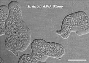
Living Entamoeba dispar amoebae under phase-contrast microscopy, showing their irregular pseudopodia, unlabelled except for the species caption

2. Entamoeba histolytica

note

  • This parasite inhabits the large intestine of humans.
  • It has two forms: trophozoite (active) and cyst (quiescent).
  • The cyst can survive in the environment for weeks. It is the infective form.
  • The infection is transmitted through the faecal-oral route by ingesting cysts shed with the faeces of a previous host (the slides write it foecal-oral).
  • Excystation (transformation of cysts into trophozoites) occurs in the intestine.
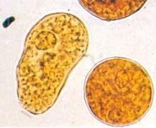
Entamoeba histolytica trophozoite, amoeboid shape on the left, beside a rounded cyst with a visible cyst wall on the right, unlabelled

Amoebiasis: how the parasite causes disease

note

Most infected people are asymptomatic carriers, but some develop a disease called amoebiasis (amoebic dysentery), which is characterised by abdominal pain, bloody diarrhoea, and lesions in the intestines.

The reason is that the parasites secrete proteases, with the help of which they enter the intestinal epithelium, where they feed on blood cells (which are often found in the amoeba cells cytoplasm), and from there, through the bloodstream, they reach other organs (liver, lungs, brain) and cause dangerous complications - abscesses, which without treatment quickly lead to death.

E. histolytica infection is estimated to kill more than 55,000 people each year.

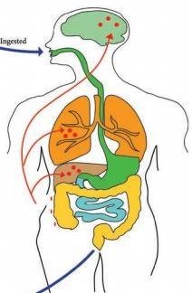
The accompanying figure traces the route: the cysts are ingested by mouth, the parasite establishes itself in the large intestine, and from there red arrows run to the liver, the lungs and the brain, which are the three sites of the abscesses.

The proteases are the key to the whole clinical picture. They are what turns a gut commensal-like organism into an invasive one, and everything downstream, the bloody diarrhoea, the ulcers, the distant abscesses, follows from the ability to digest a way through the epithelium.

Diagram tracing Entamoeba histolytica from ingestion through the gut, with red arrows running from the large intestine to the liver, lungs and brain, the sites of amoebic abscesses

Entamoeba histolytica - morphological forms

note

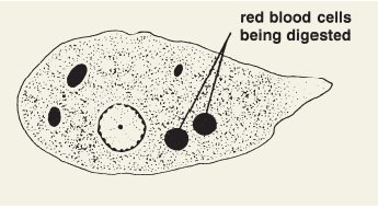
  • The trophozoite is amoeboid; ingested erythrocytes distinguish it from related commensal species. This is the single most useful diagnostic feature: a commensal amoeba does not eat blood cells.
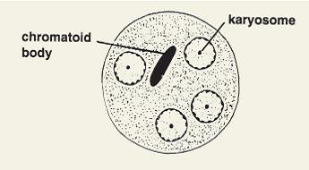
  • The cyst is spherical. When mature, it has 4 nuclei. In its cytoplasm, ribosomes form an aggregation called chromatoid body.
  • In both forms, chromatin is seen in two locations in the nucleus: at the periphery and in a central cluster called karyosome. (Do not study these details.)
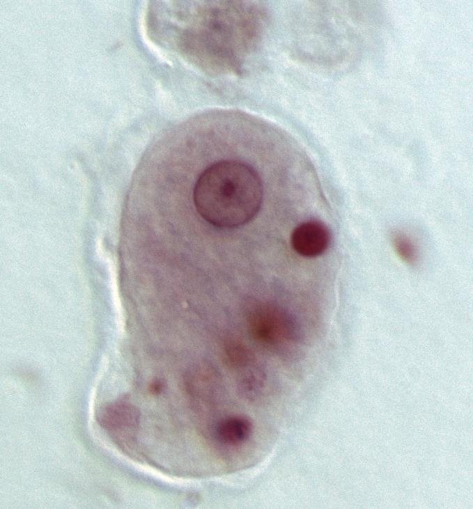
Entamoeba histolytica trophozoite in a stained smear with darkly stained ingested erythrocytes in its cytoplasm and a nucleus with a central karyosome
Mature Entamoeba histolytica cyst drawing labelled chromatoid body and karyosome, four nuclei each with a central karyosome

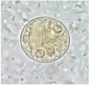
The drawings label the red blood cells being digested inside the trophozoite, and the chromatoid body and the karyosome inside the cyst. The micrographs show a trophozoite with engulfed erythrocytes, a cyst, and a trophozoite and a cyst side by side in which the cyst wall is visible.

Micrograph of an Entamoeba histolytica cyst, round with faint internal granularity, unlabelled
Entamoeba histolytica trophozoite and cyst side by side in a stained smear, the cyst wall clearly visible around the rounded cyst
TrophozoiteCyst
Stateactivequiescent
Shapeamoeboid, with pseudopodiaspherical, with a cyst wall
Nucleione4 when mature
Contentsingested erythrocytes, which distinguish it from commensal specieschromatoid body, an aggregation of ribosomes
Nuclear chromatinperipheral, plus a central karyosomeperipheral, plus a central karyosome
Rolefeeds, multiplies, invadesthe infective form; survives in the environment for weeks
Trophozoite drawing labelled red blood cells being digested, with a nucleus shown alongside the ingested cells in the cytoplasm

Entamoeba histolytica - life cycle

note

The contaminated food is fruit and vegetables with cysts on their surface.

The cycle in the drawing runs:

  • Ingestion in contaminated food and water.
  • The mature cyst reaches the intestine.
  • Excystation: one trophozoite with four nuclei emerges, divides three times and each nucleus divides once, to produce eight trophozoites from each cyst.
  • The trophozoites migrate to the large intestine.
  • Trophozoites multiply by binary fission.
  • Encystation, giving first the immature cyst and then the quadrinucleate cyst.
  • Noninvasive infection: cysts exit the host in the stool.
  • Invasive infection: trophozoites invade the intestinal mucosa, and then spread through the bloodstream, infecting sites such as the liver, brain, and lungs.

So the cycle has two branches from the same starting point: the parasite either stays in the lumen and is passed on as cysts, or invades, which is the branch that produces disease.

Entamoeba histolytica life cycle diagram: ingestion, mature cyst, excystation into eight trophozoites, migration to the large intestine, binary fission, encystation, noninvasive cysts exiting in stool or invasive spread through the bloodstream to liver, brain and lungs

The sexual process

note

Like the metamonads, E. histolytica has genes related to recombination, and it is speculated that it undergoes meiosis during the conversion from the uninucleate trophozoite to quadrinucleate cyst; however, the sexual process has not yet been studied.

The suspicion is reasonable on the face of it: a single nucleus becoming four is the sort of event meiosis would explain, and the genes are there. But the lecture is careful to call it speculation, and so should you.

Mature Entamoeba histolytica cyst with four nuclei, chromatoid body and karyosome labelled, the quadrinucleate stage the parasite is thought to reach by meiosis

3. Other species of Entamoeba in humans

note

Entamoeba dispar

  • Nonpathogenic species in the large intestine.
  • Similar to E. histolytica but never invades the intestinal tissue to feed on blood cells.
  • Trophozoite and cyst forms.

Entamoeba coli

  • Nonpathogenic, in the large intestine.
  • Does not move much by its pseudopodia and keeps its round shape.
  • Trophozoite and cyst forms.

Entamoeba gingivalis

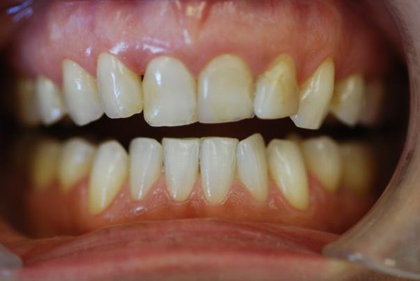
  • Found between the gingival pockets and near the base of the teeth.
  • In 95% of people with gum disease and in 50% of people with healthy gums.
  • Direct transmission by kissing or by sharing eating utensils.
  • Trophozoite and cyst forms.
SpeciesWherePathogenic?Distinguishing featureForms
*Entamoeba histolytica*large intestineyes - amoebiasis, amoebic dysentery, abscesses of liver, lungs and brain; more than 55,000 deaths a yearingested erythrocytes in the trophozoite; secretes proteases and invadestrophozoite and cyst
*Entamoeba dispar*large intestinenolooks like E. histolytica but never invades the tissue to feed on blood cellstrophozoite and cyst
*Entamoeba coli*large intestinenomoves little by pseudopodia, keeps its round shapetrophozoite and cyst
*Entamoeba gingivalis*gingival pockets, base of the teethnoin 95% of people with gum disease, 50% with healthy gums; spread by kissing and shared utensilstrophozoite and cyst

E. gingivalis is of particular interest to dental students, both because of how common it is and because the lecture points to a video of it phagocytosing a polymorphonuclear leucocyte.

Living Entamoeba dispar amoebae under phase-contrast microscopy, irregular pseudopodia, labelled only with the species caption
Close-up of teeth and gums showing plaque and gum inflammation, the site where Entamoeba gingivalis lives in the gingival pockets

The most important things to know

note

  • Amoeboid protozoans move and feed by pseudopodia; *Entamoeba* means "internal amoeba", and its members are commensals or intestinal parasites.
  • *Entamoeba histolytica* lives in the large intestine and has two forms: the trophozoite (active) and the cyst (quiescent). The cyst is the infective form and survives weeks in the environment; transmission is faecal-oral, from cysts on fruit and vegetables and in contaminated water.
  • Excystation in the intestine gives eight trophozoites from one quadrinucleate cyst; encystation returns the parasite to the quadrinucleate cyst that leaves in the stool.
  • Most infected people are asymptomatic carriers. Amoebiasis (amoebic dysentery) gives abdominal pain, bloody diarrhoea and intestinal lesions, because the parasite secretes proteases, enters the epithelium, feeds on blood cells, and spreads through the bloodstream to the liver, lungs and brain, where abscesses are fatal without treatment. More than 55,000 deaths a year.
  • Morphology: the trophozoite is amoeboid and contains ingested erythrocytes, which distinguish it from the commensal species; the mature cyst is spherical with 4 nuclei and a chromatoid body of aggregated ribosomes; chromatin lies at the nuclear periphery and in a central karyosome.
  • E. histolytica carries recombination genes and may undergo meiosis in the conversion of the uninucleate trophozoite into the quadrinucleate cyst, but the sexual process has not been studied.
  • The other human species are nonpathogenic: *E. dispar* (like histolytica but never invades), *E. coli* (barely moves, stays round) and *E. gingivalis* (in the gingival pockets, in 95% of people with gum disease and 50% with healthy gums, spread by kissing and shared utensils). All of them have trophozoite and cyst forms.
Mature Entamoeba histolytica cyst labelled chromatoid body and karyosome, with four nuclei visible, summarising the cyst's distinguishing structure

All 105 Medical Biology topics · exam study support, not clinical guidance. SuperMed is not affiliated with the Medical University of Sofia.

Start free. No card needed.

Every account starts free, with Medical Ethics, Urology and Computer Science open in full and free topics in Cytology, Latin Language, Medical Chemistry, Medical Physics, Medical Biology, Anatomy, Biophysics, Medical Psychology, MCS (Disaster Medicine), Physiology, Biochemistry, Social Medicine, Medical Genetics, Pharmacology, Pathophysiology, Microbiology, Internal Medicine, Nephrology, Cardiology, Pulmonology, Gastroenterology, Internal Medicine (Clinical), Obstetrics and Gynecology, Physiotherapy and Rehabilitation, Dental Materials, Preclinics of Prosthetic Dental Medicine, Medical Psychology (Dental Medicine), SMME (Social Medicine & Medical Ethics), Preclinics of Pediatric Dental Medicine, Hygiene and Epidemiology, General Surgery, Preclinics of Conservative Dental Medicine, Preclinics of Oral and Maxillofacial Medicine, Dental Public Health, Dental Prophylaxis, Diagnostic Imaging, Dental Allergology, Dental Implantology, Physiotherapy, Cytology & Histology, Medical Chemistry, Medical Physics, Anatomy I, Biophysics and Medical Biology. Super opens the rest. Super is €14.99 a month.

Create a free account