


Medical Biology · Year 1 · Medical University of Sofia

82

Phylum Apicomplexa. Toxoplasma gondii

Free notes for topic 82 of the Medical Biology syllabus, open without an account. Written by a senior student against the syllabus question and checked line by line by a second student before publishing. How content is made

Updated

What this topic covers

note

The phylum Apicomplexa (Sporozoa) and the apical complex that gives it its name; the parasitophorous vacuole the parasite builds for itself inside the host cell; the two ways an apicomplexan solves the problem of passing the apical complex on to its daughters, endodyogeny and endopolygeny; the alternation of merogony, gamogony and sporogony; and then the phylum's commonest human parasite, Toxoplasma gondii - its three infective stages, its definitive and intermediate hosts, its life cycle in the cat and in the intermediate host, all the routes by which it reaches a human, the disease it causes, who is at risk from it, and the changes it makes to the behaviour of the animal it infects. The lecture opens with Lennart Nilsson's photograph of red blood cells bursting after infection with the malaria parasite, which is where the phylum's other great genus, Plasmodium, is going.
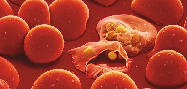
Scanning electron micrograph of red blood cells, one burst open by an emerging malaria parasite, several round golden parasite forms spilling out

1. Phylum Apicomplexa (Sporozoa)

note

They all are intracellular parasites of animals. There are no free-living apicomplexans; every member of the phylum spends the working part of its life inside somebody else's cell.

A special structure at one of the poles is named apical complex. That is a group of cytoskeletal structures and associated membrane organelles.

The apical complex is involved in:

  • attachment to the host cell;
  • penetration into the cell;
  • rearrangement of the cell organelles;
  • and in parasite proliferation.

The apical complex consists of:

  • conoid - a hollow organelle shaped like a truncated cone;
  • apical ring made of contractile actin fibres;
  • rhoptries - contain enzymes that are released during the penetration;
  • micronemes - secrete proteins capable of interacting with host cell receptors;
  • dense granules - secrete proteins capable of modifying the post-translational protein trafficking within the host cell and to maintain the integrity of the parasitophorous vacuole.

Apicoplast - a relict, nonphotosynthetic plastid found in Apicomplexa. It is the remnant of an alga that an ancestor of the group once swallowed, and it is not part of the apical complex despite the similar name.

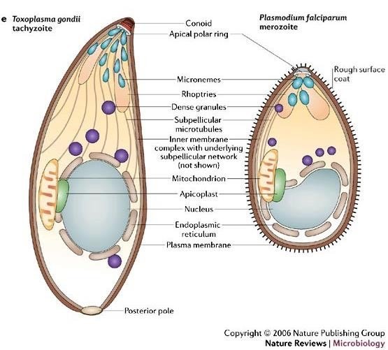
Toxoplasma gondii tachyzoite and Plasmodium falciparum merozoite side by side, both labelled conoid, apical polar ring, micronemes, rhoptries, dense granules, subpellicular microtubules, inner membrane complex, mitochondrion, apicoplast, nucleus, endoplasmic reticulum, plasma membrane, posterior pole, merozoite also showing a rough surface coat

The apicomplexan cell, drawn out

note

The lecture puts a Toxoplasma gondii tachyzoite and a Plasmodium falciparum merozoite side by side, and labels the same parts on both. Read from the apical (front) end backwards: conoid, apical polar ring, micronemes, rhoptries, dense granules, subpellicular microtubules, the inner membrane complex with its underlying subpellicular network, then mitochondrion, apicoplast, nucleus, endoplasmic reticulum, plasma membrane, and at the back the posterior pole. The Plasmodium merozoite carries in addition a rough surface coat.

The two cells are drawn to the same plan because they are the same kind of cell. Everything that is specialised sits at the front, pointing at the host cell membrane the parasite is about to go through.

Toxoplasma gondii tachyzoite and Plasmodium falciparum merozoite side by side, both labelled conoid, apical polar ring, micronemes, rhoptries, dense granules, subpellicular microtubules, inner membrane complex, mitochondrion, apicoplast, nucleus, endoplasmic reticulum, plasma membrane, posterior pole, merozoite also showing a rough surface coat

The parasitophorous vacuole

note

During penetration, the parasites create for themselves a specialised compartment, the parasitophorous vacuole, to survive within host cells.

The proteins secreted by the parasite make the host cell relocate its endoplasmic reticulum, Golgi complex and mitochondria around the parasitophorous vacuole in order to provide energy and all necessary molecules to the parasite.

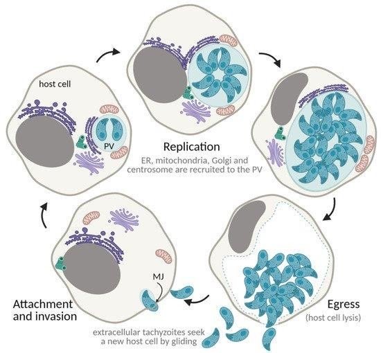
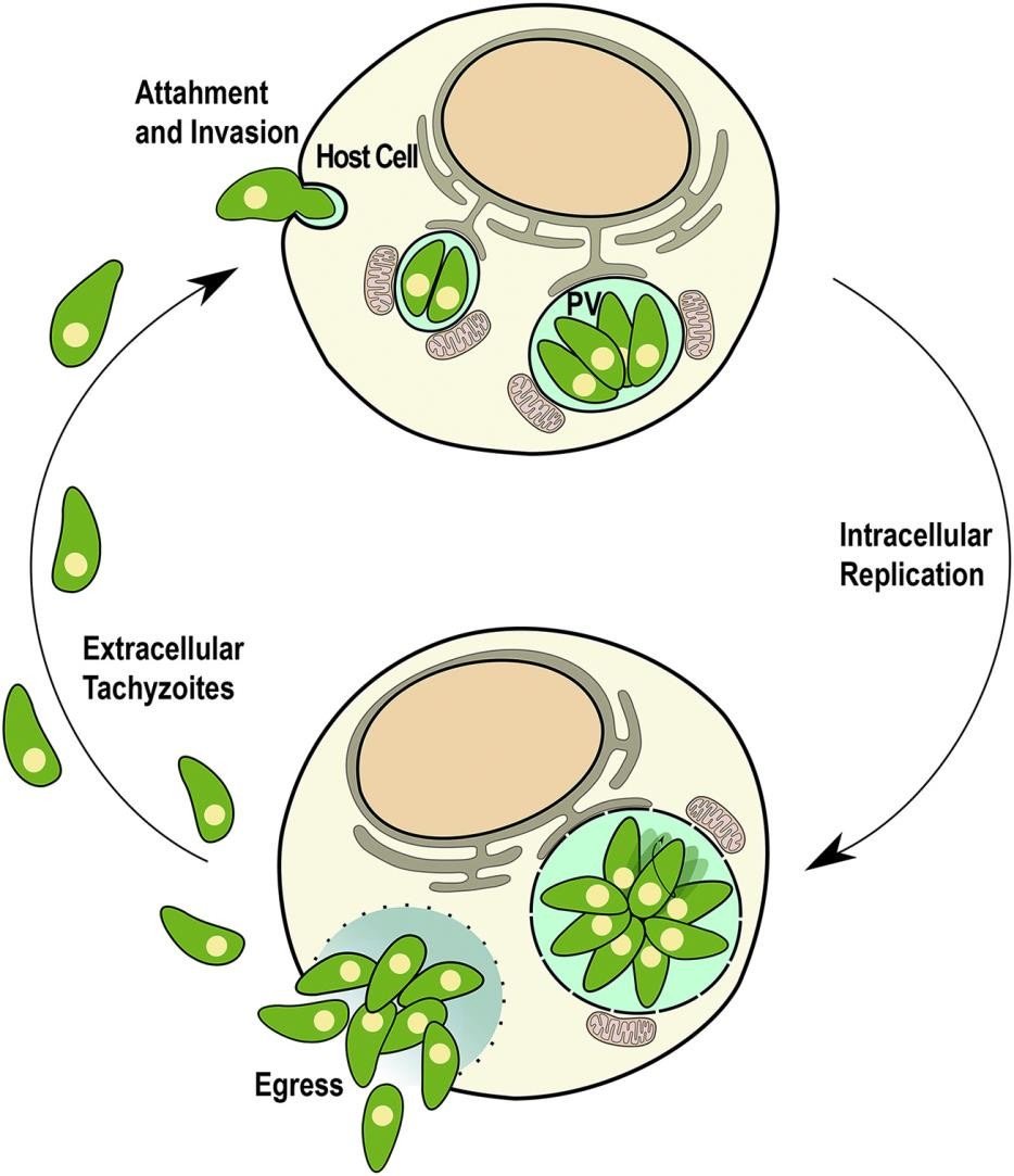
The lecture's diagram runs the whole sequence round in a circle: attachment and invasion, with the moving junction (MJ) forming between parasite and host membrane; then replication, during which ER, mitochondria, Golgi and the centrosome are recruited to the PV; then egress, which is host cell lysis; and finally the extracellular tachyzoites seek a new host cell by gliding. The abbreviations used are ER for endoplasmic reticulum, PV for parasitophorous vacuole and MJ for moving junction.

So the parasite does not simply hide in the cell. It rebuilds the cell around itself into a feeding chamber.

Cycle of the parasitophorous vacuole (PV) round a host cell: attachment and invasion forming a moving junction (MJ), replication with ER, mitochondria, Golgi and centrosome recruited to the PV, egress by host cell lysis, then extracellular tachyzoites gliding to a new host cell

The apical complex imposes division specificity

note

Transmission of the apical complex to daughter cells is problematic. A dividing cell has one apical complex and needs to end up with two, and the structure is too complicated to be built at short notice. There are two solutions:

A) Endodiogeny (in Toxoplasma) - the mother cell retains its shape and apical complex until the end, and two daughter cells with the same polarity are formed in it.

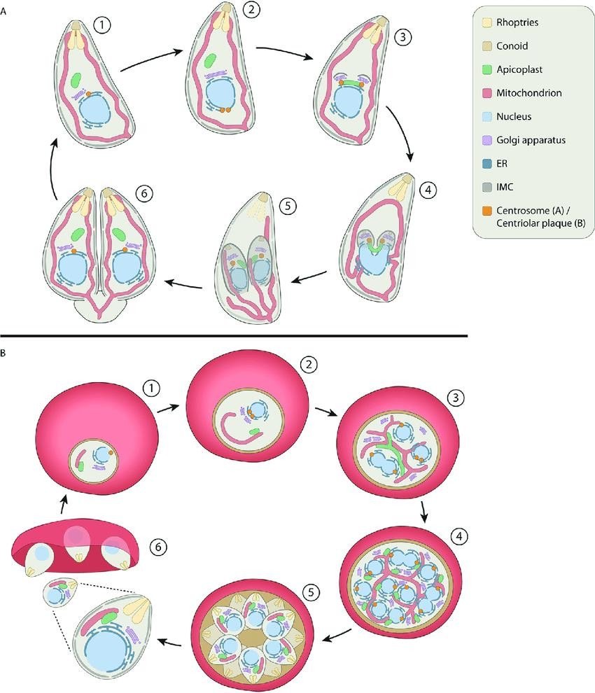
B) Endopolygeny - the mother cell loses its shape, polarity and apical complex, but since it is not profitable to take such a dramatic step often, the division is multiple: more than one replication occurs and multiple daughter cells are formed in the mother cell, which acquire polarity anew.

When the division of the nucleus occurs long before the formation of the cells themselves, endopolygeny is called schizogony (in Plasmodium).

In both endodio- and endopolygeny, the daughter cells grow, gradually incorporating the body and organelles of the mother. The mother is not discarded; she is eaten from the inside by her own daughters.

Two division schemes labelled with a shared key (rhoptries, conoid, apicoplast, mitochondrion, nucleus, Golgi, ER, IMC, centrosome or centriolar plaque): panel A endodyogeny, mother keeps her shape while two daughters form inside her, panel B endopolygeny or schizogony, mother rounds up and loses polarity while many daughters form inside her

Apicomplexa - reproduction

note

Sexual and asexual reproduction alternate:

  • merogony (schizogony) is the name for asexual reproduction by mitosis (most often multiple); the resulting cells are generally called "merozoites", but in different species you may encounter different names;
  • gamogony (gametogenesis): formation of gametes;
  • sporogony: the zygote divides by meiosis and then by mitosis, producing sporozoites, and they are an infectious stage for humans.

This is a haploid-diploid cycle. The parasite is haploid for almost the whole of its life. The lecture's scheme names the players at each turn: the sporozoite (1n) starts merogony (schizogony), which yields the merozoite, also called meront or schizont (1n); gamogony then produces the micro- and macrogamonts (1n); their fusion gives the zygote (2n), the only diploid cell in the cycle, and sporogony returns the parasite to 1n.

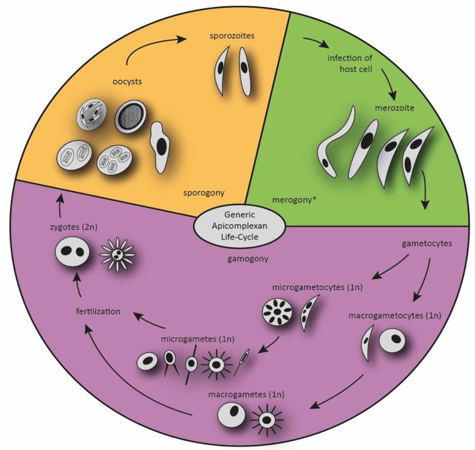
The generic apicomplexan cycle drawn in the lecture runs: sporozoites infect the host cell and become merozoites by merogony; some merozoites become gametocytes, which mature into microgametocytes (1n) and macrogametocytes (1n) in gamogony; these give microgametes (1n) and macrogametes (1n); fertilisation (1n to 2n) produces zygotes (2n); the zygotes become oocysts, in which sporogony (2n to 1n) makes new sporozoites.

Generic apicomplexan life cycle wheel: sporozoites infect the host cell and become merozoites by merogony, gametocytes mature into microgametocytes and macrogametocytes by gamogony, giving microgametes and macrogametes, fertilisation forms the zygote, which becomes oocysts undergoing sporogony back to sporozoites

2. *Toxoplasma gondii*

note

Obligate intracellular parasite infecting almost all warm-blooded animals. One of the most common human parasites.

Definitive hosts - all kinds of cats (family Felidae). In the intestine of cats Toxoplasma reproduces sexually.

Intermediate hosts - warm-blooded animals, including human. In intermediate hosts just asexual multiplication occurs.

That single fact explains everything else about the parasite's biology: sex happens only in a cat, so every other host the parasite ever occupies is a way of getting back into a cat.

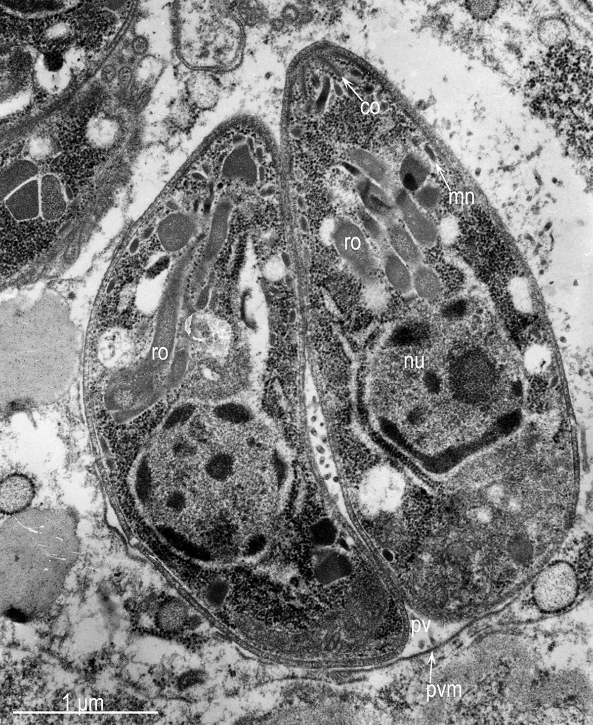
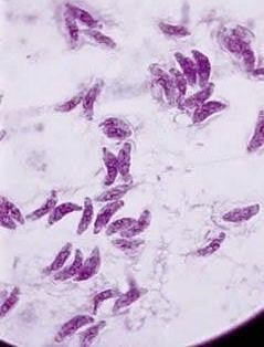
The electron micrograph in the lecture shows two Toxoplasma cells inside a host cell, labelled co for conoid, ro for rhoptries, nu for nucleus and mn for micronemes, with a 1 μm scale bar. The stained preparation beside it, the lecturer notes, is the one you can see at our lab classes: crescent-shaped purple cells scattered across the field.

Electron micrograph of two Toxoplasma gondii tachyzoites inside a host cell, labelled co for conoid, ro for rhoptries, nu for nucleus and mn for micronemes, with a 1 micrometre scale bar
Light micrograph of a Giemsa-stained smear showing numerous crescent-shaped purple Toxoplasma gondii tachyzoites scattered across the field

Endodyogeny of *Toxoplasma*

note

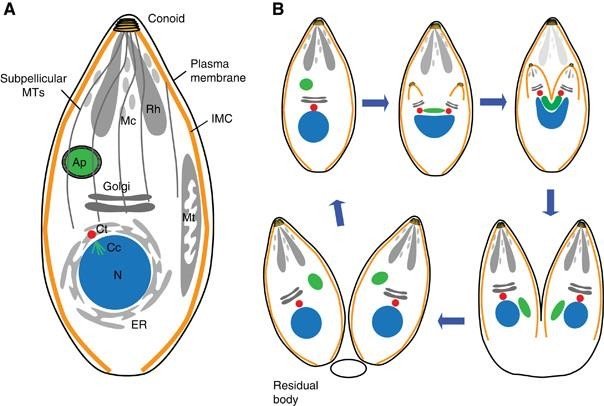
Toxoplasma divides by endodyogeny, illustrated in the lecture from The Fine Structure and Reproduction of Toxoplasma gondii, Harley et al, 1968, and in a modern scheme beside it. The scheme labels the parasite's parts as conoid, subpellicular microtubules, plasma membrane, Mc (micronemes), Rh (rhoptries), IMC (inner membrane complex), Ap (apicoplast), Golgi, N (nucleus), ER and Mt (mitochondrion), and then follows two daughters being built inside the mother until the mother's remains are left behind as a residual body.

Modern scheme of Toxoplasma endodyogeny, panel A a single parasite labelled conoid, subpellicular microtubules, plasma membrane, Mc micronemes, Rh rhoptries, IMC, Ap apicoplast, Golgi, N nucleus, ER, Mt mitochondrion, panel B six-step sequence of two daughters forming inside the mother, ending with a residual body left behind

Three infective stages

note

Three stages of the cycle can infect humans:

  • A) tachyzoitea form that divides rapidly and is found in the first days after infection;
  • B) bradyzoitedivides slowly and is characteristic of the later stages of infection (chronic infection); forms clusters (tissue cysts) mainly in muscle and nerve tissue, but other locations are also possible;
  • C) sporozoiteproduced only in cats and excreted in their faeces.

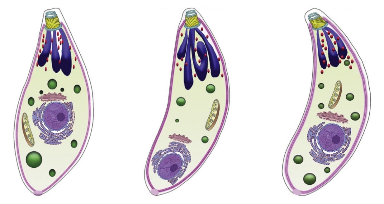
All three are drawn as the same crescent-shaped cell with the same organelles; what separates them is where they are and how fast they divide.

Three crescent-shaped apicomplexan cells drawn identically with conoid, rhoptries, micronemes, mitochondrion and nucleus, representing the unlabelled A, B and C infective stages, tachyzoite, bradyzoite and sporozoite

3. Pathways of transmission

note

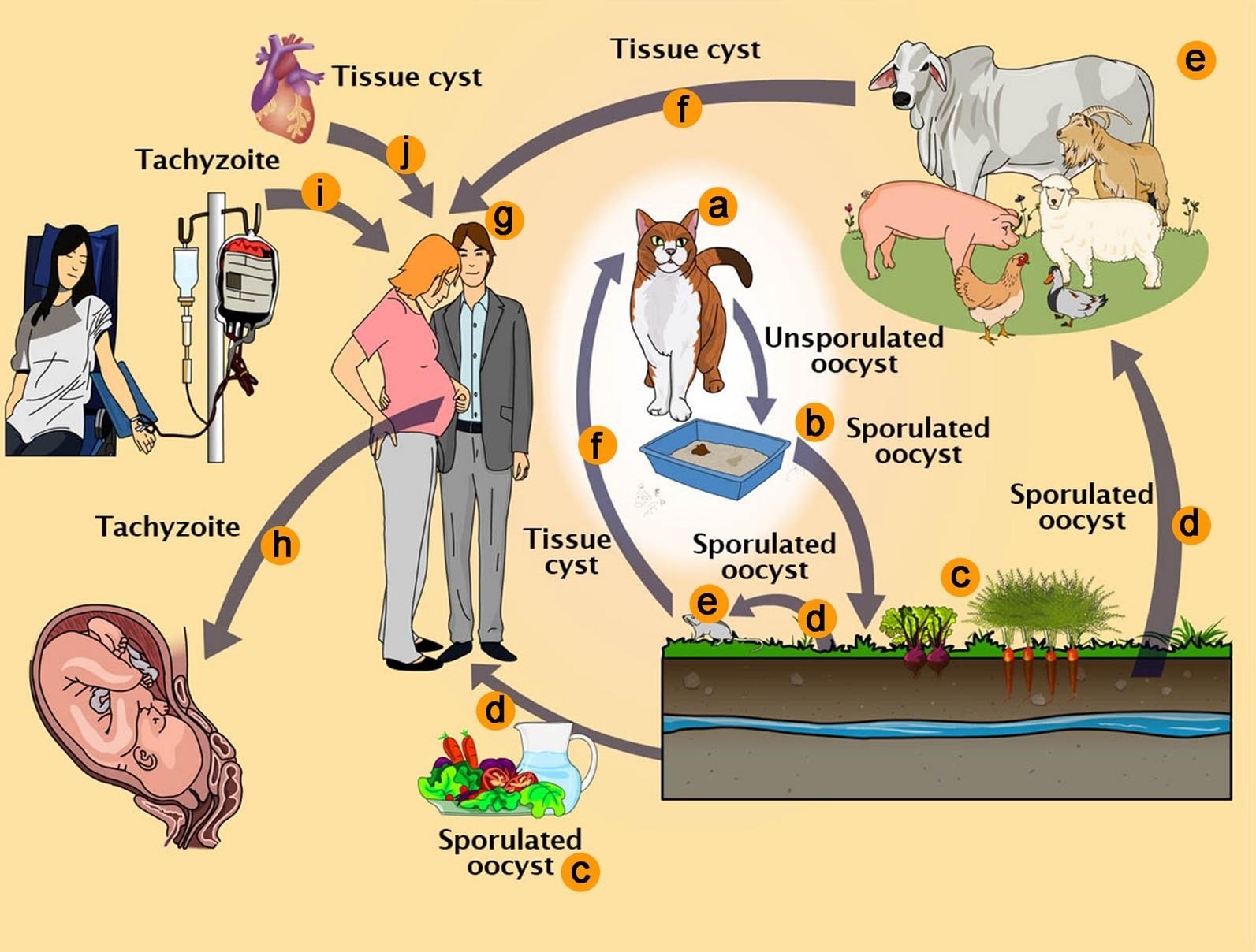
The lecture labels the transmission diagram from a to j, and states that the pathways which infect humans are d, f, h, i and j:

  • a. Feline definitive host (cat).
  • b. Unsporulated oocysts in cat faeces.
  • c. Food contaminated with sporulated oocysts.
  • d. Oocysts may be ingested by intermediate hosts via water or raw vegetables.
  • e. Intermediate hosts (e.g. cattle, sheep, poultry and swine).
  • f. Ingestion of tissue cysts in uncooked meat.
  • g. Intermediate hosts (humans).
  • h. Tachyzoites transmitted through the placenta to the foetus.
  • i. Transmission by blood transfusion, and organ transplant (j).

Grouped by what a person actually does, that is: swallowing oocysts from soil, water or unwashed vegetables; eating undercooked meat that contains tissue cysts; and three routes that bypass the gut entirely, the placenta, a transfusion and a transplanted organ.

Toxoplasma gondii transmission diagram lettered a to j: cat shedding unsporulated oocysts (b) that sporulate (c), sporulated oocysts ingested via water or vegetables (d) by intermediate hosts (e) or humans, tissue cysts in meat eaten by cat (f) or human (g), tachyzoites crossing the placenta (h), transfusion (i) and organ transplant (j) into humans

4. Life cycle of *Toxoplasma gondii* in the cat

note

With the meat of the prey, the cat ingests tissue cysts containing bradyzoites. This transmission route, in which the parasite uses a predator-prey relationship between two hosts, is called trophic.

Then, in order:

  • The cyst wall is digested in the stomach and intestines, liberating the bradyzoites.
  • Bradyzoites invade intestinal epithelial cells (enterocytes).
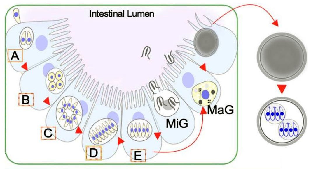
  • In the enterocytes bradyzoites divide by schizogony giving rise to merozoites (A to E in the drawing).
  • Merozoites differentiate into macro (MaG) and micro (MiG) gametes.
  • Microgametes penetrate cells containing macrogametes to fertilise them. This produces zygotes released as unsporulated oocysts.
  • In the environment, they undergo meiosis and one mitosis and become sporulated, containing 2 sporocysts, each with 4 sporozoites.

So one oocyst carries eight sporozoites, and it becomes infective only after it has spent time outside the cat.

Toxoplasma life cycle in the cat intestinal epithelium: bradyzoites (A) divide by schizogony through stages B to E into merozoites, which differentiate into microgametes (MiG) and macrogametes (MaG), fertilisation gives an unsporulated oocyst that sporulates outside the host into two sporocysts each with four sporozoites

Oocysts, tissue cysts and merozoites as they actually look

note

The lecture shows the stages photographed rather than drawn.

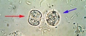
An unsporulated oocyst and a sporulated oocyst are shown side by side in a faecal preparation, the sporulated one already containing its two sporocysts. A short film of the same material runs oocyst, excystation, tachyzoites, host cell invasion in sequence, the tachyzoites visible as small crescents swarming over a cultured cell, and another clip follows the divisions inside the cat's intestinal epithelium.

Merozoites in cat intestine are shown in a histological section, filling the enterocytes. A second section is captioned Toxoplasma (arrows) seen in the lung of a pig, which is an intermediate host.

In brain tissue, tissue cysts are labelled, each packed with bradyzoites, at a scale of 20 μm; a further section shows one such cyst lying inside a skeletal muscle fibre.

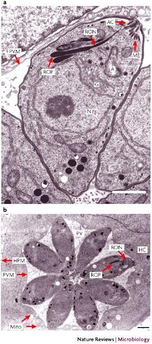
Electron micrographs of the tachyzoite inside its vacuole are labelled AC (apical complex), ROP (rhoptries), RON (rhoptry necks), MJ (moving junction), PVM (parasitophorous vacuole membrane), PV (parasitophorous vacuole), HPM (host plasma membrane), HC (host cell), N (nucleus), A (apicoplast), G (Golgi) and Mito (mitochondria). One panel catches a parasite halfway through the host cell membrane at the moving junction; another shows eight daughter cells arranged as a rosette inside a single parasitophorous vacuole.

Electron micrographs of Toxoplasma gondii tachyzoites, panel a a single parasite entering a host cell labelled AC apical complex, ROP rhoptries, RON rhoptry necks, MJ moving junction, PVM parasitophorous vacuole membrane, N nucleus, A apicoplast, G Golgi, panel b eight daughter cells arranged as a rosette in one vacuole labelled PV, HPM host plasma membrane, HC host cell, Mito mitochondria
Light micrograph of a faecal preparation with an unsporulated oocyst, red arrow, next to a sporulated oocyst already showing its two sporocysts, blue arrow

5. Life cycle of *Toxoplasma gondii* in the intermediate host

note

Tachyzoites: rapid growth and division. Soon, the immune response inactivates the parasites, and they are turned into bradyzoites.

Bradyzoites: clusters enclosed by irregular wall (called cysts or pseudocysts); the cysts can be developed in any tissue, but mainly in muscle and nervous tissue.

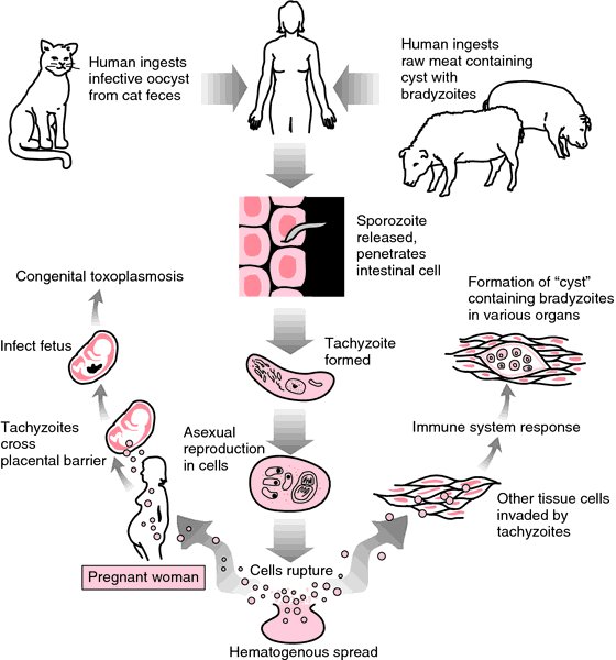
The diagram of the cycle in a human runs as follows. A person ingests an infective oocyst from cat faeces, or raw meat containing a cyst with bradyzoites. The sporozoite is released and penetrates an intestinal cell; the tachyzoite is formed; asexual reproduction in cells follows until the cells rupture and there is haematogenous spread. Other tissue cells are invaded by tachyzoites; the immune system responds, and there is formation of "cysts" containing bradyzoites in various organs. In a pregnant woman, tachyzoites cross the placental barrier and infect the foetus, which is congenital toxoplasmosis.

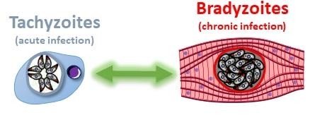
The lecture's summary picture pairs the two forms directly: tachyzoites, acute infection, drawn as a rosette inside a vacuole in a single cell, against bradyzoites, chronic infection, drawn as a dense cyst inside a muscle fibre.

Toxoplasma life cycle in the intermediate host: human ingests oocyst from cat faeces or raw meat with bradyzoite cysts, sporozoite penetrates intestinal cell, tachyzoite forms and spreads haematogenously after cells rupture, tissue cysts with bradyzoites form under immune pressure, and in a pregnant woman tachyzoites cross the placenta causing congenital toxoplasmosis
Paired diagram of tachyzoites, acute infection, as a rosette inside a vacuole in one cell, against bradyzoites, chronic infection, as a dense cyst inside a striated muscle fibre

*Toxoplasma gondii* in host cells

note

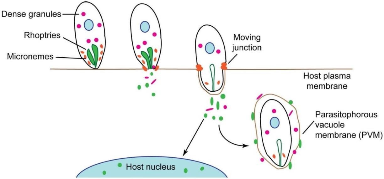
LEFT: Toxoplasma attaches to the host cell membrane and secretes microneme contents. The parasite actively penetrates the host cell membrane, secreting from rhoptries and dense granules as it enters the cell and establishes the parasitophorous vacuole, derived primarily from the host cell plasma membrane.

RIGHT: The vacuole pinches off from the host cell plasma membrane and migrates towards the nucleus, recruiting mitochondria and endoplasmic reticulum, and becomes associated with cytoskeletal components. The parasite divides exponentially and then egresses, resulting in the lysis of the host cell.

The left-hand drawing labels dense granules, rhoptries and micronemes in the parasite, the host plasma membrane, the moving junction where the two membranes meet, the parasitophorous vacuole membrane (PVM), and the host nucleus towards which secreted proteins travel. The right-hand drawing runs the cycle as attachment and invasion, intracellular replication, egress and back to extracellular tachyzoites.

Note the order of secretion, because it is the sequence of the invasion itself: micronemes first, to stick; rhoptries and dense granules next, to build the vacuole and to reprogramme the cell.

Sequence of Toxoplasma attaching to and crossing the host plasma membrane, labelled dense granules, rhoptries, micronemes and a moving junction, secreted proteins travelling to the host nucleus while the parasite becomes wrapped in the parasitophorous vacuole membrane (PVM)
Cycle diagram of attachment and invasion of a host cell forming a PV, intracellular replication of tachyzoites inside the vacuole, egress by host cell lysis, and extracellular tachyzoites seeking a new host cell

Tachyzoites, bradyzoites and the switch between them

note

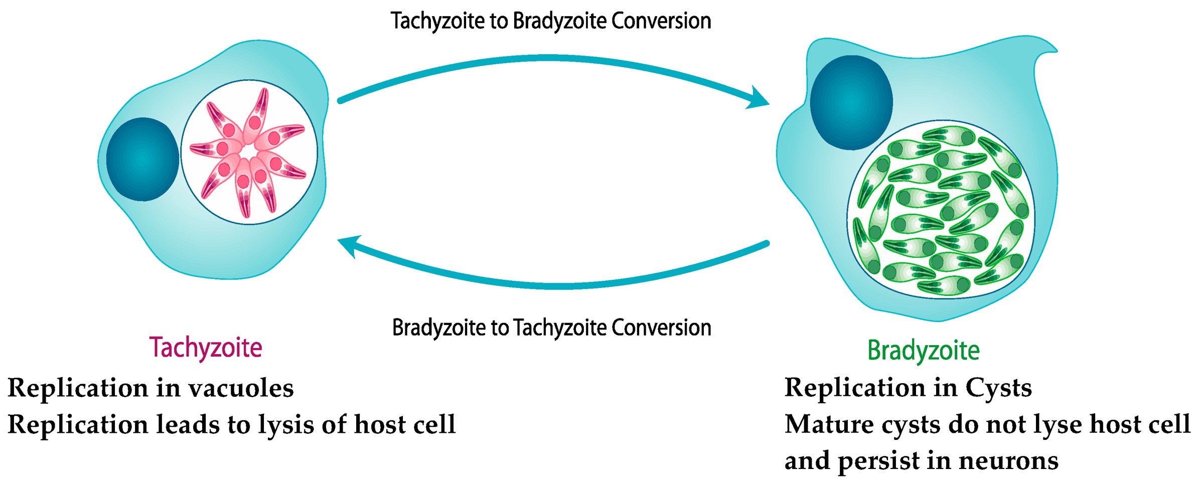
Tachyzoites divide rapidly in the vacuole and lyse the host cell, most often 36 to 72 hours after infection.

Bradyzoites divide slowly or are quiescent. They are found in intracellular tissue cysts that do not lyse the host cells and remain there for the life of the host.

Once developed, the immune response easily destroys the tachyzoites, but the bradyzoites are latent and persist. Later, from time to time, some bradyzoites turn back into tachyzoites. A healthy immune system will kill them, but if the immunity is weakened, they will survive and the disease will revert to its acute phase.

The lecture draws this as a two-way arrow: tachyzoite to bradyzoite conversion and bradyzoite to tachyzoite conversion, with the tachyzoite characterised by replication in vacuoles and replication leading to lysis of the host cell, and the bradyzoite by replication in cysts and by mature cysts that do not lyse the host cell and persist in neurons.

Tachyzoite to bradyzoite and bradyzoite to tachyzoite conversion diagram: tachyzoite, pink rosette in a vacuole, replication in vacuoles and lysis of host cell, versus bradyzoite, green cyst, replication in cysts, mature cysts do not lyse host cell and persist in neurons

The entire life cycle in one picture

note

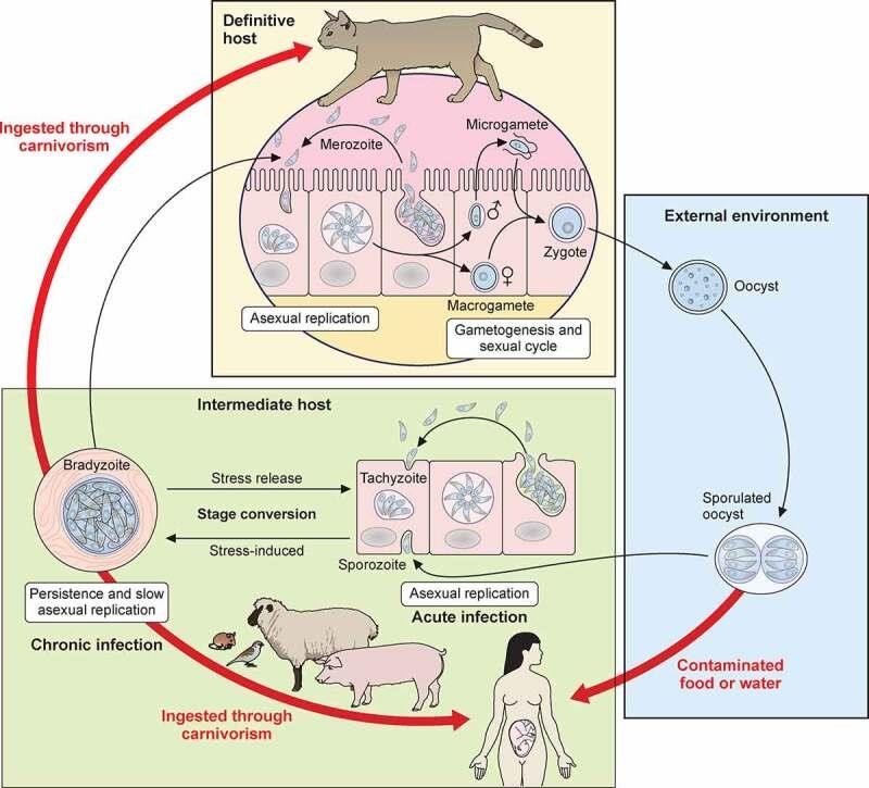
The summary diagram puts the three compartments together. In the definitive host, the cat, the parasite runs asexual replication in the intestinal epithelium, giving merozoites, then gametogenesis and the sexual cycle, giving microgametes and macrogametes and then a zygote. In the external environment the oocyst becomes a sporulated oocyst. In the intermediate host the sporozoite starts asexual replication as the tachyzoite of acute infection; stage conversion is stress-induced in one direction and by stress release in the other, and the bradyzoite represents persistence and slow asexual replication in chronic infection. Two red arrows mark how the parasite is acquired: ingested through carnivorism, and from contaminated food or water.

Full Toxoplasma life cycle across three compartments: definitive host cat with asexual replication and gametogenesis producing a zygote, external environment where the oocyst sporulates, and intermediate host where stress-induced and stress-release stage conversion links the tachyzoite of acute infection to the bradyzoite of chronic infection, red arrows for ingestion through carnivorism and through contaminated food or water

6. Toxoplasmosis

note

Toxoplasmosis may cause flu-like symptoms in some people, but most affected people never develop signs and symptoms.

In the chronic phase, the immune system eliminates the tachyzoites and leaves only inactive tissue cysts; these remain for the rest of life. If for some reason the immunity ceases to be effective, the bradyzoites can turn into tachyzoites with impunity, and thus acute toxoplasmosis occurs again.

Who is at risk for serious problems during severe toxoplasmosis?

  • Fetuses and infants born to mothers who are newly infected during or just before pregnancy.
  • Persons with severely weakened immune system: individuals with AIDS, those taking certain types of chemotherapy, those who have recently received an organ transplant and very aged people.

In these patients, the immunity is unable to keep the bradyzoites inactive and they turn into tachyzoites.

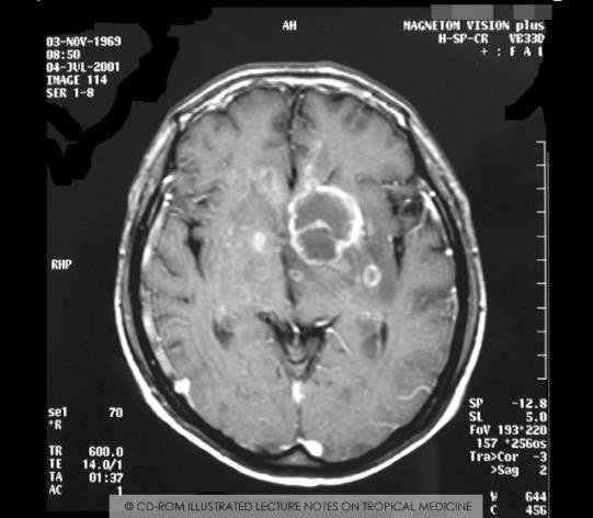
The lecture illustrates the point with an MRI of the brain showing cerebral toxoplasmosis in an AIDS patient, in which a reactivated focus appears as a ring-shaped lesion.

Two things follow for practice. The dangerous patient is not the one who is newly infected but the one whose immunity fails years later; and the dangerous new infection is the one acquired in pregnancy, because it is the tachyzoite that crosses the placenta.

Axial MRI of the brain in an AIDS patient with cerebral toxoplasmosis, showing a ring-enhancing reactivated lesion in the right hemisphere

7. *Toxoplasma* changes the behaviour of its host

note

Toxoplasma changes the behaviour of its intermediate host to its advantage:

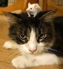
  • change in the behaviour of the infected rodent: it ceases to be afraid of the smell of cat urine and even of the cat itself;
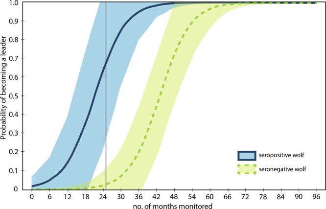
  • leadership qualities in wolves - infected wolves become more willing to take risks and are therefore more likely to become pack leaders.

The wolf data are plotted in the lecture as the probability of becoming a leader against the number of months monitored: the curve for seropositive wolves rises steeply from the start and is essentially complete by about 30 months, while the curve for seronegative wolves does not begin to rise until about 36 months.

The rodent case is the parasite's own interest made visible. A rat that is not afraid of cats is a rat that gets eaten, and being eaten by a cat is exactly how Toxoplasma reaches the only host in which it can reproduce sexually.

A white mouse sitting fearlessly on the head of a cat, illustrating the loss of fear of cats in a Toxoplasma-infected rodent
Graph of probability of becoming a pack leader against months monitored: seropositive wolves rise steeply and plateau by about 30 months, seronegative wolves do not begin rising until about 36 months

The most important things to know

note

  • Apicomplexa (Sporozoa) are all intracellular parasites of animals, defined by the apical complex at one pole: conoid, apical ring of contractile actin fibres, rhoptries (enzymes for penetration), micronemes (proteins binding host cell receptors) and dense granules (proteins that modify host protein trafficking and maintain the parasitophorous vacuole). The apicoplast is a relict, nonphotosynthetic plastid.
  • The parasite builds a parasitophorous vacuole and forces the host cell to gather its ER, Golgi and mitochondria around it.
  • Because the apical complex is hard to inherit, division is either endodyogeny (two daughters inside an intact mother, as in Toxoplasma) or endopolygeny (mother loses polarity, many daughters); endopolygeny with early nuclear division is schizogony, as in *Plasmodium*.
  • Reproduction alternates merogony (schizogony) - gamogony - sporogony; only the zygote is diploid; sporozoites are the infectious stage for humans.
  • *Toxoplasma gondii* is an obligate intracellular parasite of almost all warm-blooded animals and one of the commonest human parasites. Definitive hosts are cats (Felidae), in whose intestine it reproduces sexually; intermediate hosts, including humans, support only asexual multiplication.
  • Its three infective stages are the tachyzoite (fast, early, acute), the bradyzoite (slow, in tissue cysts in muscle and nerve tissue, chronic) and the sporozoite (made only in cats, shed in faeces).
  • In the cat: tissue cysts eaten with prey (trophic transmission) - bradyzoites released - schizogony in enterocytes - merozoites - micro- and macrogametes - zygote - unsporulated oocyst in the faeces - sporulation outside the host into 2 sporocysts with 4 sporozoites each.
  • Humans are infected by oocysts in water or on raw vegetables, tissue cysts in undercooked meat, transplacentally, by transfusion and by organ transplant.
  • Tachyzoites lyse the host cell 36 to 72 hours after infection; bradyzoites in cysts do not, and persist for the life of the host. Reactivation follows loss of immunity.
  • Most infections are asymptomatic or flu-like. The people at risk are foetuses and infants of mothers newly infected in or just before pregnancy and the severely immunosuppressed (AIDS, chemotherapy, recent transplant, the very old), in whom cerebral toxoplasmosis can occur.
  • The parasite alters host behaviour: infected rodents lose their fear of cat urine and of cats, and infected wolves take more risks and more often become pack leaders.
Full Toxoplasma life cycle across three compartments: definitive host cat with asexual replication and gametogenesis producing a zygote, external environment where the oocyst sporulates, and intermediate host where stage conversion links the tachyzoite of acute infection to the bradyzoite of chronic infection

All 105 Medical Biology topics · exam study support, not clinical guidance. SuperMed is not affiliated with the Medical University of Sofia.

Start free. No card needed.

Every account starts free, with Medical Ethics, Urology and Computer Science open in full and free topics in Cytology, Latin Language, Medical Chemistry, Medical Physics, Medical Biology, Anatomy, Biophysics, Medical Psychology, MCS (Disaster Medicine), Physiology, Biochemistry, Social Medicine, Medical Genetics, Pharmacology, Pathophysiology, Microbiology, Internal Medicine, Nephrology, Cardiology, Pulmonology, Gastroenterology, Internal Medicine (Clinical), Obstetrics and Gynecology, Physiotherapy and Rehabilitation, Dental Materials, Preclinics of Prosthetic Dental Medicine, Medical Psychology (Dental Medicine), SMME (Social Medicine & Medical Ethics), Preclinics of Pediatric Dental Medicine, Hygiene and Epidemiology, General Surgery, Preclinics of Conservative Dental Medicine, Preclinics of Oral and Maxillofacial Medicine, Dental Public Health, Dental Prophylaxis, Diagnostic Imaging, Dental Allergology, Dental Implantology, Physiotherapy, Cytology & Histology, Medical Chemistry, Medical Physics, Anatomy I, Biophysics and Medical Biology. Super opens the rest. Super is €14.99 a month.

Create a free account