Medical Biology · Year 1 · Medical University of Sofia

04

Recognition and self-assembly of macromolecules. Cytoskeleton

Free notes for topic 04 of the Medical Biology syllabus, open without an account. Written by a senior student against the syllabus question and checked line by line by a second student before publishing. How content is made

Updated

What this topic covers

note

This topic draws on two lectures. From the self-assembly lecture: how biopolymers are recognised and self-assembled; examples of self-assembly of nucleic acid complexes, of small and large quaternary protein complexes, of nucleoprotein complexes and of lipoprotein complexes; what lipid rafts are and how they form; and how medicine benefits from the self-assembly of biopolymers. From the cytoskeleton lecture: the three types of cytoskeletal fibre, how each assembles, the motor proteins that use them, centrioles, cilia and flagella, and the diseases and drugs that act on the system.
Intermediate filament assembly: monomer with N- and C-terminal heads, coiled-coil dimer, staggered tetramer, unit-length filament (ULF) and the mature intermediate filament

1. What self-assembly of macromolecules is

note

Life would not have been possible without the ability of macromolecules to recognise each other specifically and to bind spontaneously, forming supramolecular complexes.

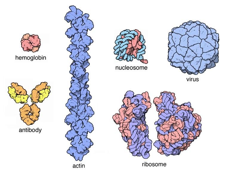
Self-assembled macromolecular complexes shown side by side: haemoglobin, an antibody, an actin filament, a nucleosome, a ribosome and a virus capsid

This recognition is based on spatial affinity: correspondence between the two participating molecules in shape and surface charges. The comparison is to a lock and key.

The self-assembled complexes are held together by non-covalent bonds - ionic, hydrogen, Van der Waals, hydrophobic. Though every single bond is weak, their large number guarantees reliable binding.

The most important specific interactions are between:

  • nucleic acids;
  • proteins;
  • proteins and nucleic acids;
  • proteins and lipids.

2. Complementary binding of nucleic acids

note

Complementary recognition and binding of nucleotides allows replication and transcription, the informational basis of life.

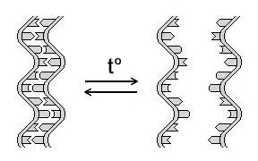
Heating above 70 °C causes denaturation of DNA, that is, separation of the two strands, but the process is reversible.

When the solution is cooled, DNA strands find their complementary partners and pair by hydrogen bonds, restoring the double-stranded structure. This process is called annealing, and it has two named cases:

Denaturation and renaturation of double-stranded DNA on heating (t°) and cooling, the strands shown unlabelled
  • If the paired strands derive from one and the same molecule, the annealing is called renaturation or reassociation.
  • Pairing is also possible between two strands from different DNA molecules with full or partial complementarity, for example during homologous recombination. Annealing of this type is often called hybridisation.

Why the base ratio decides the strength of the bond

note

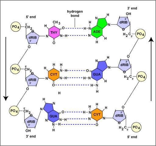
  • A-T is two hydrogen bonds.
  • G-C is three hydrogen bonds.

The consequence is a design rule the genome obeys:

  • DNA regions that need a stronger connection between the two strands require more G-C pairs;
  • DNA regions that need a weaker connection require more A-T pairs.
Hydrogen bonding in DNA base pairs: thymine to adenine with two hydrogen bonds and cytosine to guanine with three, sugar-phosphate backbone labelled dRIB and PO4

RNAs also hybridise

note

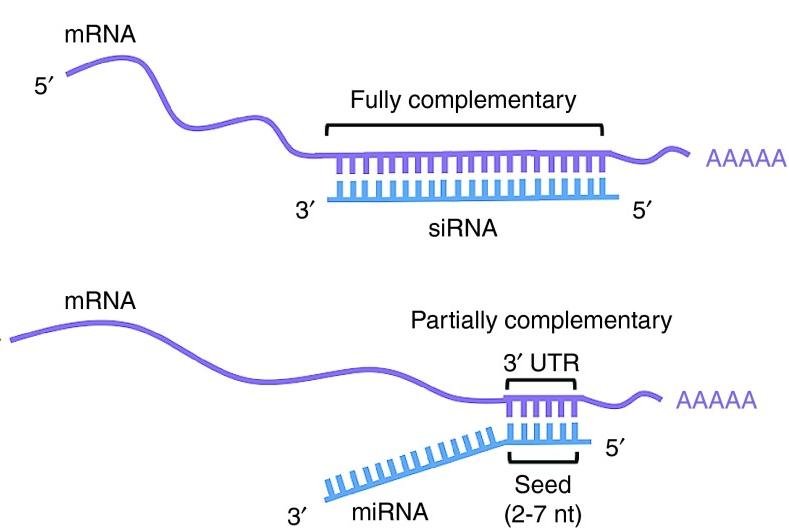
The same rule operates outside DNA. The example is the hybridisation of microRNA (miRNA) and small interfering RNA (siRNA) with the 3' untranslated region of an mRNA - a recognition event that decides whether that message is translated at all.

mRNA hybridising with a fully complementary siRNA and with a partially complementary miRNA at the 3' UTR, the seed region of 2 to 7 nt labelled

3. Quaternary structure of proteins

note

Proteins recognise and bind other proteins, or other molecules of the same protein, in the formation of quaternary structure.

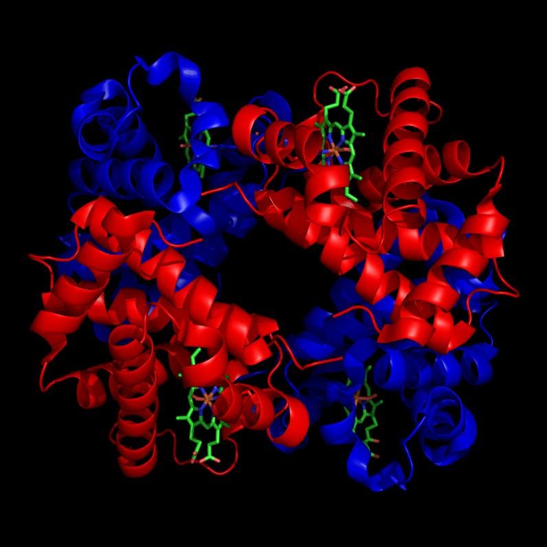
Small quaternary complexes, such as haemoglobin, consist of two or several subunits. In these complexes the number of subunits is fixed. The whole is often called a molecule, even when it is not supported by covalent bonds.

Haemoglobin tetramer: two alpha and two beta globin subunits, red and blue, each carrying a green haem group

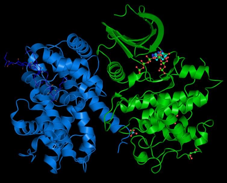
Many enzymes are quaternary complexes including catalytic and regulatory subunits. For example, the cyclin-dependent kinases (CDKs) which control the cell cycle are activated by regulatory subunits called cyclins.

A cyclin-dependent kinase, blue, bound to its regulatory cyclin subunit, green, a two-subunit quaternary enzyme complex

Large quaternary complexes

note

Large quaternary complexes are formed by many protein subunits, and the number is often indefinite. They include:

  • cytoskeletal structures;
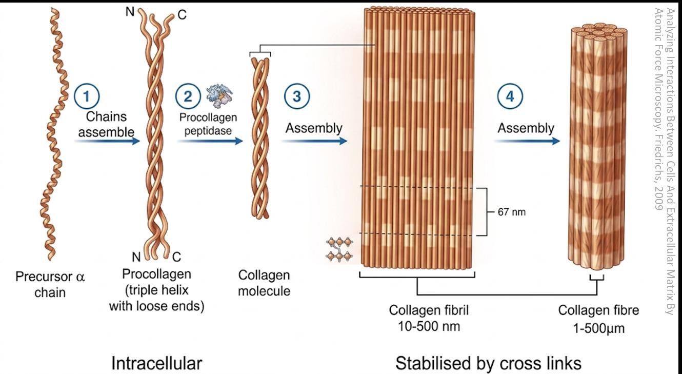
  • fibres of the extracellular matrix, for example the self-assembly of a collagen fibre;
  • capsids of some viruses, partially assembling before the nucleic acid enters.
Self-assembly of a collagen fibre: precursor alpha chain, procollagen triple helix, collagen molecule, then collagen fibril and collagen fibre stabilised by cross links

The distinction from the small complexes is worth holding: a fixed number of parts makes a molecule, an indefinite number makes a structure.

Actin and myosin filaments in muscle

note

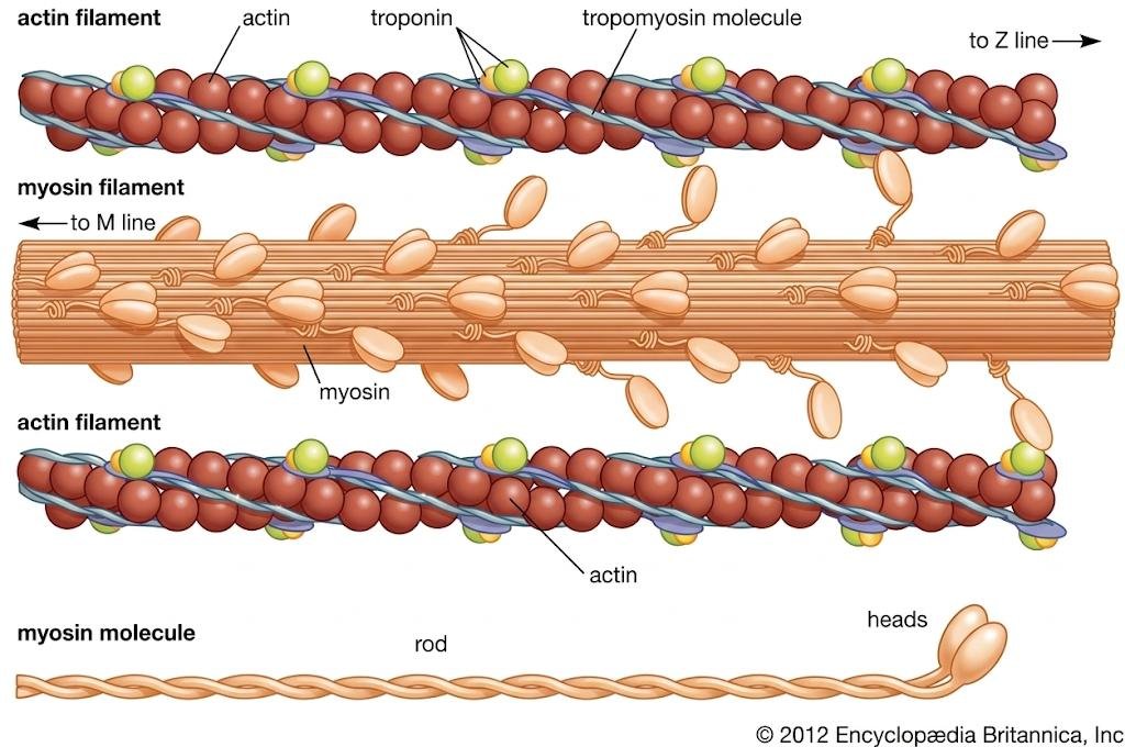
The muscle filaments are the standard example of large quaternary assembly, and two families of additional proteins ride on them:

  • Tropomyosins integrate into the actin filaments and have a role in regulating their action.
  • Troponins also regulate muscle contraction.
Actin and myosin filaments of a muscle sarcomere, labelled actin, troponin, tropomyosin molecule, myosin filament, and the myosin molecule's rod and heads

4. Nucleoproteins

note

Nucleic acids and proteins bind together, forming complexes called nucleoproteins:

  • deoxyribonucleoproteins (DNP) for DNA;
  • ribonucleoproteins (RNP) for RNA.

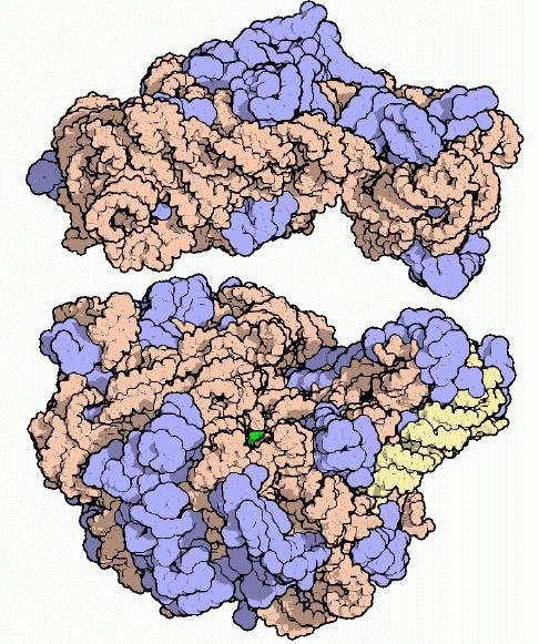
The examples that follow are the nucleosome, the ribosome, sperm chromatin, and - of course - the viruses.

The ribosome, a ribonucleoprotein: proteins in blue, and the large and small subunit rRNAs in peach and yellow

The nucleosome

note

Nucleosomes, about 10 nm in diameter, are DNA-protein complexes. They are separated by several dozen base pairs of linker DNA, and the array of nucleosomes along the DNA is called "beads on a string".

The histones involved are H2A, H2B, H3 and H4.

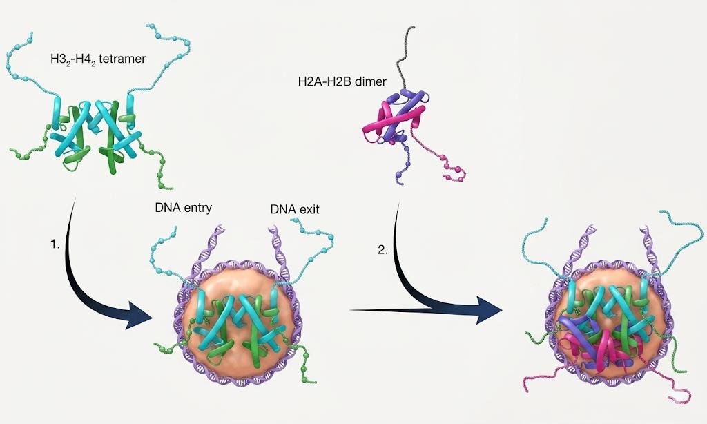
The order of assembly in vivo matters: the H3-H4 tetramers form and bind DNA first, and then two H2A-H2B dimers are added to complete the nucleosome.

Nucleosome assembly: an H3-H4 tetramer binds DNA first at the entry and exit sites, then two H2A-H2B dimers are added to complete the nucleosome

The ribosome

note

The ribosome is the most important RNP.

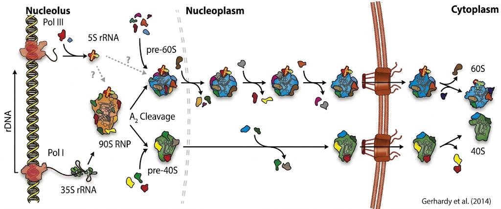
Ribosome biogenesis is a multistep process. In bacteria it requires the coordinated synthesis, processing and folding of three rRNAs, and the translation, folding, modification and binding of about 50 ribosomal proteins.

Eukaryotic ribosome maturation, in outline: during transcription the new rRNA binds early trans-acting factors, and the 35S rRNA forms the 90S preribosome. Cleavage at the A2 site separates the pre-60S subunit and the pre-40S subunit, which undergo independent maturation pathways in the nucleoplasm and eventually in the cytoplasm. Final maturation in the cytoplasm allows the two subunits to join and form the functional 80S ribosome. The details of this pathway are an illustration and are not to be learned.

Eukaryotic ribosome maturation from nucleolus through nucleoplasm to cytoplasm: the 90S RNP undergoes A2 cleavage into pre-60S and pre-40S, ending as mature 60S and 40S subunits

Sperm chromatin

note

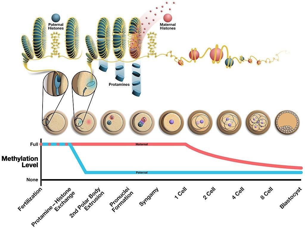
During sperm differentiation, histones are replaced by protamines. They package DNA very tightly into toroid-shaped structures.

In the zygote, the protamines of the male pronucleus are replaced by histones - the packaging is reversed as soon as the genome has to be read again.

Histone to protamine exchange in sperm chromatin and its reversal after fertilisation, with maternal and paternal DNA methylation tracked from fertilisation to the blastocyst

Self-assembly of the tobacco mosaic virus

note

This is the clearest demonstration that the nucleic acid can direct the shape of the protein shell.

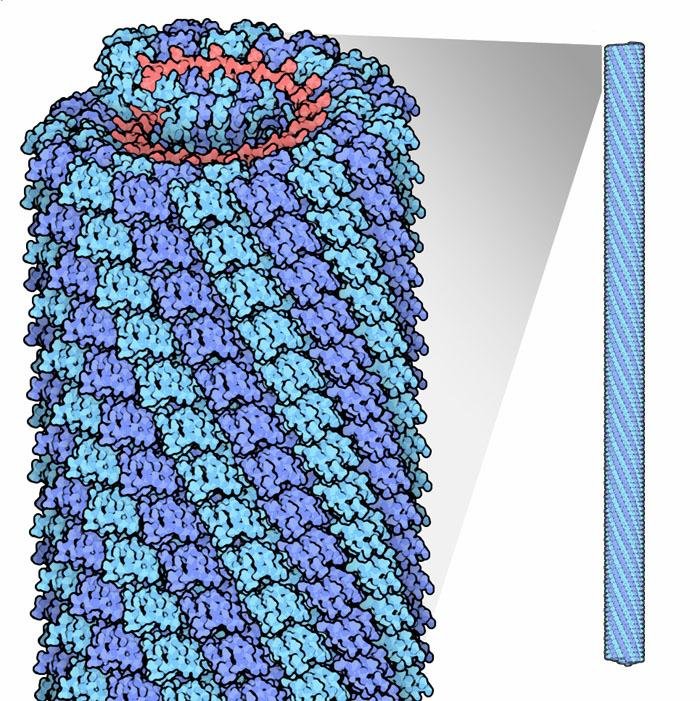
  1. First, capsid proteins form a two-layer disk.
  2. Then RNA joins. This forces the protein molecules to rearrange into a helix.
  3. More protein subunits stack on this structure, and the cylindrical capsid is elongated until the RNA is covered.

The virus therefore measures its own genome: the shell is exactly as long as the thing inside it.

Self-assembly of the tobacco mosaic virus capsid: the helical protein coat elongating around the RNA core, with a cross-section of the tube shown at right

Self-assembly of phages

note

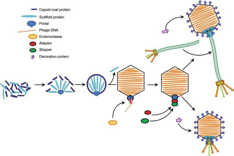
  1. Multiple copies of the capsid/scaffold complex bind the portal protein to form the procapsid.
  2. The scaffold proteins are ejected, and DNA is packaged into the procapsid, which expands to the size of the mature capsid.
  3. The head completion proteins, the stopper and the adaptor, bind to the portal complex, preventing DNA leakage.
  4. Decoration proteins bind to the capsid.
  5. The tail, assembled separately or after DNA packaging, is attached, and the final infectious phage is produced. In some cases a preassembled tail attaches.
Phage self-assembly: capsid coat and scaffold proteins form the procapsid around the portal, DNA is packaged, adaptor, stopper and decoration proteins are added, then the tail attaches

5. Proteins and lipids: the biomembranes

note

Membrane proteins are vital components of biological membranes as transporters, receptors and structural supports.

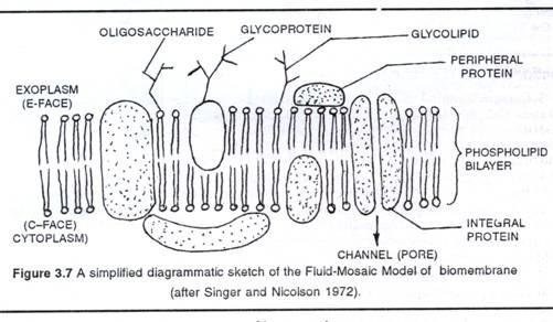
The original idea for the structure of biomembranes was the fluid mosaic model of Singer and Nicolson (1972). One correction is worth noting: in fact, very few proteins stay half-immersed in the lipid bilayer, as that model drew them.
Singer and Nicolson fluid mosaic model of the biomembrane: phospholipid bilayer with integral and peripheral proteins, a channel pore, and glycoprotein and glycolipid oligosaccharides

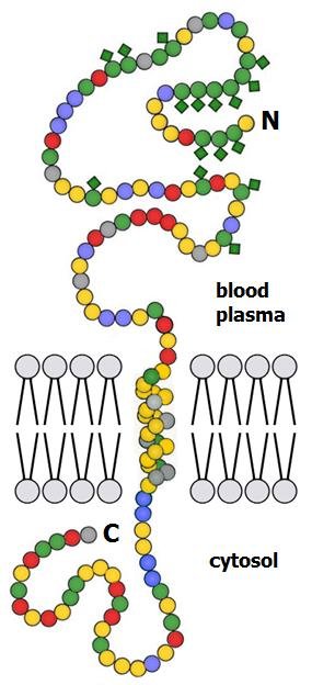
Typical membrane proteins have at least one transmembrane domain of about 20 hydrophobic amino acids. It forms an alpha helix crossing the lipid bilayer, anchoring the protein while the polypeptide chain is still being synthesised. Some proteins are instead anchored to the membrane by being bound to a lipid. Glycophorin A is the standard illustration.

Glycophorin A, a single-pass transmembrane glycoprotein with its N-terminus in the blood plasma and C-terminus in the cytosol, amino acid residues unlabelled

Cholesterol controls membrane fluidity

note

Cholesterol influences membrane fluidity by forming strong interactions with phospholipids.

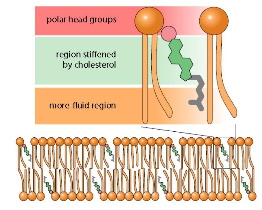
Like the phospholipids, cholesterol in the bilayer orients with its polar head group facing the aqueous environment and its non-polar region facing the membrane interior.

One of cholesterol's functions is to reduce the fluidity of the membrane, which it does using its rigid steroid ring structure. But the effect is temperature-dependent, and it works in both directions:

  • At high temperatures, these interactions stiffen the membrane and interfere with phospholipid mobility.
  • At low temperatures, the flexible, non-polar tail of cholesterol interferes with the tight packing of adjacent phospholipid chains.

Cholesterol is therefore a buffer rather than simply a stiffener: it opposes whatever the temperature is trying to do.

Cholesterol stiffening a phospholipid bilayer: polar head groups, the region stiffened by cholesterol, and the more fluid region, with cholesterol molecules along the membrane

Lipoproteins

note

Proteins bind to lipids outside membranes as well, forming lipoproteins. Their main function is the transport and turnover of lipids.

In our blood:

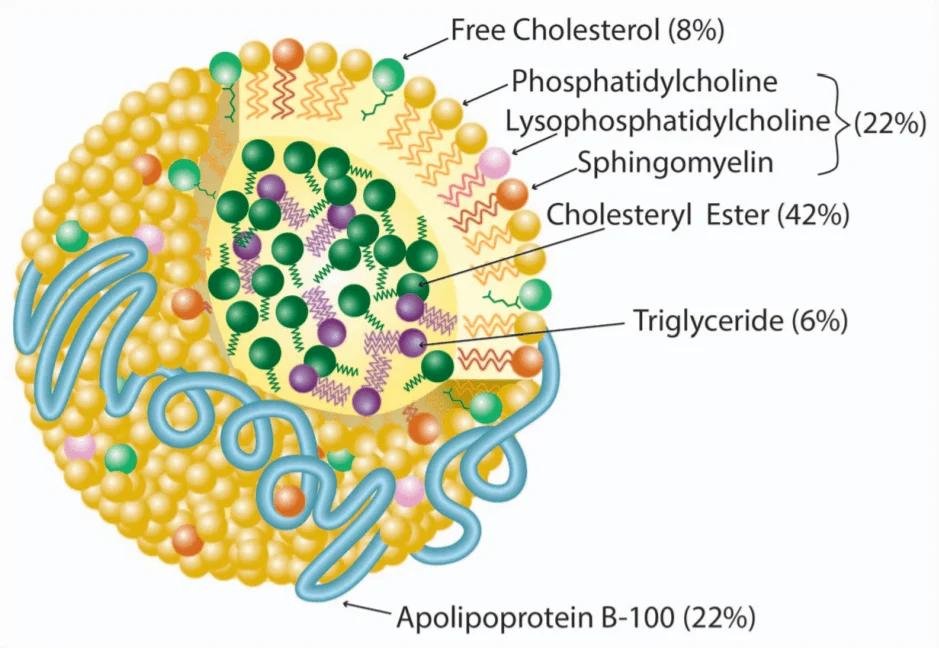
  • low-density lipoproteins (LDL), the "bad cholesterol", carry cholesterol from the liver to other tissues;
  • high-density lipoproteins (HDL), the "good cholesterol", transport it from the tissues to the liver.
Composition of an LDL particle: free cholesterol, phosphatidylcholine, lysophosphatidylcholine and sphingomyelin, a cholesteryl ester and triglyceride core, and apolipoprotein B-100 on the surface
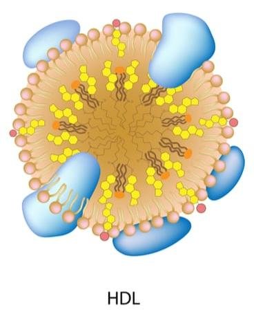
HDL particle: a phospholipid and apolipoprotein shell around a cholesteryl ester and triglyceride core, with more surface apolipoprotein than LDL

The protein components of these particles are called apolipoproteins. The structure of LDL and HDL particles is the same, but HDL contain more proteins on the surface.

The pathological role. If there is too much cholesterol included in LDL, not all of it can be used by the tissue. The remaining quantity accumulates on artery walls and causes atherosclerosis.

Lipid rafts

note

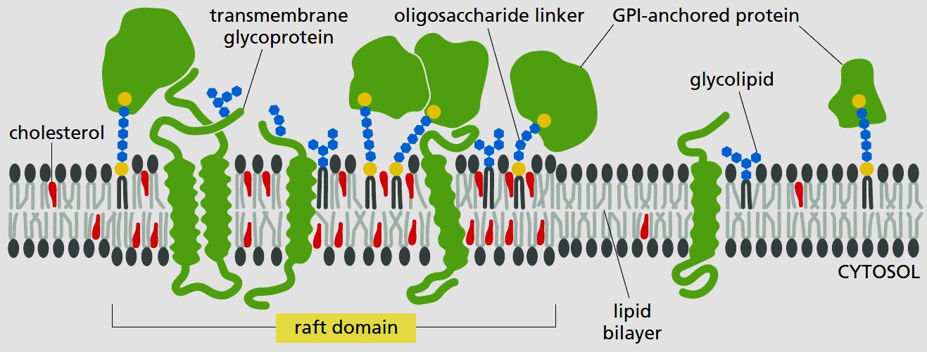
Lipid rafts are microdomains in the plasma membrane.

They contain combinations of glycosphingolipids, cholesterol and protein receptors.

GPI (glycosylphosphatidylinositol) anchors are relevant here: GPI are lipid anchors for many cell-surface proteins.

Lipid raft domain in the plasma membrane: cholesterol, transmembrane glycoprotein, oligosaccharide linker, GPI-anchored protein and glycolipid either side of the raft

6. When abnormal structure causes abnormal self-assembly

note

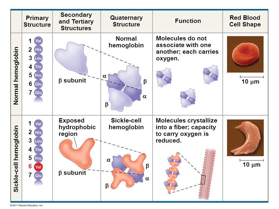
Haemoglobin is the example. A single amino acid change in the β-globin subunit leads to abnormal folding in its secondary, tertiary and quaternary structure. The assembly is totally wrong.

The change: valine replaces glutamate at position 6, on the surface of the beta chain.

The result: reduced capacity to carry oxygen, and an abnormal shape of the erythrocytes.

Normal versus sickle-cell haemoglobin: valine replacing glutamate at position 6, the exposed hydrophobic region, quaternary structure, and the resulting red cell shape

Melittin disturbs the self-assembly of the membrane

note

The venom of the hymenoptera is a mixture of low molecular weight chemicals, proteins, and small to medium peptides acting together. The venom produces prolonged pain, inflammation, and local tissue damage.

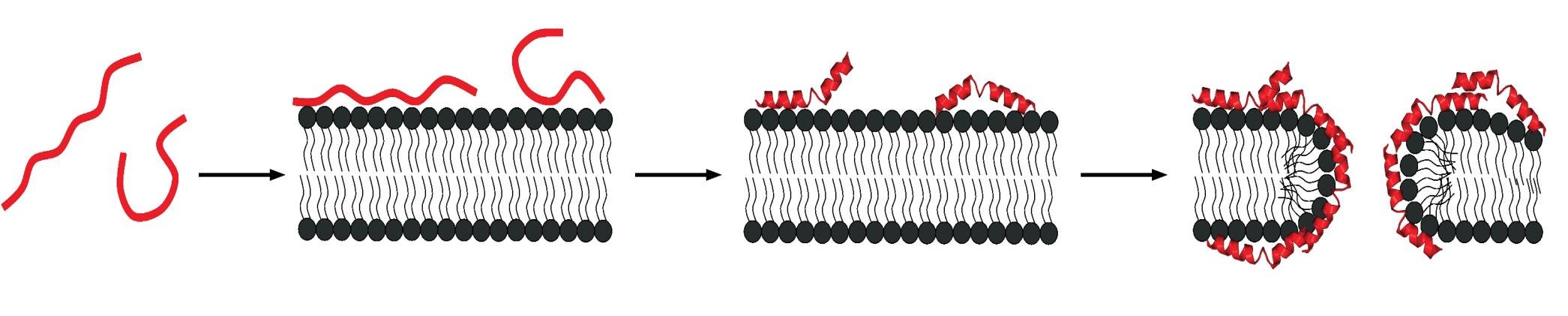
Melittin is the active molecule of apitoxin, bee venom. It forms small pores on the cell membrane. Once melittin binds to the membrane, toroid-shaped pores are formed, and the pore-forming effect causes the release of pro-inflammatory cytokines.
Melittin peptide monomers binding the plasma membrane, forming surface helices that assemble into toroid-shaped pores through the bilayer

Dystrophin

note

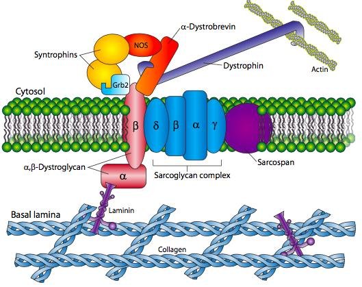
The cell membrane is unstable without protein contacts that connect the cytoskeleton and the extracellular matrix.

The protein dystrophin has that function in muscle cells, and mutation in this protein leads to muscular dystrophy.

Dystrophin-glycoprotein complex linking the actin cytoskeleton to the basal lamina: dystrophin, syntrophins, alpha-beta-dystroglycan, the sarcoglycan complex, sarcospan, laminin and collagen

7. How medicine benefits from self-assembly

note

Self-assembled complexes of natural macromolecules are used as an innovative approach to deliver medicines to a certain tissue, with great potential for precise medicine aimed at overcoming tissue barriers in order to reach specific target cells.

For example, the technique can be used for encapsulating drugs in nanoparticles, which are systematically released and precisely directed to targeted sites.

The advantages:

  • efficient drug transport;
  • reduced risk of side effects;
  • extended drug action time.

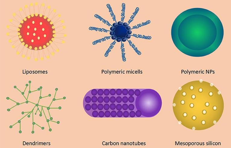
The natural structures used in drug delivery nanoparticles are biocompatible and biodegradable: polysaccharides such as hyaluronic acid, proteins such as collagen and silk fibroin, and membrane vesicles.

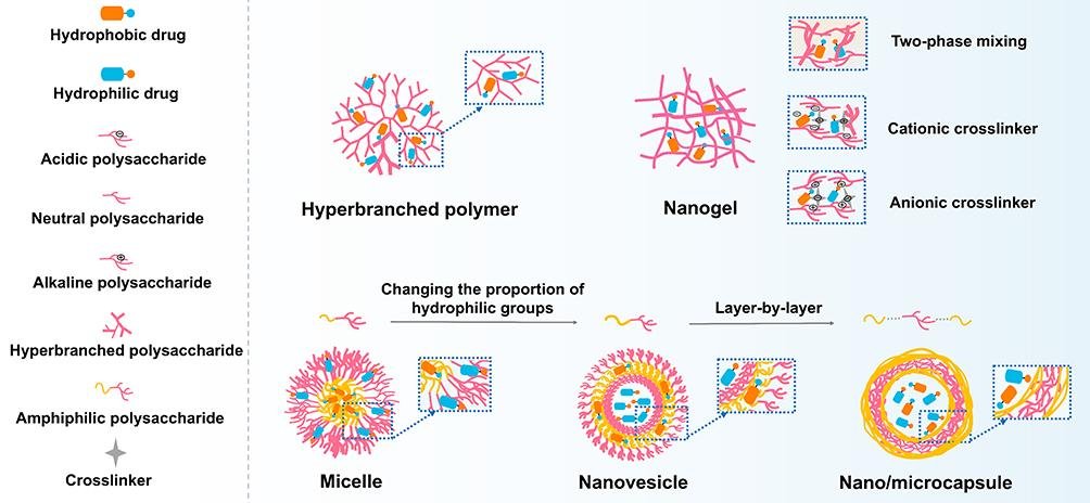
Types of drug delivery nanocarrier: liposomes, polymeric micelles, polymeric nanoparticles, dendrimers, carbon nanotubes and mesoporous silicon

Natural polysaccharide self-assembly, and how nanoparticles cross barriers

note

Some natural polysaccharides are hyperbranched polymers, such as plant glycogen. Phytoglycogen nanoparticles possess a high level of water retention, low viscosity, and exceptional stability of aqueous dispersion. They can effectively accommodate and disperse insoluble substances, improving their solubility and permeability.

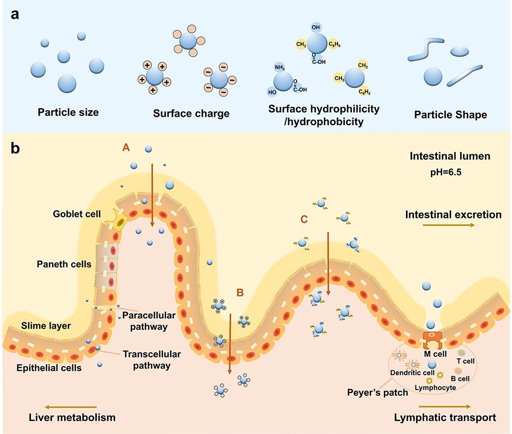
Why the particles get through. Self-assembled nanoparticles help medications penetrate the oral and gastrointestinal barrier because they have appropriate:

Nanoparticle properties that let them cross barriers, by size, surface charge and hydrophilicity, and their route through the intestinal epithelium by paracellular, transcellular and M cell pathways to lymphatic transport
  • size;
  • shape;
  • electric charge;
  • hydrophilicity or hydrophobicity.

Nano-platforms of this kind are used for the development of different nano-drugs and delivery systems in clinical cancer care, and the same properties let nanoparticles overcome a range of other biological barriers in precision medicine.

Nanocarrier types grouped as lipid-based, self-assembled and inorganic, including liposome, nanogel, micelle, virosome, polymer and metal nanoparticles

8. The cytoskeleton: the three main types of fibre

note

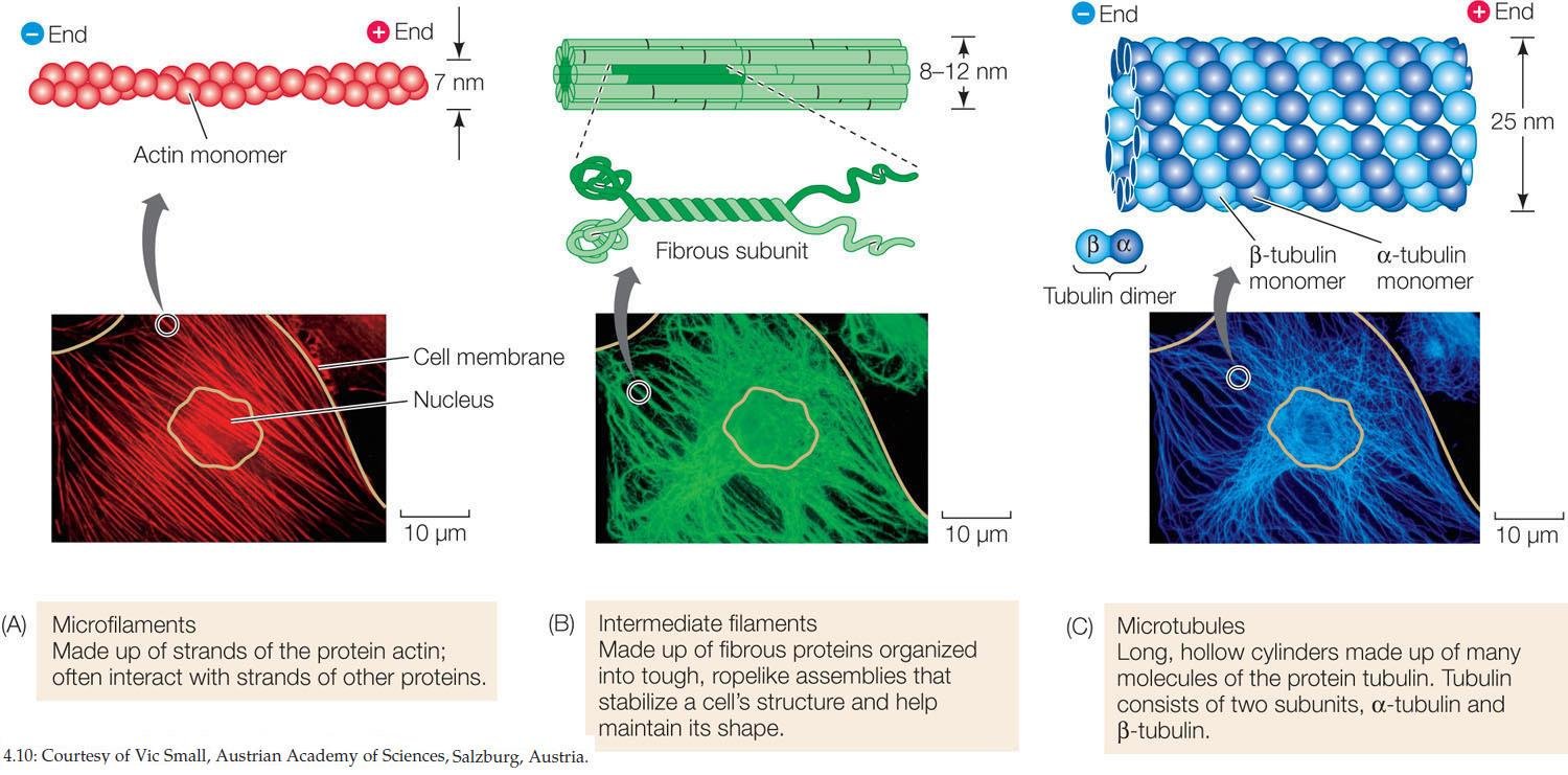
The cytoskeleton includes microtubules, microfilaments and intermediate filaments.

The first two systems are found in all eukaryotes, while intermediate filaments are characteristic of animals.

TypeDiameterStructureProteins
Microfilaments6 nmHelix appearing as if made of 2 protofilamentsActin (globular)
Intermediate filaments10 nmBundle of tetramers made of dimersIntermediate filament proteins (fibrous): nuclear - lamins; cytoplasmic and tissue-specific - keratins (epithelia), vimentin (mesenchyme), desmin (muscle), neurofilament proteins (axons of neurons), glial fibrillary acidic protein (glial cells)
Microtubules25 nmHollow tube of 13 protofilaments made of αβ-tubulin dimersα- and β-tubulin (globular)
The three cytoskeletal fibres with fluorescence micrographs: microfilaments (actin, red), intermediate filaments (ropelike bundles, green) and microtubules (alpha and beta tubulin, blue)

Microtubule composition, assembly and polarity

note

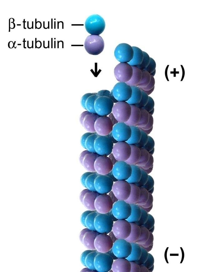
  • Microtubules self-assemble from dimers composed of alpha and beta tubulin, using energy from GTP.
  • They are hollow tubes with a diameter of 25 nm.
  • It was initially thought that tubulin dimers form a simple helix; it is now known that their arrangement is more complex, with a seam.
  • As a rule, cytoskeletal proteins assemble end-to-end into long thin strings called protofilaments, which then form bundles. The microtubule is composed of 13 protofilaments.
  • During assembly, or polymerisation, dimers are added only to one of the ends. That end is called plus (+), and the other minus (-).
A microtubule protofilament built from alternating alpha- and beta-tubulin dimers, with the plus end and minus end labelled

Microtubule dynamics

note

Some microtubules - in flagella, cilia and axons - are very stable.

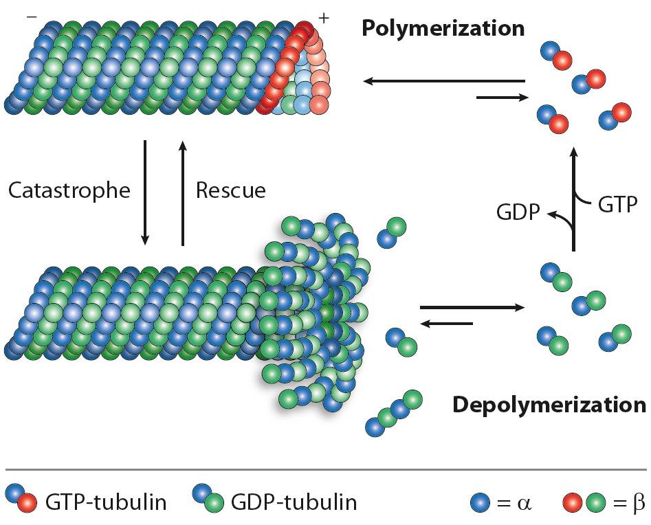
Microtubules in the cytoplasm, however, have a property called dynamic instability. This means their (+) end most of the time grows by the addition of tubulin dimers, but occasionally collapses, splitting into protofilaments that later depolymerise to dimers.

Dynamic instability is regulated. It is increased when the cell starts division, and as a result the microtubule network characteristic of non-dividing cells is replaced by a mitotic spindle.

Energy from GTP is used: tubulin is a GTPase that hydrolyses GTP to GDP.

Microtubule polymerisation and depolymerisation: GTP-tubulin adds at the plus end, catastrophe and rescue switch between growing and shrinking, GTP is hydrolysed to GDP

Microtubule nucleation and the centrosome

note

Once there is a microtubule, even a short one, new dimers of α- and β-tubulin are added at the (+) end. But how do the first dimers assemble together?

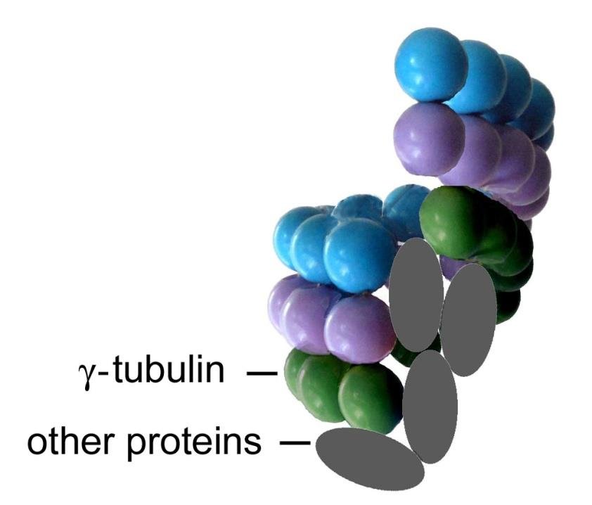
They bind to a ring complex containing a third type of tubulin, γ tubulin, along with some other proteins. The formation of a new microtubule is called nucleation, and sites containing γ-tubulin can nucleate microtubules and are therefore called microtubule-organising centres.

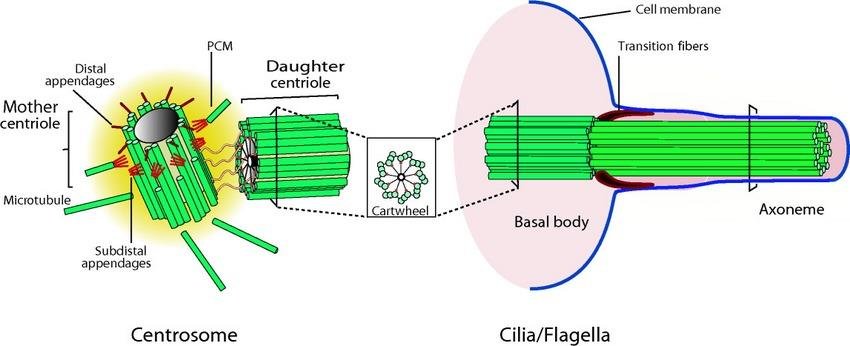
Cells of animals and most unicellular eukaryotes have one dominant microtubule-organising centre, called the centrosome. It consists of two microtubular cylinders called centrioles and the surrounding amorphous pericentriolar material containing γ-tubulin, from which the microtubules radiate.

A gamma-tubulin ring complex with other proteins nucleating a new microtubule from alpha- and beta-tubulin dimers

Microtubule-based transport

note

When needed, membrane organelles and chromosomes bind to microtubules and move along them as if on rails.

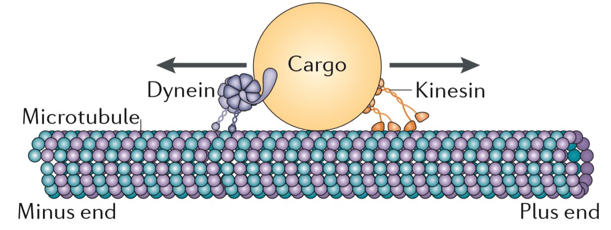
This is mediated by motor proteins, using energy from ATP to change their shape in a regular pattern. They decorate the cargo particle, then bind to the microtubule and force the cargo to slide along.

There are two groups of microtubule-associated motor proteins:

  • kinesins, which move towards the (+) end;
  • dyneins, which move towards the (-) end.
Motor proteins on a microtubule: dynein moving cargo towards the minus end and kinesin moving it towards the plus end

The structure of centrioles: 9 x 3

note

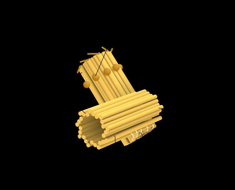
The centriole is a cylinder formed by 9 triplets of parallel microtubules.

Throughout most of the cell cycle the cell has 2 centrioles, perpendicular to each other.

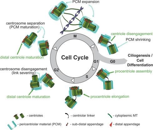
Before mitosis, the centrosome and centrioles duplicate at the same time as DNA replicates. This happens by the formation of a new (daughter) centriole perpendicular to each old (mother) centriole.

Three-dimensional model of a centriole, a cylinder built from triplet microtubules
Centrosome and centriole cycle through the cell cycle: centriole disengagement, PCM shrinking and expansion, procentriole assembly, elongation and distal centriole maturation

Two types of microtubule nucleation

note

The centriole can take part in nucleation in two ways, and the distinction is easy to lose.

  1. In the cytoplasm, microtubules originate from γ-tubulin ring complexes at the centrosome periphery, that is in the pericentriolar material in the vicinity of the centrioles. This way the centrioles are not directly involved in nucleation. Rather, they help the cell keep track of its centrosome.
  2. In eukaryotic flagella and cilia, which have an axoneme of microtubules, the basal body of each flagellum or cilium is a centriole, which directly nucleates the peripheral microtubules of the axoneme.
Centrosome with mother and daughter centriole, cartwheel and subdistal and distal appendages, compared with a basal body nucleating the axoneme, transition fibres labelled

The motility of flagella and cilia

note

Motor proteins can move not only organelles along microtubules but also microtubules along other microtubules.

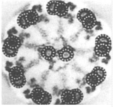
The axoneme of flagella and cilia is formed by 2 central and 9 doublets of peripheral microtubules: the 9 x 2 + 2 arrangement.

Electron micrograph cross-section of an axoneme showing the 9 times 2 plus 2 arrangement of peripheral doublet and central microtubules

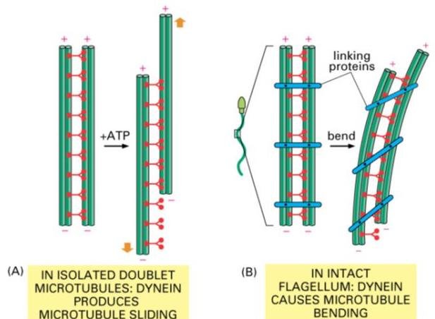
Each peripheral doublet carries dynein projections, the "arms", reaching the next doublet. Using ATP, this dynein moves one of the doublets along the other. The result is bending, that is movement, of the flagellum.

Dynein-driven microtubule doublet sliding with ATP in an isolated doublet, versus dynein bending an intact flagellum through linking proteins

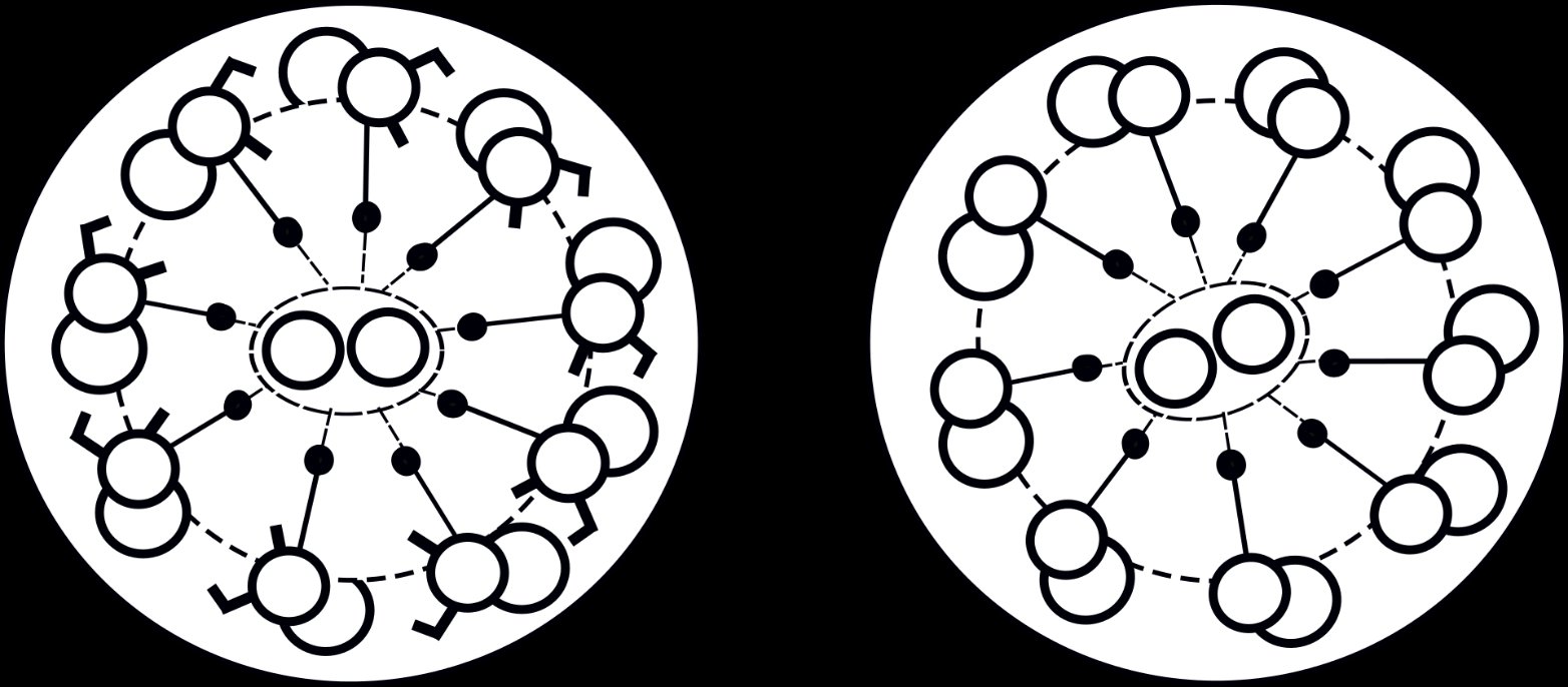
If one or both dynein arms are absent as a result of a mutation, the axonemes are immotile. The condition is called primary ciliary dyskinesia, and it is characterised by recurrent airway infections and male infertility.

Primary ciliary dyskinesia and Kartagener syndrome

note

Primary ciliary dyskinesia (PCD) is caused by an autosomal recessive mutation. It blocks the action of cilia and flagella - the cilia covering the respiratory tract (lower and upper, sinuses, Eustachian tube, middle ear), the fallopian tube, and the flagella of spermatozoa.

Cross-section of a normal axoneme with dynein arms on each doublet, compared with an axoneme in Kartagener's syndrome that lacks the arms

PCD leads to very frequent respiratory infections, infertility, and abnormal position of the internal organs. This combination of symptoms is called Kartagener syndrome, also known as ciliary dyskinesia syndrome.

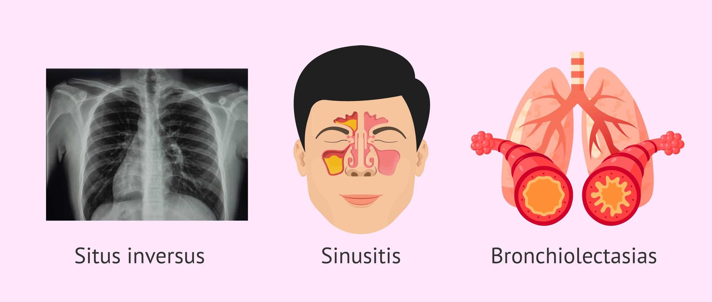
The Kartagener syndrome triad: situs inversus on a chest X-ray, sinusitis, and bronchiectasis of the lungs

The diagnosis of Kartagener syndrome is based on the identification of a triad of clinical symptoms: situs inversus, sinusitis, and bronchiectasis.

In the airway, mucus accumulates in the small airway and the dilated airway produces bronchiectasis. In reproduction, the flagellum affected by the syndrome means the sperm is not able to reach the egg, and the affected hair cells of the fallopian tubes hinder egg displacement.

Situs inversus and how cilia decide left from right

note

Situs inversus is congenital, and in it the major visceral organs are mirrored from their normal positions. It can be complete, with everything reversed, or partial, with only some organs reversed.

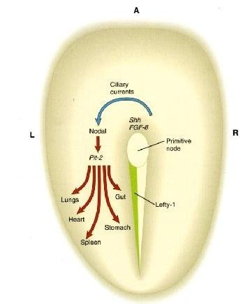
How the asymmetry is normally established. The left-right asymmetry of the body is directed by a cascade of asymmetrically expressed signals, and the process starts during gastrulation, long before overt asymmetric morphogenesis.

  • These signals include the asymmetric expression of key genes, here Shh and FGF8, on the left side of the embryo.
  • This triggers expression of specific proteins adjacent to a structure known as Hensen's node, at the rostral end of the primitive streak, and subsequently in a broad domain throughout the left side.
  • As a result, on the left side the genes are expressed and the organs develop on the correct side.
Embryo diagram of left-right asymmetry: Shh and FGF8 signals at the primitive node drive asymmetric Pitx2 expression and correct organ placement of lungs, heart, stomach, gut and spleen

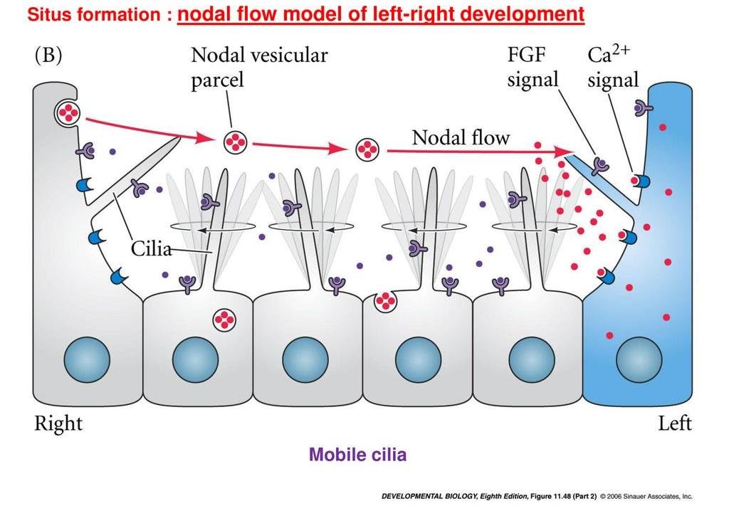
The link to cilia. The cells on the surface of the primitive node have cilia that beat, producing a nodal flow which carries nodal vesicular parcels and generates a calcium signal on one side. If the cilia are defective, Shh can randomly end up on the right side, resulting in reversal of symmetry - liver on the left, spleen on the right.

Nodal flow model: motile cilia at the node drive nodal vesicular parcels from right to left, triggering an FGF signal and a calcium signal on the left side cell

That is why a disease of dynein arms produces a mirror-image body: the same beating cilia that clear the airway also told the embryo which side was which.

9. Microfilaments

note

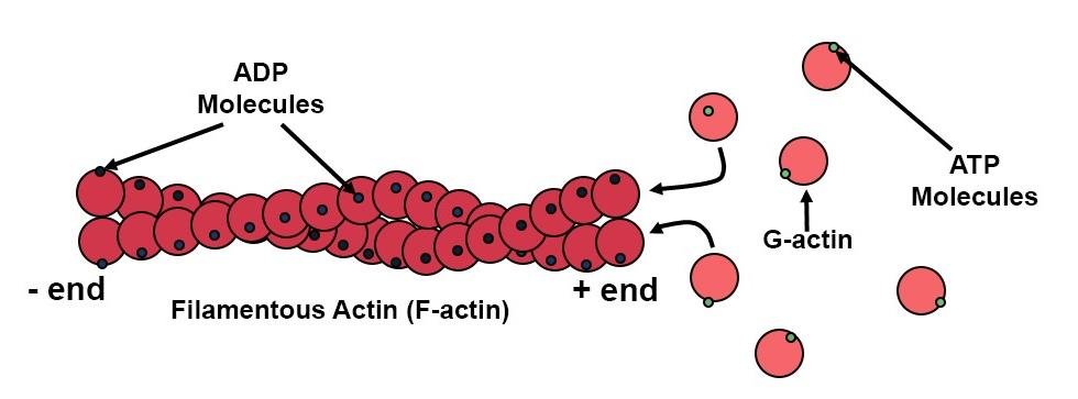
Microfilaments are assembled from actin using ATP energy.

  • The single soluble actin molecules are called globular (G-) actin; those included in microfilaments are called fibrillar (F-) actin.
  • Microfilaments have a diameter of 6 to 8 nm.
  • Actin molecules are arranged in a helix. The microfilament can be described either as a single helix or as two interwound protofilaments.
  • Microfilaments, like microtubules, grow at only one of their ends. It is called (+) or "barbed", while the other is (-) or "pointed".

The actin cytoskeleton requires interactions with other proteins to do anything useful.

Filamentous (F-) actin built from globular (G-) actin monomers using ATP, ADP-bound subunits shown in the filament, with the minus and plus ends labelled

Microfilament nucleation

note

Microfilaments are dynamic structures with relatively short life spans, except in myofibrils, where they are stable.

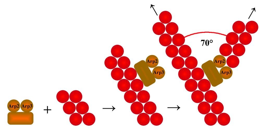
Microfilament nucleation is mediated by a protein complex called Arp2/3, whose role is analogous to that of the γ-tubulin ring complex for microtubules.

The organisational difference from microtubules matters: there is no central structure analogous to the centrosome. New microfilaments usually arise as branches of old ones.

Arp2/3 complex nucleating a branched actin filament at a 70° angle from an existing filament

Myosins

note

Microfilaments are responsible for the transport of membrane organelles, as microtubules are, and also for amoeboid movements, cytoses and cell shape changes.

The motor proteins called myosins use ATP to move towards the (+) end.

Each myosin has:

  • a motility-generating globular domain, the "head", with ATPase activity;
  • a long fibrous alpha-helical domain, the "tail".

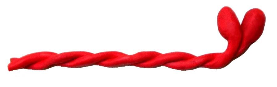
Tails wind together in pairs. This structural motif, the coiled coil, is common in fibrous proteins.

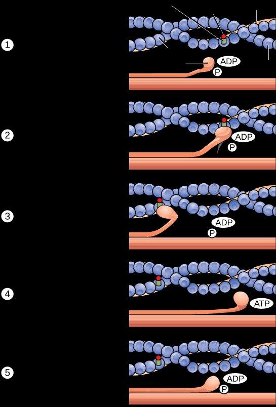
Myosin is unusual in being both a fibrous protein and an enzyme, since in principle enzymes are globular proteins. It is an ATPase that breaks down ATP to ADP and converts the chemical energy into mechanical activity. Every time myosin attaches to ATP its shape changes, and this causes a "walking" movement along the actin filaments. Myosin is therefore both an enzyme and a motor protein.

A pair of myosin molecules: two alpha-helical tails wound together as a coiled coil, ending in two globular heads
Five-step myosin walking cycle along an actin filament, the head cycling between ADP, ADP with phosphate, and ATP-bound states

Microfilament-based motility and contraction

note

Myosin, like dynein, can move not only organelles along microfilaments but also microfilaments along other microfilaments.

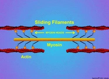
For this purpose a number of myosin doublets stack together and form long fibres sliding along microfilaments. The result is contraction of the actomyosin complex.

This mechanism is developed to perfection in striated muscles, but it is present also in other muscles and in non-muscle cells. In animal cell division, the splitting of the daughter cells, cytokinesis, is based on a contractile ring of microfilaments.

Sliding filament mechanism: myosin heads walk along actin filaments of opposite polarity, pulling the two ends of the sarcomere together

10. Intermediate filaments

note

  • They are named intermediate because their diameter is about 10 nm, less than in microtubules and more than in microfilaments.
  • Their components are fibrous proteins with the coiled coil motif. These subunits form dimers and then tetramers without requiring energy.
  • They provide mechanical strength but are not fit for transport, because they have no polarity - their two ends are identical.
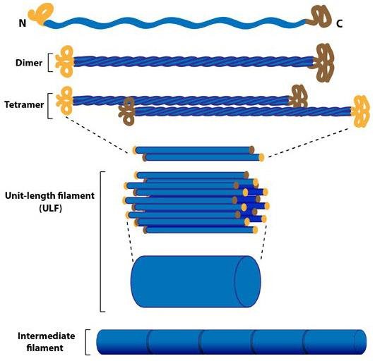
Intermediate filament assembly: monomer with N- and C-terminal heads, coiled-coil dimer, staggered tetramer, unit-length filament (ULF) and the mature intermediate filament

The lack of polarity is due to the antiparallel, head-to-tail, assembly of their dimers into tetramers. That single structural fact is what separates them functionally from the other two systems: no polarity means no direction, and no direction means no motor can walk along them.

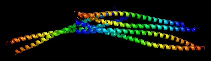
Six molecules of vimentin bound together is the standard picture of an intermediate filament in formation.

Molecular model of six vimentin molecules bound together, an intermediate filament forming

The types of intermediate filament

note

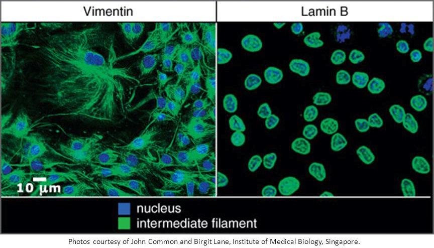
The most widespread type, found in all animals, are the lamins. They are located in the nucleus and form the innermost layer of the nuclear envelope, called the nuclear lamina.

During the evolution of vertebrates, duplication and divergence of the lamin genes produced the genes for the cytoplasmic intermediate filaments.

The cytoplasmic intermediate filaments are tissue-specific:

  • keratins in epithelia;
  • vimentin in connective tissue;
  • desmin in muscles;
  • glial fibrillary acidic protein in neuroglia;
  • neurofilament proteins in neurons.

Because they are tissue-specific, they are also diagnostically useful: which intermediate filament a cell makes says what kind of cell it is.

Immunofluorescence comparing cytoplasmic vimentin intermediate filaments with nuclear lamin B, nuclei counterstained blue

Progeria

note

Progeria means "rapid ageing".

It is a very rare condition, caused by mutations in the lamin genes.

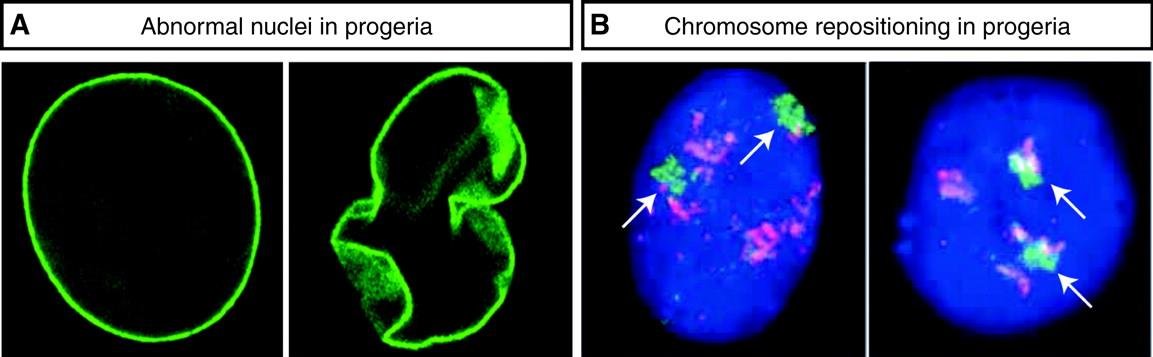
  • The nuclear shape is abnormal.
  • The control of gene activity is disturbed.
  • Patients die of heart disease at an average age of 14.5 years.
Progeria: abnormal, folded nuclear shape (green nuclear lamina) compared with a normal round nucleus, and repositioned chromosome territories, arrowed, in progeria nuclei

Viruses use the host cytoskeleton

note

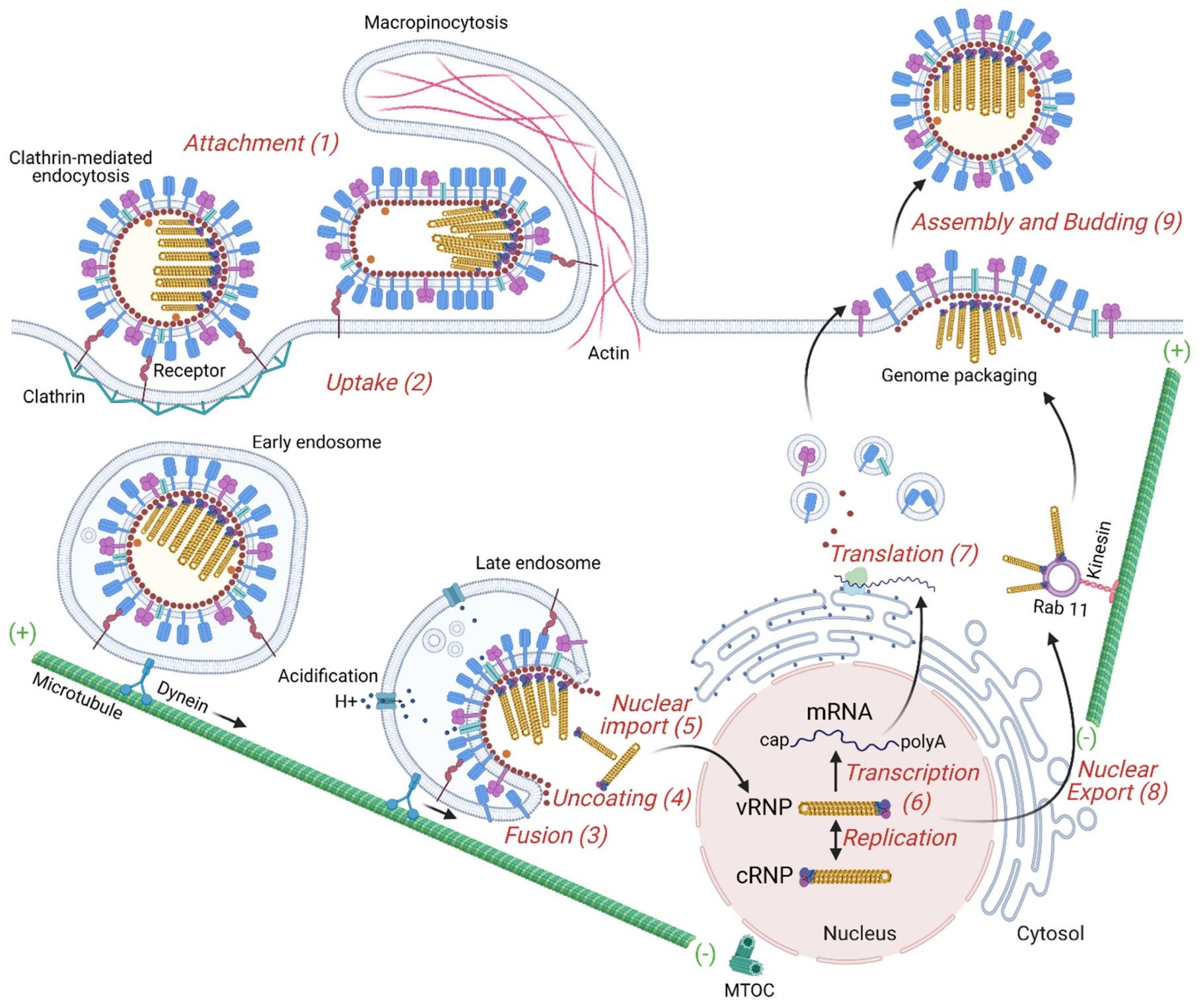
Viruses use the host cell cytoskeleton for the assembly of new viral copies. The worked example is the influenza virus, which uses host cell pathways during uncoating.

Influenza virus life cycle using the host cytoskeleton: clathrin-mediated attachment and uptake, endosomal fusion and uncoating, dynein transport to the nucleus, then kinesin transport, genome packaging and budding

11. Drugs and toxins that act on the cytoskeleton

note

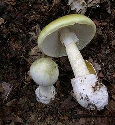
Phalloidin is a toxin that blocks microfilament function, produced in *Amanita phalloides. It is a *bicyclic heptapeptide, lethal after a few days when injected into the bloodstream. It **binds and stabilises filamentous actin (F-actin) and prevents the depolymerisation of actin fibres.

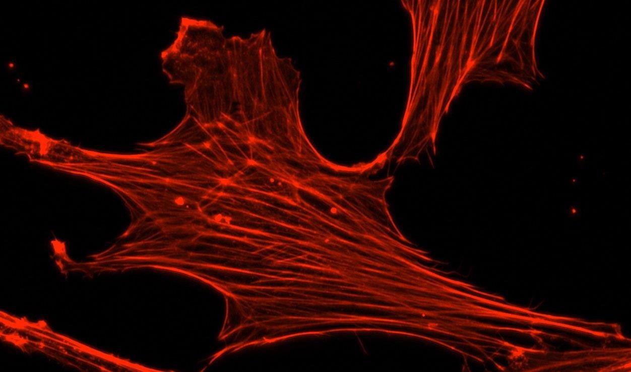
Several other small molecules bind to actin either during polymerisation or during depolymerisation, with varying effects on filament dynamics: cytochalasins (fungal metabolites), jasplakinolide and latrunculin (from a marine sponge). The names are not to be learned. All these molecules can be used as molecular markers to label actin structures in the cell - single actin filaments stained with phalloidin-rhodamine fluoresce under UV light.

Actin stress fibres in a cell stained with phalloidin-rhodamine, fluorescing under UV light
Amanita phalloides, the death cap mushroom that produces phalloidin

Taxol and colchicine

note

Taxol, also called paclitaxel, is applied as a chemotherapy medication and is naturally produced by the yew (Taxus) plant. It blocks the function of microtubules.

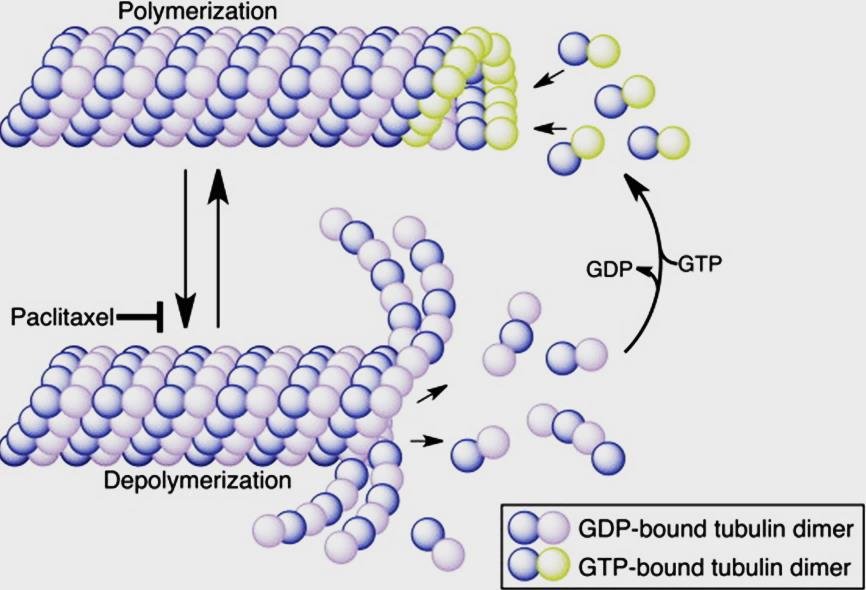
The mechanism is worth the detail. Molecular motors such as kinesin bind to the outer surface of the microtubule, while Taxol binds to β-tubulin along the interior of the microtubule. That prevents microtubule depolymerisation.

Microtubule polymerisation and depolymerisation from GDP- and GTP-bound tubulin dimers, with paclitaxel blocking depolymerisation

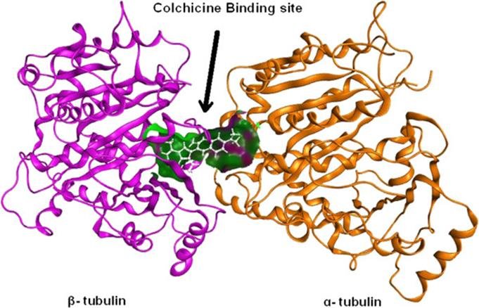
Colchicine does the opposite: it blocks microtubule polymerisation by binding to tubulin, at a site between the α and β subunits. It comes from *Colchicum autumnale*.

Colchicine binding site between beta-tubulin, magenta, and alpha-tubulin, orange

Its applications are of two quite different kinds:

  • Colchicine inhibits multiple pro-inflammatory mechanisms, and is used to reduce the symptoms of gout and other situations involving very strong inflammatory pain.
  • Its ability to arrest cell division is used for karyotype analysis.

The most important things to know

note

  • Self-assembly is recognition by shape and surface charge, held by many weak non-covalent bonds. Nothing in the cell would exist without it.
  • Annealing has three names by case: renaturation (same molecule), hybridisation (different molecules), denaturation is the reverse and is reversible above 70 °C.
  • Small quaternary complexes have a fixed subunit number; large ones do not.
  • Nucleosome assembly order: H3-H4 tetramer first, then two H2A-H2B dimers.
  • Tobacco mosaic virus shows the nucleic acid dictating the shell: RNA joining a protein disk forces it into a helix.
  • LDL carries cholesterol out to tissues, HDL brings it back to the liver; the excess left on artery walls is atherosclerosis. Same structure, HDL has more surface protein.
  • Cholesterol opposes the temperature: it stiffens a warm membrane and loosens a cold one.
  • Sickle haemoglobin is valine for glutamate at position 6 of the beta chain, on the surface - a single change that wrecks the whole assembly.
  • Three fibres: microfilaments 6 nm (actin), intermediate 10 nm (fibrous proteins), microtubules 25 nm (13 protofilaments of αβ-tubulin).
  • Kinesin walks to (+), dynein to (-) on microtubules; myosin walks to (+) on microfilaments.
  • Centriole is 9 x 3; axoneme is 9 x 2 + 2. Missing dynein arms give primary ciliary dyskinesia, whose triad with situs inversus is Kartagener syndrome - because nodal cilia are what tell the embryo left from right.
  • Intermediate filaments have no polarity, so they bear load but carry nothing. Lamins are the ancestral type; the cytoplasmic ones are tissue-specific, and lamin mutation causes progeria.
  • Phalloidin stabilises actin. Taxol stabilises microtubules from the inside. Colchicine prevents their polymerisation - and is used both for gout and for karyotyping.
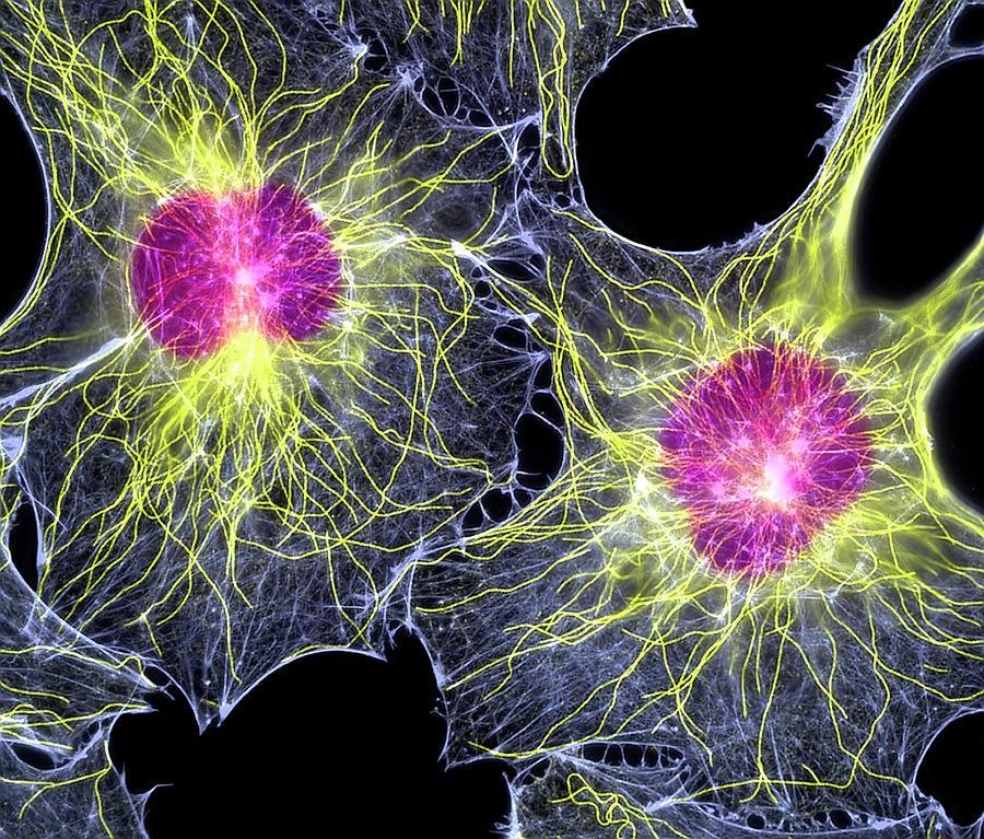
Fluorescence micrograph of the cytoskeleton in two cells: microtubules radiating yellow-green from pink centrosomes, with actin and intermediate filaments in the surrounding network

All 105 Medical Biology topics · exam study support, not clinical guidance. SuperMed is not affiliated with the Medical University of Sofia.

Start free. No card needed.

Every account starts free, with free topics in Cytology and Medical Biology. Super opens the rest. Super is €15 a month.

Create a free account