Medical Biology · Year 1 · Medical University of Sofia

05

Transcription

Free notes for topic 05 of the Medical Biology syllabus, open without an account. Written by a senior student against the syllabus question and checked line by line by a second student before publishing. How content is made

Updated

What this topic covers

note

The general characteristics of transcription and the enzyme that carries it out; how the nucleotides are selected and where the energy comes from; the three stages and the transcription unit; the role of topoisomerases and why that matters for antibiotics; transcription in prokaryotes, including the two kinds of terminator; transcription in eukaryotes, the three RNA polymerases and the transcription factors; what transcription looks like down the microscope; the effect on nucleosomes; why the death cap is deadly; and transcription in mitochondria.
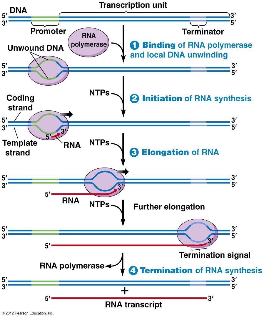
Transcription unit diagram with four numbered steps: RNA polymerase binding and local DNA unwinding, initiation, elongation with NTPs added to the growing RNA, and termination releasing the RNA transcript

1. General characteristics

note

  • The function of DNA is just to be a store for genetic information.
  • The genes are expressed after their transcription into RNA.
  • Definition: transcription is a process of RNA synthesis using a particular DNA segment as a template.
  • The copied DNA segment has specific beginning and end sequences, which define the first and the last point of transcription.
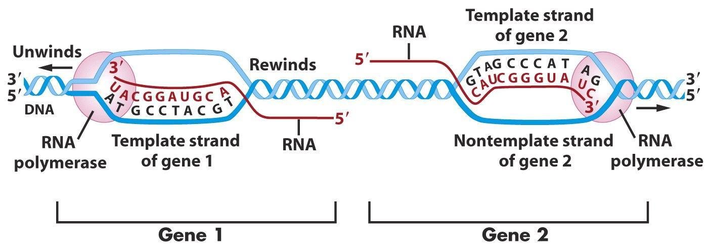
  • Each DNA strand is template for some genes and complementary (coding) for other genes.
  • The template DNA and the synthesised RNA are antiparallel and complementary, pairing A-U and G-C.
  • The direction of RNA synthesis is 5' to 3'.
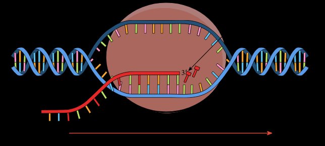
RNA polymerase, orange, moving along a DNA double helix, opening a transcription bubble and extending a red RNA strand in the direction of the arrow

2. The enzyme

note

The enzyme is DNA-dependent RNA polymerase (RNApol, RNAP). It has a complex and dynamic quaternary structure, and in shape it resembles a crab claw.

It has several functions:

  • separates the two DNA strands within a short region;
  • uses one of them as a template;
  • catalyses the formation of covalent phosphodiester bonds between nucleotides;
  • participates in the regulation of gene activity.

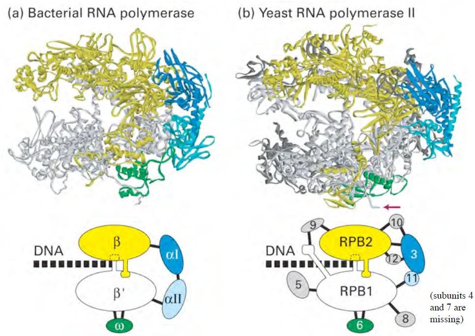
The bacterial core enzyme, in Thermus aquaticus, is built from the subunits α, α, β, β' and ω. The eukaryotic core RNAP II, in yeast, is built from RPB1, RPB2, RPB3, RPB11 and others.

A reminder on the chemistry: an ester bond forms between an alcohol and an acid, as acetic acid and ethanol give ethyl acetate and water. In the same way a phosphodiester bond forms between a pentose and a phosphate.

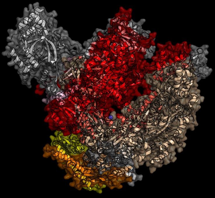
Surface structure of RNA polymerase coloured by subunit, its two lobes forming a crab claw shape that clamps around the DNA
Subunit structures of bacterial RNA polymerase and yeast RNA polymerase II compared, main subunits (beta, beta prime, alpha for bacteria, RPB1 to RPB3 for yeast) coloured and labelled

The enzyme does not select the nucleotides

note

Adding a nucleotide to the RNA chain is a two-step process, and only the second step is enzymatic.

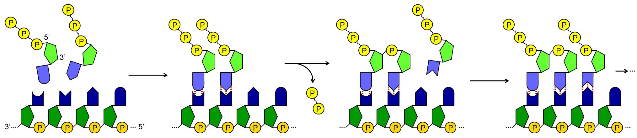
  1. The incoming ribonucleotide spontaneously binds to a complementary DNA nucleotide, by hydrogen bonds between the two bases.
  2. Then the RNA polymerase covalently binds its phosphate, with a phosphodiester bond, to the ribose of the previous ribonucleotide.

Hence transcription is a typical template biopolymer synthesis, in which the monomers forming the product are selected not by an enzyme but by a pre-existing biopolymer, the template. This is the same division of labour as in replication, and it is what makes accuracy possible at all: an enzyme choosing between four similar molecules would be far less reliable than base pairing.

Two-step nucleotide addition: an incoming ribonucleotide first pairs spontaneously with its complementary base, then RNA polymerase forms the covalent phosphodiester bond

Energy, and the fate of the pyrophosphate

note

Energy is provided by the substrates themselves, the ribonucleotides, which are nucleoside triphosphates: ATP, UTP, GTP and CTP.

  • Only one of the phosphate groups stays in the RNA product.
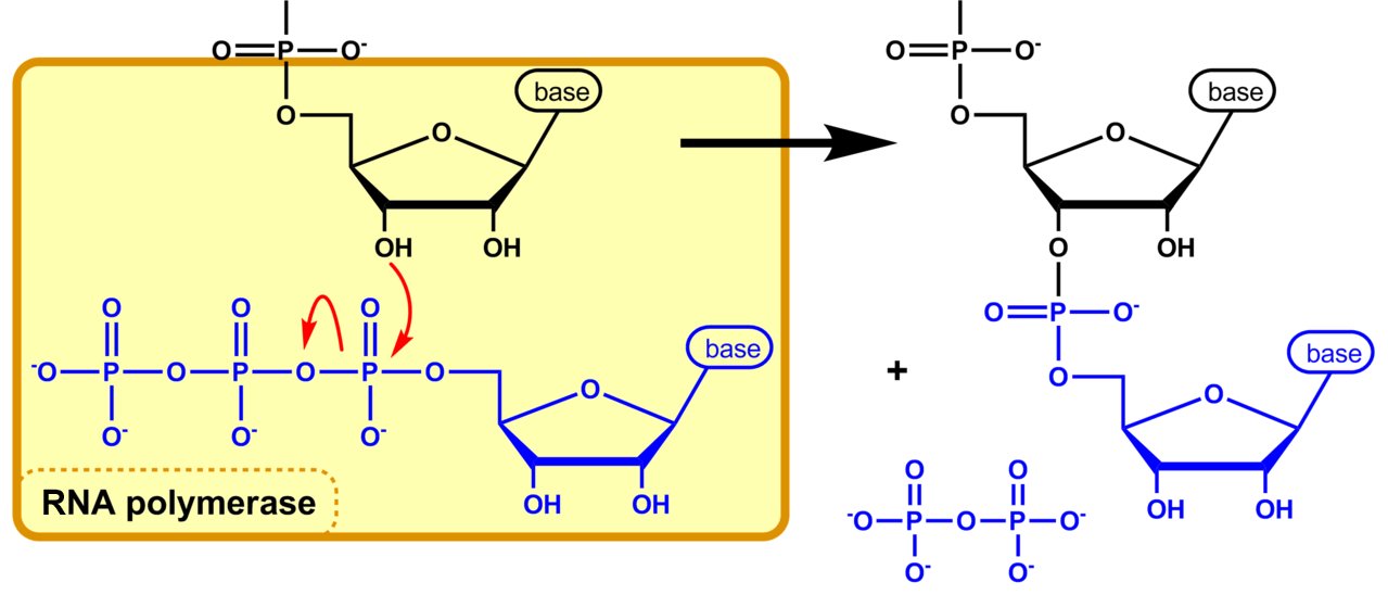
  • The RNA polymerase breaks the high-energy bond between it and the next phosphate and uses the energy of that bond.
  • Pyrophosphate is released as a by-product.

Then another enzyme, inorganic pyrophosphatase, hydrolyses the pyrophosphate without using the energy of the bond. That looks wasteful and is not: this removal of the by-product shifts the reaction in the needed direction. Destroying the product of a reversible reaction is how the cell makes it irreversible.

Reaction mechanism of nucleotide addition by RNA polymerase: attack on the phosphate forms the new phosphodiester bond and releases pyrophosphate

Direction of synthesis

note

  • The RNA polynucleotide chain is synthesised in the 5' to 3' direction, and the DNA template strand is read 3' to 5'.
  • The template strand is determined case by case. Each DNA strand is template for some genes and complementary for others.
DNA unwinding and rewinding at two adjacent genes, RNA polymerase using the lower strand as template for gene 1 and the upper strand as template for gene 2

3. The stages

note

Initiation.

  • The RNA polymerase recognises a specific nucleotide sequence, the promoter, which is the signal to start transcription. In eukaryotes the recognition requires additional proteins.
  • After attachment to the promoter, the enzyme opens the DNA strands and forms a phosphodiester bond between the first two ribonucleotides.
  • The first transcribed nucleotide is called the start site.

Elongation.

  • More complementary nucleotides come to the DNA template, and the RNA polymerase joins them with covalent bonds.
  • Enzymes called topoisomerases relax the DNA supercoiling before and after the RNA polymerase.

Termination.

  • The enzyme recognises a specific nucleotide sequence, the terminator, which is the signal to end transcription.
Transcription unit diagram with four numbered steps: RNA polymerase binding and local DNA unwinding, initiation, elongation with NTPs added to the growing RNA, and termination releasing the RNA transcript

The transcription unit

note

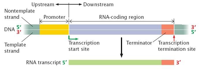
  • A transcription unit is a DNA segment being transcribed into an RNA molecule.
  • It includes three critical regions: a promoter, an RNA-coding region, and a terminator.
  • One transcription unit may contain one or more genes. Hence a single promoter may control several genes - which is the structural basis of the operon.

In sequence: binding of RNA polymerase and local DNA unwinding, then elongation of the RNA, then termination of RNA synthesis at the termination signal, releasing the RNA transcript.

Transcription unit on DNA: promoter, RNA coding region and terminator marked, with the transcription start site, transcription termination site and resulting 5' to 3' RNA transcript below

4. Topoisomerases

note

Topoisomerases are enzymes that change DNA supercoiling by breaking and rejoining one or both strands of the DNA molecule.

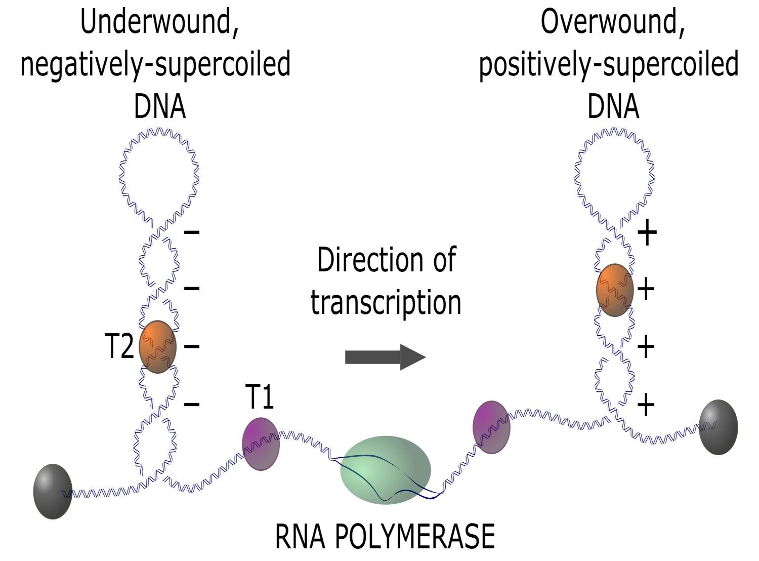
Why transcription needs them. When RNA polymerase moves along DNA, it causes negative and positive supercoiling of the double helix before and after itself respectively. This leads to tension in the sugar-phosphate backbone. Topoisomerases are therefore needed both before and after the open segment of the double helix.

Temporary covalent bonds are formed between the topoisomerase and the broken DNA strand.

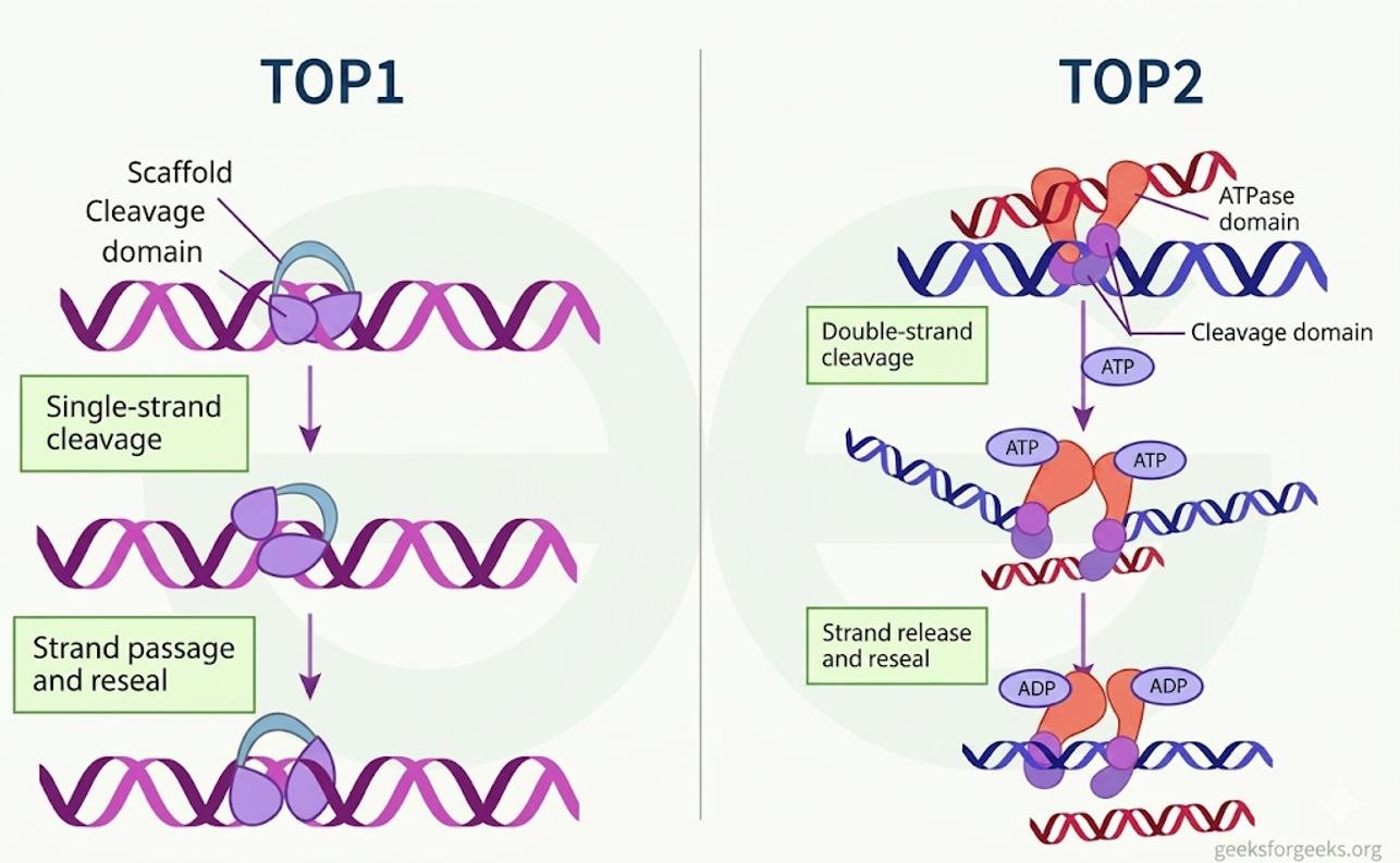
  • Topoisomerase type 1 (TOP1) makes single-strand breaks.
  • Topoisomerase type 2 (TOP2) makes double-strand breaks.
DNA anchored at both ends becomes underwound and negatively supercoiled behind RNA polymerase and overwound and positively supercoiled ahead of it, topoisomerases T1 and T2 marked at each side
TOP1 makes a single-strand cleavage in DNA and reseals it after strand passage, while TOP2 makes an ATP dependent double-strand cleavage and reseal

Why this matters for antibiotics: the quinolones

note

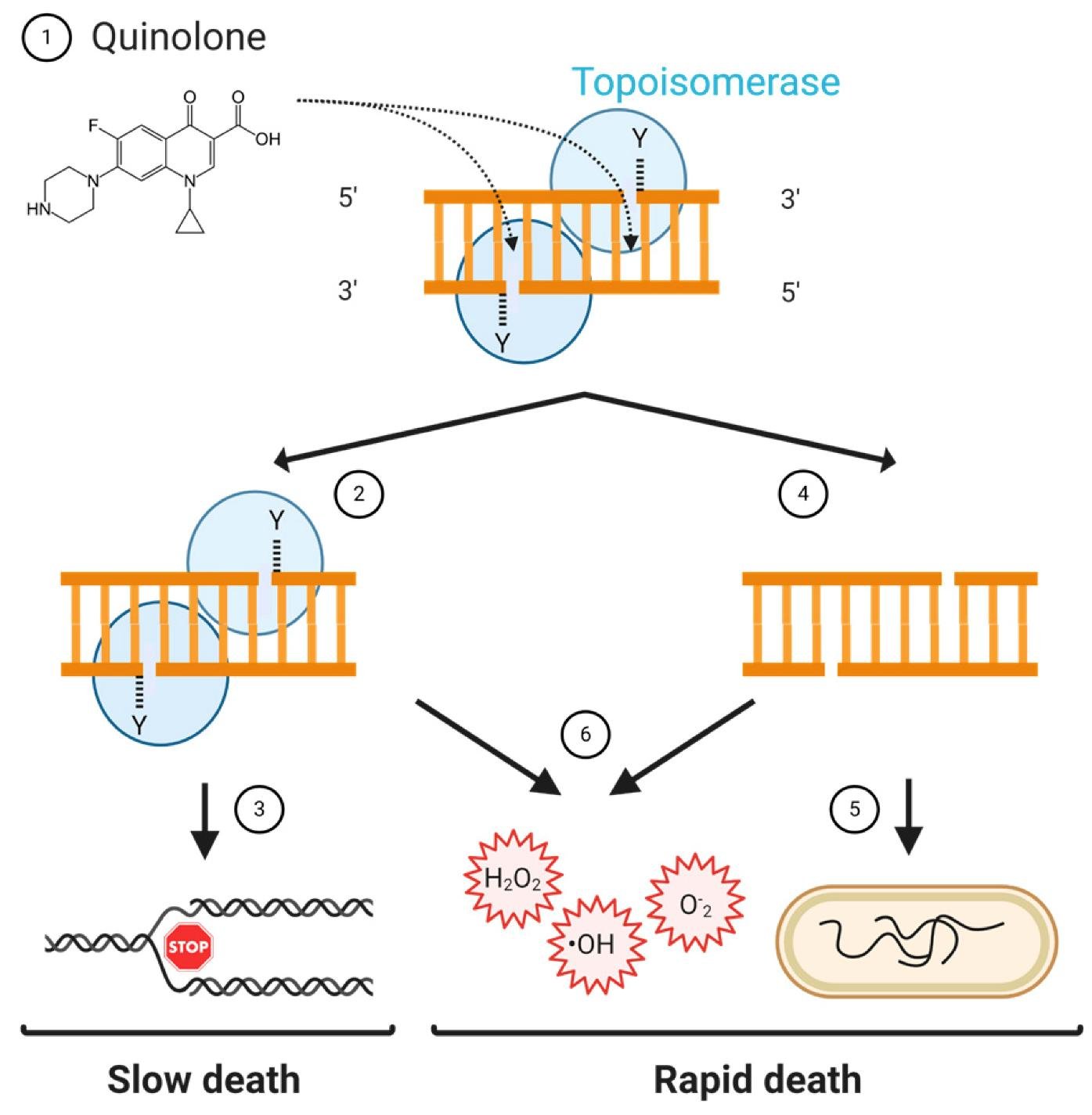
Understanding the actions of drugs requires knowing the mechanisms of cellular processes. The quinolones are a group of antibiotics that are inhibitors of bacterial topoisomerase type 2, also called gyrase.

The sequence of events, and note that it has two quite different speeds:

  1. Quinolones block gyrase after it has obtained a double-stranded DNA breakage.
  2. The gyrase can remain bound to the DNA, and this makes bacterial transcription and replication impossible.
  3. This kills bacteria relatively slowly.
  4. If instead the gyrase is removed from the DNA and
  5. the double-stranded break is not repaired, the bacterial chromosome will be broken - and this kills bacteria very quickly.
  6. The action of the antibiotic can also lead to an accumulation of reactive oxygen species, which kill bacteria.

This knowledge is used both for the correct application of existing antibiotics and for the development of new antibiotic variants.

Quinolone blocking bacterial topoisomerase after it cuts double stranded DNA, leading either to slow bacterial death or, if the enzyme detaches and the break stays unrepaired, to rapid death

5. Transcription in prokaryotes

note

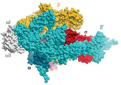
  • The prokaryotic RNA polymerase has 6 subunits: two identical α, plus β, β', ω and σ. In this complete form it is called the holoenzyme.
  • It binds random DNA segments, and when it hits a promoter it can initiate transcription. There is no search mechanism; there is only repeated sampling until it lands on the right sequence.
  • Prokaryotic promoters are located between positions +1 and -40.
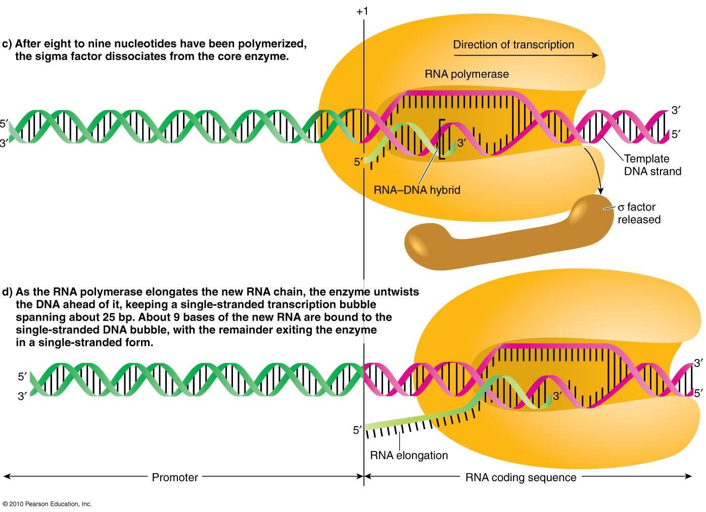
  • σ is necessary just for initiation, in order to recognise the promoter, and then it dissociates.
  • During elongation only the core enzyme (α₂ββ'ω) works.
  • Together with the core enzyme, a small open DNA segment of 17 nucleotides moves along the transcription unit towards its 3' end.
  • The terminators are included in the gene. All of them contain palindromes, or symmetrical sequences - for example CAATTG, whose right half is complementary to its left half.
Surface structure of the bacterial RNA polymerase holoenzyme with subunits alpha I, alpha II, beta, beta prime, omega and sigma coloured and labelled

Initiation is therefore: the holoenzyme binds to the promoter and separates the two DNA strands locally to expose the template strand. Elongation follows once detachment of the σ factor allows the core enzyme to move along the DNA and transcribe.

RNA polymerase elongating the RNA-DNA hybrid within a single stranded transcription bubble after the sigma factor has dissociated from the core enzyme

Rho-independent terminators

note

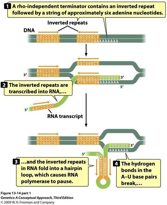
  • They contain a palindrome rich in C-G base pairs.
  • The palindrome is followed by multiple A residues, so the RNA finishes with oligo-U.
  • The connection between A and U is weak, so the RNA detaches from DNA spontaneously, and the CG-rich palindrome forms a stem-loop. G and C are connected by triple hydrogen bonds, which makes the loop very stable.
  • This termination is called intrinsic or rho-independent.

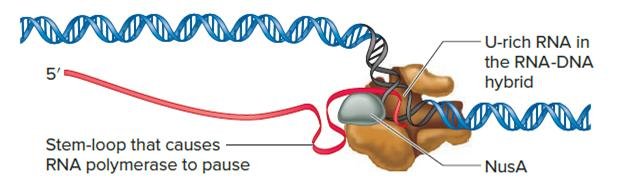
The mechanism in one sentence: the stem-loop causes the RNA polymerase to pause, and while it pauses, the weakly bound U-rich sequence in the RNA-DNA hybrid is not able to hold the hybrid together. Termination occurs.

Rho-independent terminator mechanism: inverted repeats in DNA are transcribed into RNA, fold into a hairpin loop, and the weak A-U hydrogen bonds break to release the transcript
A stem-loop in the nascent RNA causes RNA polymerase to pause, with NusA protein bound and a U-rich sequence in the RNA-DNA hybrid unable to hold it together

Rho-dependent terminators

note

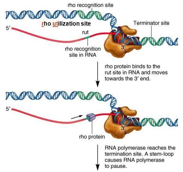
These make the RNA polymerase pause, but for its detachment the help of a protein called the ρ (rho) factor is needed.

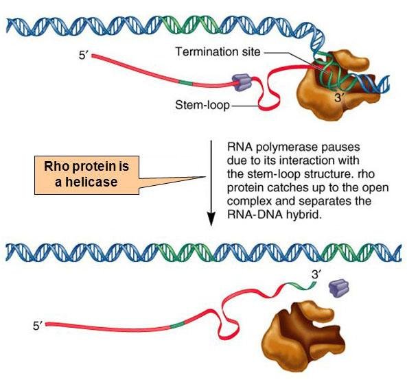
Rho is a helicase, that is, it can separate polynucleotide double strands using ATP energy.

  1. The rho factor binds to a region upstream of the terminator, called the rho utilisation site (rut).
  2. It then crawls along the RNA.
  3. When rho reaches the RNA-DNA hybrid, it uses its helicase activity to detach the RNA from the DNA.
Rho protein binds the rho utilisation site in the RNA and moves towards the 3' end while RNA polymerase pauses at a stem-loop and terminator site
Rho protein, a helicase, catches up to RNA polymerase paused at the stem-loop and separates the RNA-DNA hybrid, releasing the RNA transcript

6. Transcription in eukaryotes

note

There are three nuclear RNA polymerases (I, II and III). They have a similar structure and some common subunits but use different promoters.

  • RNApol I synthesises the 45S precursor of the 18S, 5.8S and 28S rRNAs.
  • RNApol II makes mRNA and most snRNA.
  • RNApol III makes tRNAs, 5S rRNA and other small RNAs.

Other differences from prokaryotes:

  • Each gene has its own promoter and terminator - rather than one promoter serving several genes.
  • Promoters are similar to prokaryotic promoters, but RNApol cannot recognise them directly and needs the help of other proteins.
  • During transcription, the RNA polymerase causes detachment of histone H1 and temporary relaxation of the nucleosome structure.
  • Terminators are poorly studied. In protein-coding genes it is known that if the sequence AAUAAA appears in the mRNA, that is the signal for the end of the transcript. An enzyme recognises AAUAAA and cuts the chain, and soon after that transcription is terminated.
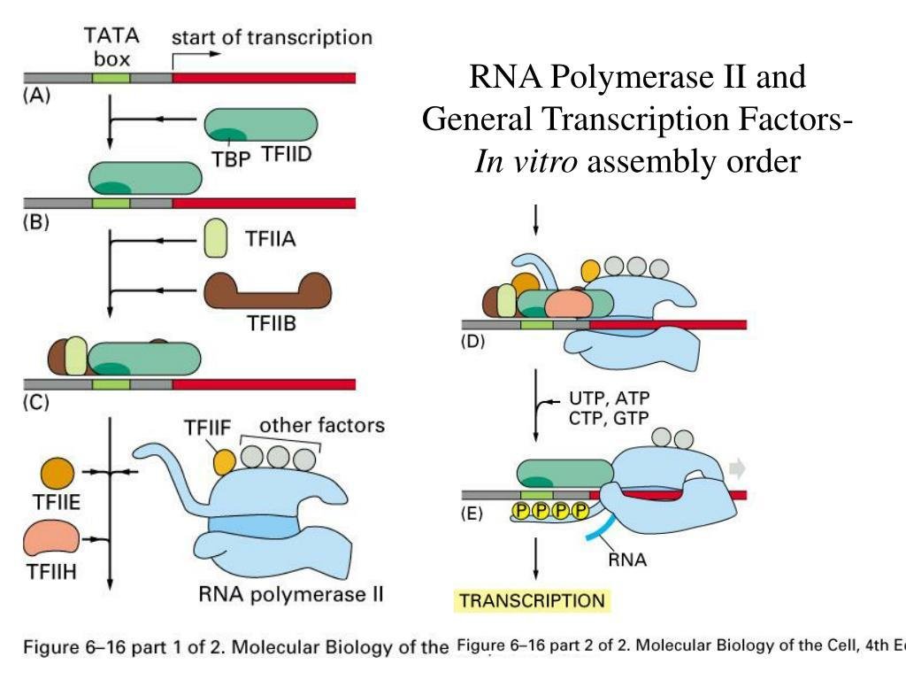
Assembly of the general transcription factors on the TATA box, TBP, TFIID, TFIIA, TFIIB, TFIIF and other factors, followed by RNA polymerase II, then TFIIE and TFIIH, before transcription begins

Initiation requires transcription factors

note

Proteins called general transcription factors (TF) recognise and bind the promoter, and then the RNA polymerase recognises them and joins.

The polymerase, in other words, does not read the DNA; it reads the proteins that have read the DNA.

Some of the general transcription factors are homologous to the bacterial σ factor - the same job, done by a separate protein rather than by a detachable subunit.

Assembly of the general transcription factors on the TATA box, TBP, TFIID, TFIIA, TFIIB, TFIIF and other factors, followed by RNA polymerase II, then TFIIE and TFIIH, before transcription begins

Termination and the making of the 3' end

note

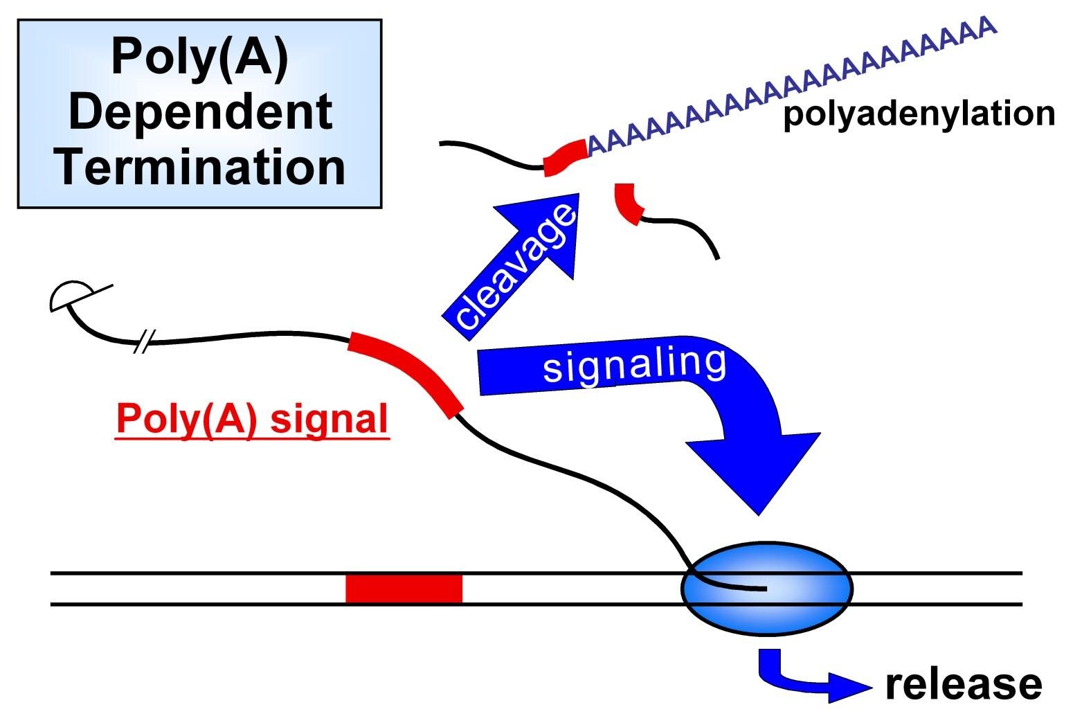
In protein-coding genes, the sequence AAUAAA is a signal to the polymerase that has just synthesised it to stop transcription a little downstream and release itself from the DNA.

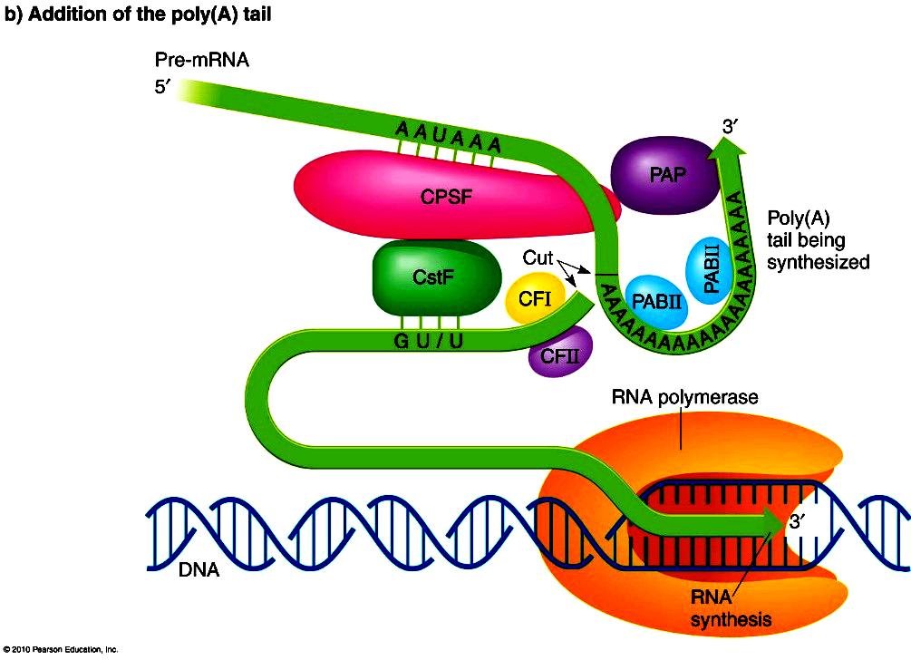
It is also a signal for cleavage of the transcript by an endonuclease. As a result the 3' end of the mRNA is generated, and about 200 A residues will be added to it during processing.

The reaction requires a protein complex containing:

  • a specificity factor;
  • an endonuclease;
  • poly(A) polymerase (PAP), which adds about 200 A residues processively to the 3' end.
Poly(A) dependent termination: the poly(A) signal downstream of a gene triggers cleavage and polyadenylation signalling and release of RNA polymerase
Poly(A) polymerase, PAP, synthesising the poly(A) tail onto the cleaved 3' end of the pre-mRNA, with CPSF, CstF and CFI bound at the cleavage site

Seeing transcription

note

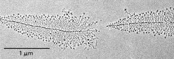
Electron micrographs of rRNA genes being transcribed in the nucleolus show transcription directly, and the picture is worth being able to read.

  • Each "Christmas tree" is one transcription unit. The top is the start site and the base of the tree is the terminator.
  • The growing transcripts are the branches. Their elongation shows the direction of transcription: short at the start, long at the end.
  • The dots forming the trunk are numerous RNApol I molecules.
  • The bigger dots at the branch tips are proteins involved in rRNA processing, and possibly some ribosomal proteins - processing begins before transcription has finished.
Electron micrograph of rRNA genes in a Christmas tree pattern, each tree one transcription unit with progressively longer growing RNA transcripts along its length

Transcription and nucleosomes

note

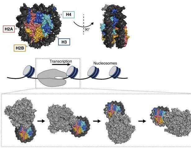
As the RNA polymerase transcribes through a nucleosome, it temporarily peels the DNA from the surface of the histone octamer.

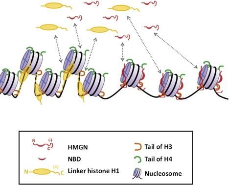
During transcription the H1 histone is detached from the 30 nm chromatin fibre, and the RNA polymerase displaces the nucleosome octamers for a while.

The chromatin is not an obstacle that has to be removed in advance; it is opened locally and closed again behind the enzyme.

Nucleosome core histones H2A, H2B, H3 and H4 with their structure, and a diagram of RNA polymerase transcribing through a series of nucleosomes
Nucleosomes with linker histone H1, HMGN protein and the tails of histones H3 and H4 diagrammed along a chromatin fibre

Why the death cap is deadly

note

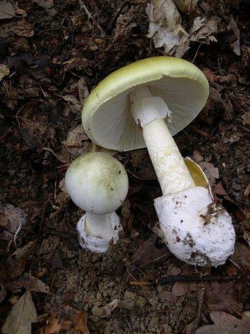
The peptide toxin α-amanitin comes from the death cap and its relatives: Amanita phalloides, and Amanita virosa, called the destroying angel.

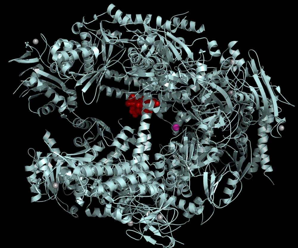
It inhibits RNAP II, and to a lesser degree RNAP III, by blocking its movement along the DNA.

Structurally, α-amanitin binds to RNAP II near the active site, which is marked by a magnesium atom.

The clinical logic follows from which polymerase is hit: blocking RNAP II stops the production of all messenger RNA, so the cell cannot make any new protein at all.

A death cap mushroom, Amanita phalloides, the source of the toxin alpha-amanitin
Ribbon structure of RNA polymerase II with alpha-amanitin, red, bound near the active site

7. Transcription in mitochondria and chloroplasts

note

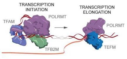
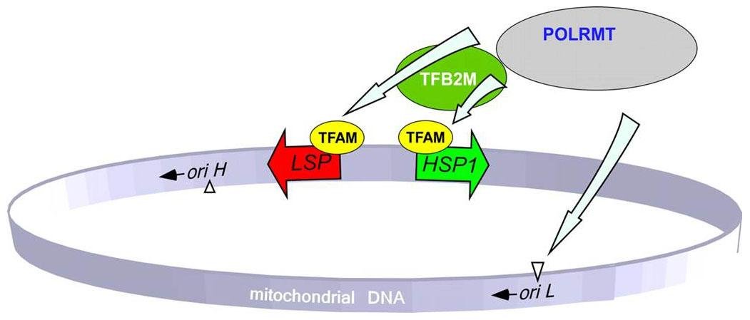
  • The RNA polymerase is different from the nuclear enzymes, and much simpler. In human mitochondria it is called POLRMT.
  • The human mitochondrial genome has just two promoters - the light strand promoter and the heavy strand promoter - and the resulting two long transcripts are cut into separate RNAs.

So the mitochondrion does not regulate its genes one at a time. It transcribes almost everything it has in two pieces and then cuts them up.

Mitochondrial transcription initiation with TFAM, TFB2M and POLRMT at the promoter, followed by elongation with POLRMT and TEFM
Circular mitochondrial DNA with the light strand promoter, LSP, and heavy strand promoter, HSP1, bound by TFAM, TFB2M and POLRMT near the origins of replication

The most important things to know

note

  • Transcription is template synthesis: base pairing selects the nucleotide spontaneously, and the polymerase only forms the phosphodiester bond.
  • Energy comes from the substrates themselves - the triphosphates - and the reaction is driven forwards by hydrolysis of the released pyrophosphate.
  • RNA grows 5' to 3'; the template is read 3' to 5'; either DNA strand can be the template, gene by gene.
  • A transcription unit is promoter + coding region + terminator, and one promoter can serve several genes in prokaryotes.
  • Topoisomerases are needed on both sides of the polymerase, because it generates positive supercoiling ahead and negative behind. TOP1 cuts one strand, TOP2 cuts two, and the quinolones poison bacterial TOP2 (gyrase).
  • Prokaryotes: holoenzyme = core + σ. σ is only for initiation. Promoters lie between +1 and -40, and the open bubble is 17 nucleotides.
  • Two terminator types: rho-independent (GC-rich stem-loop plus oligo-U, self-releasing) and rho-dependent (rho is an ATP-driven helicase that boards at rut and chases the polymerase down the RNA).
  • Eukaryotes: RNApol I → 45S rRNA precursor; RNApol II → mRNA and most snRNA; RNApol III → tRNA, 5S rRNA. Each gene has its own promoter, and the polymerase needs general transcription factors, some homologous to σ.
  • AAUAAA is both the termination signal and the cleavage signal that creates the 3' end for polyadenylation.
  • α-amanitin from Amanita blocks RNAP II, so no mRNA is made at all.
  • Mitochondria have one simple polymerase and only two promoters, producing two long transcripts that are then cut up.
Transcription unit diagram with four numbered steps: RNA polymerase binding and local DNA unwinding, initiation, elongation with NTPs added to the growing RNA, and termination releasing the RNA transcript

All 105 Medical Biology topics · exam study support, not clinical guidance. SuperMed is not affiliated with the Medical University of Sofia.

Start free. No card needed.

Every account starts free, with free topics in Cytology and Medical Biology. Super opens the rest. Super is €15 a month.

Create a free account