



Cytology · Year 1 · Medical University of Sofia

34

Cell cycle. Cell division. Mitosis. Endomitosis. Amitosis

Free notes for topic 34 of the Cytology syllabus, open without an account. Written by a senior student against the syllabus question and checked line by line by a second student before publishing. How content is made

Updated

The short version

note

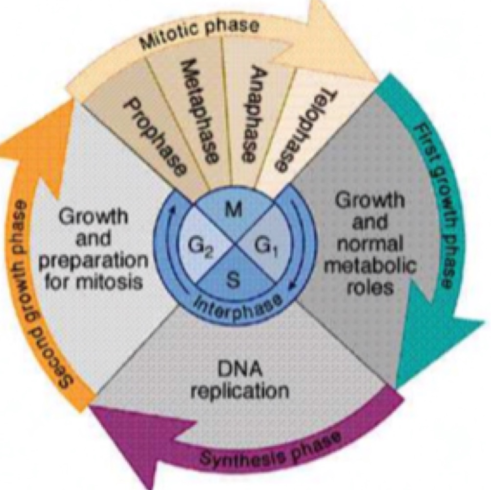
The cell cycle is the series of events that takes place in the cell as it grows and divides. Its duration is 15 to 20 h and it has 2 phases: mitosis, the M phase, lasting 1 h, and interphase, everything else.

Interphase is G₁, presynthetic, about 10 h and the longest period; S, synthetic, 8 h, when DNA is replicated; and G₂, postsynthetic, 2 to 4 h, preparing for mitosis. G₀ is a specialised resting stage in which cells exit the cell cycle, permanently in neurons and heart muscle cells.

Three checkpoints halt progression until conditions are favourable: G₁, G₂ and M.

Mitosis has four stages: prophase, metaphase, anaphase, telophase, followed by cytokinesis. Endomitosis and amitosis are two special forms.
1. The cell cycle

note

The cell cycle is a series of events that takes place in the cell as it grows and divides.

  • Its duration is 15 to 20 h.
  • It has 2 phases: mitosis, the M phase, lasting 1 h, and interphase.

The phases of interphase

note

PhaseDurationWhat happens
G₁, presyntheticabout 10 hTime gap between mitosis and DNA replication. The longest period: the cell grows, and protein synthesis occurs
S, synthetic8 hSynthesis phase: DNA replication, and the beginning of centrosome duplication
G₂, postsynthetic2 to 4 hTime gap between DNA replication and mitosis. Preparation for mitosis: proteins required for mitosis accumulate

G₀

note

G₀ phase is a specialised resting stage, an inactive stage in which cells exit the cell cycle when they are not actively preparing to divide.

It is usually a temporary stage, but some cells remain permanently: neurons and heart muscle cells.

That permanence has a consequence worth carrying forward. A tissue whose cells sit in G₀ for good cannot replace what it loses, which is why damage to the heart and to nerve tissue does not heal the way damage to skin does.

Diagram of the cell cycle showing the mitotic phase with prophase, metaphase, anaphase and telophase, and interphase divided into the first growth phase, DNA replication in the synthesis phase, and the second growth phase preparing for mitosis

2. Cell cycle checkpoints

note

Checkpoints halt progression of the cell cycle to the next stage until conditions are favourable. There are three.

The G₁ checkpoint evaluates damage to DNA and other external factors. If conditions are unfavourable, the cell is not allowed to continue to S phase.

The G₂ checkpoint ensures all chromosomes have been replicated, and that the replicated DNA is not damaged, before entering mitosis.

The M checkpoint controls whether all sister chromatids are correctly attached to spindle microtubules before entering the irreversible anaphase stage.

The word irreversible is what makes the M checkpoint matter. Once anaphase begins there is no undoing it, so everything has to be verified beforehand.

Promoters

note

Cyclin is any of a group of proteins that regulates the cell cycle by forming a complex of kinases.

3. Mitosis

note

Cell division is the process in which a parent cell divides into two or more daughter cells.

Mitosis, the M phase, is the cell cycle of somatic cells. It has four stages.

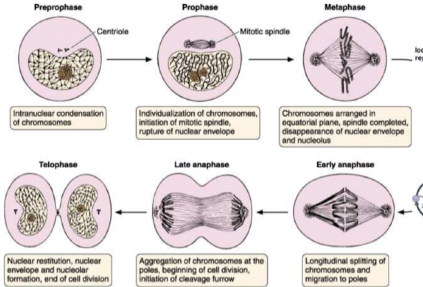
Diagram of the stages of mitosis showing preprophase, prophase, metaphase, early anaphase, late anaphase and telophase, with the centriole, mitotic spindle, chromosomes at the equatorial plane and the cleavage furrow

Prophase

note

  • Rupture of the nuclear envelope.
  • Replicated chromatin condenses into chromosomes.
  • 2 centrioles duplicate, get separated and move to opposite poles, to organise the mitotic spindle.

Metaphase

note

  • Chromosomes condense further, becoming much thicker.
  • They are arranged in the equatorial plane, and the cell is now more spherical.
  • Nucleolus and nuclear envelope disappear.
  • The spindle is completed and binds to kinetochores.

Anaphase

note

Sister chromatids are pulled toward opposite spindle poles by microtubules.

Telophase

note

Reconstruction.

  • Chromosomes revert to an uncondensed state.
  • Microtubules depolymerize.
  • Nuclear envelope and nucleolar formation.

Cytokinesis

note

A belt-like contractile ring of actin filaments with myosins develops and creates a cleavage furrow.

It progresses until the cytoplasm and its organelles are divided into two daughter cells, each with one nucleus.

4. Endomitosis

note

Endomitosis is a special form of mitosis, seen in megakaryocytes.

  • Replication of chromatids without cell division.
  • Chromosomes separate without spindle apparatus.
  • Result: a big cell with a lot of chromatids.

5. Amitosis

note

Amitosis is a special form of mitosis, seen in osteoclasts.

It is cell division by division of cytoplasm without spindle formation, without the appearance of chromosomes, and without breakdown of the nuclear envelope.

The two special forms are opposite failures of the ordinary process: endomitosis copies the chromosomes without dividing the cell, and amitosis divides the cell without properly handling the chromosomes.

All 95 Cytology topics · exam study support, not clinical guidance. SuperMed is not affiliated with the Medical University of Sofia.

Start free. No card needed.

Every account starts free, with free topics in Cytology and Medical Biology. Super opens the rest. Super is €15 a month.

Create a free account